Article
J. Braz. Chem. Soc., Vol. 26, No. 7, 1450-1456, 2015. Printed in Brazil - ©2015 Sociedade Brasileira de Química 0103 - 5053 $6.00+0.00
A
*e-mail: [email protected]
Ursane Saponins from the Stems of
Firmiana simplex
and their Cytotoxic Activity
Kyeong Wan Woo,a Sang Un Choi,b Ki Hyun Kima and Kang Ro Lee*,a
aNatural Products Laboratory, School of Pharmacy, Sungkyunkwan University, 2066 Seobu-Ro, Jangan-ku, Suwon, Gyeonggi-do, Republic of Korea
bKorea Research Institute of Chemical Technology, 305-343 Daejeon, Republic of Korea
Three new ursane triterpene saponins, together with twelve known ursane triterpenes were isolated from the stems of Firmiana simplex. The structures of the saponineswere elucidated on the basis of spectroscopic and chemical methods. The cytotoxic activity of all compoundswas evaluated in vitro against lung adenocarcinoma (A549), ovarian cancer (SK-OV-3), skin melanoma (SK-MEL-2), and colon cancer (HCT-15) human cell lines, using a sulforhodamine (SRB) assay. 23-Hydroxyursolic acidshowed cytotoxicity against the tested cell lines with IC50 values ranging
from 11.96 to 14.11 µM.
Keywords: Firmiana simplex, Sterculiaceae, ursane triterpene saponin, cytotoxicity
Introduction
Firmiana simplex W. F. Wight (synonym Firmiana platanifolia Schott et Endl), family of Sterculiaceae, known as “phoenix tree”, is distributed throughout Korea and China.1,2 Its seeds have been used as a folk medicine to treat symptoms of diarrhea and stomach disorders.3 Previous chemical investigations on this plant reported the isolation of quinones,2 flavonoids,3 and an antipsychotic neolignan.4
In the course of our continuing search for potential lead compounds from Korean traditional medicinal plants, we investigated the MeOH extract of F. simplex stems and isolated three new ursane triterpene saponins (1-3), together with twelve known ursane triterpenes (4-15) (Figure 1). All the compounds (1-15) were tested for their cytotoxic activity against the cultured human tumor cell lines lung adenocarcinoma (A549), ovarian cancer (SK-OV-3), skin melanoma (SK-MEL-2), and colon cancer (HCT-15).
Experimental
General procedures
Optical rotations were obtained on a JASCO P-1020 polarimeter. Infrared (IR) spectra were recorded on a Bruker Vector 22 IR spectrophotometer. Nuclear magnetic resonance (NMR) spectra including 1H-1H correlation spectroscopy
(COSY), distortionless enhancement by polarization transfer (DEPT), heteronuclear multiple quantum coherence (HMQC), heteronuclear multiple-bond correlation (HMBC) and nuclear Overhauser effect spectroscopy (NOESY), were recorded on a Varian UNITY INOVA 700 spectrometer operating at 700 MHz (1H) and 175 MHz (13C). High resolution fast atom bombardment mass spectrometry (HR-FABMS) was conducted using a JEOL JMS700 mass spectrometer. Preparative high performance liquid chromatography (HPLC) was performed using a Gilson 306 pump with a Shodex refractive index detector. Silica gel 60 (230-400 mesh, Merck) and reversed-phase (RP)-C18 silica gel (230-400 mesh, Merck) were used for column chromatography. A Hewlett-Packard gas chromatography (GC) system 6890 Series was equipped with a 5973 mass selective detector (MSD). The system was controlled by the Enhanced Chem Station version B.01.00 program. The capillary column used for GC was an Agilent J&W HP-5MSUI (30.0 m × 0.25 mm i.d., 0.25 µm film thickness, coated with 5% diphenyl and 95% dimethylpolysiloxane). Thin-layer chromatography (TLC) was performed using Merck precoated silica gel F254 plates and RP-18 F254s plates. Spots were detected on TLC under ultraviolet (UV) light or by heating after spraying with 10% v/v H2SO4 in EtOH.
Plant material
authenticated by one of the authors (K. R. Lee). A voucher specimen (SKKU-NPL-1209) was deposited at the herbarium of the School of Pharmacy, Sungkyunkwan University, Suwon, Korea.
Extraction and isolation
The stems of F. simplex (7.0 kg) were extracted with 80% MeOH under reflux. The filtered MeOH extract was concentrated under reduced pressure to afford a viscous concentrate (400 g), which was suspended in water (800 mL) and solvent-partitioned successively to yield hexane (24 g), CHCl3 (14 g), EtOAc (50 g), and BuOH (270 g) extracts. The CHCl3 extract (14 g) was separated over a silica gel column (230-400 mesh, 500 g) with hexane:EtOAc:MeOH (5:1:0.5, v/v) to give five fractions (C1-C5). Fraction C3 (6.5 g) was separated on a RP-C18 silica gel column (230-400 mesh, 150 g) with a gradient solvent system of MeOH:H2O (2:3, 3:2, 4:1, and 1:0, v/v) to give sixteen subfractions (C3-1-C3-16). Fraction C3-12 (110 mg) was further separated over a silica gel column with CHCl3:MeOH (20:1, v/v) elution, and further purified
through RP-C18 silica gel semi-preparative HPLC with 40% CH3CN elution, at a flow rate of 2.0 mL min-1 (Econosil RP-18 column; 250 × 10 mm; 10 µm particle size; Shodex refractive index detector) to yield 7 (3 mg, tR = 14.3 min). Fraction C3-13 (470 mg) was further separated over a silica gel column with CHCl3:MeOH (20:1, v/v) elution, and purified through a RP-C18 silica gel semi-preparative HPLC with 60% CH3CN elution, to yield 6 (6 mg, tR = 17.6 min), 14 (12 mg, tR = 19.1 min), and 15 (7 mg, tR = 21.7 min). Fraction C3-16 (120 mg) was purified through RP-C18 silica gel semi-preparative HPLC with 70% CH3CN elution, to yield 12 (6 mg, tR = 12.1 min). The EtOAc layer (18 g) was chromatographed on a RP-C18 silica gel (230-400 mesh, 300 g), eluting with a gradient solvent system of MeOH:H2O (2:3, 3:2, 4:1, and 1:0, v/v) to yield eight subfractions (E1-E8). Fraction E3 (1.9 g) was separated over a Sephadex LH-20 column with MeOH:H2O (4:1, v/v), and purified through RP-C18 silica gel semi-preparative HPLC with 30 and 40% CH3CN elution, to yield 5 (12 mg, tR = 12.1 min), 8 (3 mg, tR = 14.5 min), 10 (14 mg, tR = 16.2 min), and 11 (12 mg, tR = 19.4 min). Fraction E4 (1.0 g) was separated over a Sephadex LH-20 column with MeOH:H2O (4:1, v/v) HO
OH
HO
HO
OH HO
R3 R4 HO
R1
R2
O
OR5
4R1=α-OH, R2=α-OH, R3= CH3, R4= CH3, R5=β-D-Glc
5R1=α-OH, R2=α-OH, R3= CH
3, R4= CH2OH, R5=β-D-Glc
6R1=α-OH, R2=α-OH, R3= CH
3, R4= CH3, R5= H
7R1=α-OH, R2=α-OH, R3= CH
3, R4= CH2OH, R5= H
8R1= H, R2=α-OH, R3= CH2OH, R4= CH3, R5=β-D-Glc
9R1=α-OH, R2=β-OH, R3= COOH, R4= CH3, R5=β-D-Glc
10R1= H, R2=β-OH, R3= CH
3, R4= CH3, R5=β-D-Glc
11R1=α-OH, R2=β-OH, R3= CH
3, R4= CH2OH, R5=β-D-Glc
OH O
OH O
14 15
1R1=α-OH, R2= CH3, R3= CH3
2R1=α-OH, R2= CH3, R3= CH2OH
3R1=β-OH, R2= COOH, R3= CH 3
1' R2 R3
HO
O
R1
O
O
O HOHO
OH
O
HO HO HO
OH 9 1
2
3 4 5
6 7 8 10
11 12
13
14
15 16 17 18 19
20 21 22
23 24
25 26
27
28 29
30
1'' HO
H
H
R2
OH R1
OR3 O
12R1= H, R2=β-OH, R3= H
13R1=α-OH, R2=α-OH, R3=β-D-Glc
H
H
and purified by RP-C18 silica gel semi-preparative HPLC with 40% CH3CN elution, to yield 9 (15 mg, tR = 11.8 min). Fraction E5 (250 mg) was separated over a Sephadex LH-20 column with MeOH:H2O (4:1, v/v) and purified by RP-C18 silica gel semi-preparative HPLC with 50% CH3CN elution, to yield 4 (5 mg, tR = 15.5 min). Fraction E6 (230 mg) was separated over a Sephadex LH-20 column with MeOH:H2O (4:1, v/v), and purified by RP-C18 silica gel semi-preparative HPLC with 60% CH3CN elution, to yield 13 (5 mg, tR = 14.7 min). The BuOH extract (30 g) was separated over a silica gel column (230-400 mesh, 500 g) with CHCl3:MeOH (5:1, v/v), to give six fractions (B1-B6). Fraction B4 (8.7 g) was chromatographed on a RP-C18 silica gel eluting with a gradient solvent system of MeOH:H2O (3:7, 5:5, 7:3, and 1:0, v/v) to yield eight subfractions (B41-B48). Fraction B45 (200 mg) was purified by RP-C18 silica gel semi-preparative HPLC with 25% CH3CN elution, to yield 2 (15 mg, tR = 16.1 min). Fraction B47 (700 mg) was separated over a Sephadex LH-20 column with MeOH:H2O (4:1, v/v), and purified by RP-C18 silica gel semi-preparative HPLC with 30% CH3CN elution, to yield 1 (19 mg, tR = 22.7 min). Fraction B5 (16.4 g) was chromatographed on a RP-C18 silica gel eluting with a gradient solvent system of MeOH:H2O (2:3, 3:2, 4:1, and 1:0, v/v) to yield seven subfractions (B51-B57). Fraction B54 (700 mg) was separated over a Sephadex LH-20 column with MeOH:H2O (4:1, v/v) and purified by RP-C18 silica gel semi-preparative HPLC with 25% CH3CN elution, to yield 3 (90 mg, tR = 18.7 min).
28-O-[β-D-Glucopyranosyl-(1→6)-β
-D-glucopyranosyl]-2α,3α,19α-trihydroxy-12-en-28-ursolic acid (1)
White gum; [α]D25 –2.0 (c 0.5, MeOH); IR (KBr) vmax / cm-1 3385, 2938, 2879, 2843, 1732, 1651, 1454, 1390, 1228, 1205, 1166, 1062, 637; 1H NMR (700 MHz, CD
3OD), see Table 1; 13C NMR (175 MHz, CD
3OD), see Table 2; HRMS-FAB [M–H]– calcd. for C
42H67O15: 811.4474; found: 811.4474.
28-O-[β-D-Glucopyranosyl-(1→6)-β -D-glucopyranosyl]-2α,3α,19α,23-tetrahydroxy-12-en-28-ursolic acid (2)
White gum; [α]D
25 –1.0 (c 0.6, MeOH); IR (KBr)
vmax / cm-1 3376, 2939, 2835, 1731, 1600, 1453, 1382, 1265, 1164, 1032, 637; 1H NMR (700 MHz, CD
3OD), see Table 1; 13C NMR (175 MHz, CD
3OD), see Table 2; HRMS-FAB [M–H]– calcd. for C
42H67O16: 827.4424; found: 827.4423.
28-O-[β-D-Glucopyranosyl-(1→6)-β -D-glucopyranosyl]-2α,3β,19α-trihydroxyurs-12-ene-24,28-dioic acid (3)
White gum; [α]D
25 + 0.2 (c 0.9, MeOH); IR (KBr)
vmax / cm-1 3366, 2935, 2839, 1732, 1695, 1454, 1380, 1263,
1228, 1167, 1032, 646; 1H NMR (700 MHz, CD 3OD), see Table 1; 13C NMR (175 MHz, CD
3OD), see Table 2; HRMS-FAB [M–H]– calcd. for C
42H65O17: 841.4216; found: 841.4216.
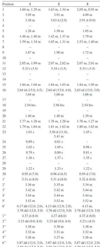
Table 1.1H NMR data in CD
3OD for compounds 1-3 (d in ppm (J in Hz))
Position 1 2 3
1 1.60 m, 1.29 m 1.63 m, 1.34 m 2.05 m, 0.93 m
2 3.95 m 3.91 m 4.09 m
3 3.38 m 3.63 d (2.0) 2.91 d (9.0)
4 – – –
5 1.28 m 1.58 m 1.05 m
6 1.46 m, 1.40 m 1.42 m, 1.37 m 1.79 m 7 1.59 m, 1.34 m 1.65 m, 1.31 m 1.53 m, 1.40 m
8 – – –
9 1.87 m 1.90 m 1.72 m
10 – – –
11 2.05 m, 1.99 m 2.07 m, 2.02 m 2.07 m, 2.01 m 12 5.33 t (3.5) 5.34 t (3.5) 5.33 t (3.5)
13 – – –
14 – – –
15 1.84 m, 1.04 m 1.84 m, 1.03 m 1.84 m, 1.05 m 16 2.64 td (13.0, 4.5),
1.64 m
2.64 td (13.0, 4.0), 1.66 m
2.63 td (13.0, 3.0), 1.66 m
17 – – –
18 2.54 brs 2.56 brs 2.54 brs
19 – – –
20 1.40 m 1.40 m 1.39 m
21 1.77 m, 1.28 m 1.76 m, 1.28 m 1.76 m, 1.27 m 22 1.79 m, 1.64 m 1.81 m, 1.62 m 1.80 m, 1.63 m 23 1.01 s 3.56 d (11.0),
3.41 m
1.45 s
24 0.89 s 0.81 s
-25 1.02 s 1.05 s 0.98 s
26 0.79 s 0.80 s 0.81 s
27 1.36 s 1.37 s 1.35 s
28 – – –
29 1.22 s 1.23 s 1.22 s
30 0.95 d (7.0) 0.96 d (6.5) 0.95 d (7.0) 1’ 5.31 d (8.0) 5.31 d (8.0) 5.32 d (8.0)
2’ 3.36 m 3.35 m 3.34 m
3’ 3.42 m 3.42 m 3.44 m
4’ 3.44 m 3.44 m 3.44 m
5’ 3.52 m 3.52 m 3.52 m
6’ 4.17 dd (12.0, 2.0), 3.78 dd (12.0, 5.0)
4.13 dd (12.0, 2.0), 3.78 dd (12.0, 5.0)
4.13 m, 3.78 dd (12.0, 5.0) 1’’ 4.37 d (8.0) 4.37 d(8.0) 4.37 d (8.0) 2’’ 3.23 dd (9.0, 8.0) 3.23 dd (9.0, 8.0) 3.23 t (8.5)
3’’ 3.38 m 3.38 m 3.38 m
4’’ 3.32 m 3.31 m 3.32 m
5’’ 3.26 m 3.27 m 3.26 m
6’’ 3.87 dd (12.0, 2.0), 3.69 dd (12.0, 5.0)
3.87 dd (12.0, 2.0), 3.69 dd (12.0, 5.0)
Acid hydrolysis of 1-3 and sugar determination
Compound 1 (2 mg) was shaken with 1 mL of 1 mol L-1 HCl for 1 h at 90 oC. After cooling, the hydrolyzate was extracted with CHCl3 and the extract was evaporated in vacuo to yield 2α,3α,19α-trihydroxyurs-12-en-28-oic acid (1a), which was identified by comparing its 1H NMR data with those reported in literature. The sugar in water
layer appeared to be glucose by co-TLC comparison (CHCl3:MeOH:H2O = 2:1:0.2, Rf value: 0.2) with a glucose standard (Aldrich), which was confirmed by gas chromatography-mass spectrometry (GC-MS) as follows. The sugars obtained from the hydrolysis of compounds 1-3 were dissolved in anhydrous pyridine (0.1 mL) and L-cysteine methyl ester hydrochloride (2 mg) was added. The mixture was stirred at 60 oC for 1.5 h. After the reaction mixture was dried in vacuo, the residue was trimethylsilylated with 1-trimethylsilylimidazole (0.1 mL) for 2 h. The mixture was partitioned between hexane and H2O (0.3 mL each). The H2O layer was neutralized by passage through an Amberlite IRA-67 column (Rohm and Haas) and was repeatedly evaporated to give D-glucose, identified by co-injection of the hydrolyzate with standard silylated samples, giving a GC-MS single peak at 9.712 min. Compounds 2 (2 mg) and 3 (5 mg) were treated using the same method to give 2α,3α,19α ,23-tetrahydroxyurs-12-en-28-oic acid (2a) and 2α,3β,19α -trihydroxy-urs-12-ene-24,28-dioic acid (3a).
2α,3α,19α-Trihydroxyurs-12-en-28-oic acid (1a) Colorless gum; 1H NMR (700 MHz, pyridine-d
5) d 5.55 (brs, 1H, CH), 4.27 (dt, 1H, J 10.0, 3.5 Hz, CHOH), 3.72 (d, 1H, J 2.5 Hz, CHOH), 3.11 (ddd, 1H, J 13.5, 13.0, 4.5 Hz, CH2), 3.01 (s, 1H, CH), 2.29 (ddd, 1H, J 13.5, 13.0, 4.0 Hz, CH2), 1.59 (s, 3H, CH3), 1.38 (s, 3H, CH3), 1.22 (s, 3H, CH3), 1.07 (d, 3H, J 6.0 Hz, CH3), 1.05 (s, 3H, CH3), 0.94 (s, 3H, CH3), 0.85 (s, 3H, CH3).
2α,3α,19α,23-Tetrahydroxyurs-12-en-28-oic acid (2a) Colorless gum; 1H NMR (700 MHz, CD
3OD) d5.31 (brs, 1H, CH), 3.89 (ddd, 1H, J 12.0, 5.0, 3.0 Hz, CHOH), 3.62 (d, 1H, J 3.0 Hz, CHOH), 3.55 (d, 1H, J 11.0 Hz, CHOH), 3.41 (d, 1H, J 11.0 Hz, CHOH), 2.58 (td, 1H,
J 13.0, 4.0 Hz, CH2), 2.53 (s, 1H, CH), 1.37 (s, 3H, CH3), 1.21 (s, 3H, CH3), 1.04 (s, 3H, CH3), 0.94 (s, 3H, CH3), 0.94 (d, 3H, J 7.0 Hz, CH3), 0.88 (s, 3H, CH3).
2α,3β,19α-Trihydroxy-urs-12-ene-24,28-dioic acid (3a) Colorless gum; 1H NMR (700 MHz, CD
3OD) d 5.51 (brs, 1H, CH), 4.69 (m, 1H, CHOH), 3.34 (d, 1H, J 9.0 Hz, CHOH), 3.02 (m, 1H, CH2), 2.33 (dd, 1H, J 13.0, 4.0 Hz, CH2), 1.68 (s, 3H, CH3), 1.65 (s, 3H, CH3), 1.39 (s, 3H, CH3), 1.11 (s, 3H, CH3), 1.06 (d, 3H, J 6.0 Hz, CH3), 1.05 (s, 3H, CH3).
Cytotoxicity assays
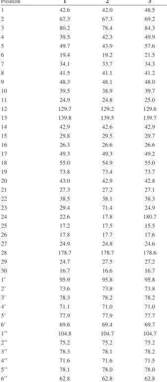
A sulforhodamine (SRB) bioassay was used to determine compound cytotoxicity against cultured human Table 2.13CNMR data in CD
3OD for compounds 1-3 (d in ppm)
Position 1 2 3
1 42.6 42.0 48.5
2 67.3 67.3 69.2
3 80.2 78.4 84.3
4 39.5 42.3 49.9
5 49.7 43.9 57.6
6 19.4 19.2 21.5
7 34.1 33.7 34.3
8 41.5 41.1 41.2
9 48.3 48.1 48.0
10 39.5 38.9 39.7
11 24.9 24.8 25.0
12 129.7 129.2 129.6
13 139.8 139.5 139.7
14 42.9 42.6 42.9
15 29.8 29.5 29.7
16 26.3 26.6 26.6
17 49.3 49.3 49.2
18 55.0 54.9 55.0
19 73.8 73.4 73.7
20 43.0 42.9 42.8
21 27.3 27.2 27.1
22 38.5 38.1 38.3
23 29.4 71.4 24.9
24 22.6 17.8 180.7
25 17.2 17.5 15.5
26 17.8 17.7 17.6
27 24.9 24.8 24.6
28 178.7 178.7 178.6
29 24.7 27.5 27.2
30 16.7 16.6 16.7
1’ 95.9 95.8 95.8
2’ 73.6 73.8 73.8
3’ 78.3 78.2 78.2
4’ 71.1 71.0 71.0
5’ 77.9 77.9 77.7
6’ 69.6 69.4 69.7
1’’ 104.8 104.7 104.7
2’’ 75.2 75.2 75.2
3’’ 78.3 78.1 78.2
4’’ 71.6 71.6 71.5
5’’ 78.1 78.0 78.0
tumor cell lines A549, SK-OV-3, SK-MEL-2, and HCT-15.5 The assays were performed at the Korea Research Institute of Chemical Technology. Doxorubicin was used as a positive control. The IC50 values of doxorubicin against A549, SK-OV-3, SK-MEL-2, and HCT-15 cell lines were 0.029, 0.036, 0.001, and 2.041 µM, respectively.
Results and Discussion
The stems of F. simplex were extracted with 80% aqueous MeOH. Chemical investigation of the extract using successive column chromatography over silica gel and Sephadex LH-20, and preparative HPLC resulted in the isolation and identification of three new ursane triterpene saponins (1-3), together with twelve known ursane triterpenes (4-15). Their structures were elucidated as follows.
Compound 1 was obtained as a colorless gum, and its molecular formula C42H67O15 was inferred from the negative HR-FABMS ion at m/z 811.4474 [M–H]–. The IR absorption bands at 3385 and 1732 cm-1 implied the presence of hydroxyl and carboxylic functionalities. The 1H NMR spectrum (Table 1) of 1 showed the signals for
an olefinic proton at dH 5.33 (t, 1H, J 3.5 Hz, H-12), two oxygenated methine protons at dH 3.95 (m, 1H, H-2) and 3.38 (m, 1H, H-3), one methine proton at dH 2.54 (brs, 1H, H-18), six tertiary methyl protons at dH 1.36 (s, 3H, H-27), 1.22 (s, 3H, H-29), 1.02 (s, 3H, H-25), 1.01 (s, 3H, H-23), 0.89 (s, 3H, H-24) and 0.79 (s, 3H, H-26), one secondary methyl proton at dH 0.95 (d, 1H, J 7.0 Hz, H-30), and two anomeric protons at dH 5.31 (d, 1H, J 8.0 Hz, H-1’) and 4.37 (d, 1H, J 8.0 Hz, H-1’’). The 13C NMR (Table 2), DEPT, and HMQC spectral data revealed forty-two signals, which included seven methyl carbon signals at dC 29.4
(C-23), 24.9 (C-27), 24.7 (C-29), 22.6 (C-24), 17.8 (C-26), 17.2 (C-25), and 16.7 (C-30), two olefinic carbon signals at dC 139.8 (C-13) and 129.7 (C-12), two oxygenated methine carbon signals at dC 80.2 (C-3) and 67.3 (C-2), eight methylene carbon signals at dC 42.6 (C-1), 38.5 (C-22), 34.1 (C-7), 29.8 (C-15), 27.3 (C-21), 26.3 (C-16), 24.9 (C-11), and 19.4 (C-6), four methine carbon signals at dC 55.0 (C-18), 49.7(C-5), 48.3 (C-9), and 43.0 (C-20), one carbonyl carbon at d 178.7 (C-28), six quaternary carbon signals at dC 73.8 (C-19), 49.3 (C-17), 42.9 (C-14), 41.5 (C-8), and 39.5 (C-4), and two anomeric carbons at
dC 104.8 (C-1’’) and 95.9 (C-1’).
The NMR data of 1 were very similar to those of the ursane 4,6 with the exception of an additional sugar moiety in 1. The linkage of the disaccharide moiety to the pentacyclic scaffold, and the attachment position between the two sugar units were assigned from the following HMBC correlations: dH 4.37 (d, J 8.0 Hz, H-1’’) to dC 69.6 (C-6’), and dH 5.31 (d, J 8.0 Hz, H-1’) to
dC 178.7 (C-28) (Figure 2).
The relative stereochemistry of the aglycone was assigned from the NOESY cross-peaks of H-2/H-25, H-3/H-24, H-5/H-9, H-9/H-27, H-24/H-25, and H-25/H-26 (Figure 3).6
The coupling constant (J 8.0 Hz) of the anomeric protons of H-1’ and H-1’’ suggested a β-orientation.7 Acid hydrolysis of 1 gave 2α,3α,19α-trihydroxyurs-12-en-28-oic acid (1a), identified by comparison of its 1H NMR spectrum data with of previously reported values,8 and D-glucose, which was identified by GC-MS.9 Thus, compound 1 was determined to be 28-O-[β-D-glucopyranosyl-(1→6)-β -D-glucopyranosyl]-2α,3α,19α-trihydroxy-12-en-28-ursolic acid.
Compound 2 was obtained as a colorless gum, and its molecular formula C42H67O16 was inferred from the negative
HO
O
HO
O
O
O HOHO
OH
O HO
HOHO OH HO
HO
O
HO
O
O
O HOHO
OH
O HO
HOHO OH HO
HO
O
HO
O
O
O HOHO
OH
O HO
HOHO OH HO
OH
1 2 3
O OH
COSY HM BC
HR-FABMS ion at m/z 827.4423 [M–H]–. The 1H and 13C NMR spectra were close to those of 1 (Tables 1 and 2).
The major differences were the disappearance of a methyl signal [dH 1.01 (s, 3H, H-23); dC 29.4] in 1, and the presence of the oxymethylene signal [dH 3.56 (d, 1H, J 11.0 Hz, H-23a), 3.41 (m, 1H, H-23b); dC 71.4] in 2. This was confirmed by the HMBC experiment showing correlations from the oxymethine proton (dH 3.56) to C-3, C-4, C-5, and C-24. The nature and position of the disaccharide moiety revealed to be the same as for compound 1, as indicated by the HMBC correlations H-1’’/C-6’ and H-1’/C-28 (Figure 2). As for compound 1, the relative configuration of 2 determined by the NOESY spectrum also indicated an ursane pentacyclic system. Acid hydrolysis of 2 gave 2α,3α,19α,23-tetrahydroxyurs-12-en-28-oic acid (2a) and D-glucose, which were identified by GC analysis and TLC comparison with authentic D-glucose.9,10 Thus, compound 2 was determined to be 28-O-[β-D-glucopyranosyl-(1→
6)-β-D-glucopyranosyl]-2α,3α,19α ,23-tetrahydroxy-12-en-28-ursolic acid.
Compound 3 was obtained as a colorless gum. The molecular formula was determined to be C42H65O17 from the deprotonated molecule [M–H]– at m/z 841.4216 in the negative-ion HR-FABMS data. The 1H and 13C NMR data of 3 were very similar to those reported for 9,11 except for the presence of an additional sugar unit The connectivities of the two sugar units were deduced by the HMBC cross peaks H-1’’/C-6’ and H-1’/C-28 (Figure 2). The relative stereochemistry of 3 was assigned by NOESY cross-peaks H-2/H-25, H-3/H-23, H-5/H-9, H-9/H-27, and H-25/H-26. Acid hydrolysis of 3 yielded 2α,3β,19α-trihydroxyurs-12-ene-24,28-dioic acid (3a) and D-glucose.9,12 Thus, compound 3 was determined to be 28-O-[β-D-glucopyranosyl-(1→6)-β -D-glucopyranosyl]-2α,3β,19α-trihydroxyurs-12-ene-24,28-dioic acid.
Compounds 4-15 were identified by comparing their 1H NMR, 13C NMR, and MS spectra with the literature
data. They were determined to be kaji-ichigoside F1 (4),6
niga-ichigoside F2 (5),13 euscaphicacid (6),14 myrianthic acid (7),15 kakisaponin A (8),16 trachelosperoside A-1 (9),12 pormolic acid-28-O-β-D-glucopyranosyl ester (10),17 niga-ichigoside F1 (11),13 23-hydroxyursolic acid (12),18 2α,3α,24-trihydroxyurs-12-en-28-oic acid-28-O-β -D-glucopyranosyl ester (13),19 arjunolic acid (14),20 and 2α,3α,23-trihydroxyursa-12,20(30)-dien-28-oic acid (15).21
Compounds 1-15 were evaluated for their cytotoxicity against A549, SK-OV-3, SK-MEL-2, and HCT-15 human tumor cell lines using the SRB assay.5 Compound 12 was cytotoxic against all the tested human cell lines with IC50 values of 11.96, 13.24, 14.11, and 12.27 µM, respectively, whereas the other compounds were inactive (IC50 > 30 µM).
Conclusions
This is the first study investigating the cytotoxic activities of triterpene derivatives (1-15) isolated from
Firmiana simplex. Among them, compound 12, which showed a significant cytotoxicity against the human tumor cell lines, could be a potentially valuable source for the development of anti-tumor agents.
Supplementary Information
Supplementary data are available free of charge at http:// jbcs.sbq.org.br as a PDF file.
Acknowledgments
This research was supported by the Basic Science Research Program through the National Research Foundation of Korea (NRF), funded by the ministry of Education, Science and Technology (2013R1A1A2A10005315). We are thankful to the Korea Basic Science Institute (KBSI) for the measurements of NMR and MS spectra.
References
1. Zhang, L. X.; Song, J. H.; Shen, J. T.; Tan, G. J.; Li, S. S.; Ding, F.; J. Phytopathol. 2013, 161, 128.
2. Bai, H.; Li, S.; Yin, F.; Hu, L.; J. Nat. Prod. 2005, 68, 1159. 3. Seetharaman, T. R.; Fitoterapia 1990, 61, 373.
4. Son, Y. K.; Lee, M. H.; Han, Y. N.; Arch. Pharmacal Res. 2005, 28, 34.
5. Skehan, P.; Storeng, R.; Scudiero, D.; Monks, A.; Mcmahon, J.; Vistica, D.; Warren, J. T.; Bokesch, H.; Kenney, S.; Boyd, M. R.; J. Natl. Cancer Inst. 1990, 82, 1107.
6. Yean, M. H.; Kim, J. S.; Hyun, Y. J.; Hyun, J. W.; Bae, K.; Kang, S. S.; Nat. Prod. Sci. 2012, 43, 107.
Figure 3. Key NOESY correlations of compound 1. H
OH HO
H
H
H
H
1
NOESY
HO O
H
7. Woo, K. W.; Moon, E. J.; Park, S. Y.; Kim, S. Y.; Lee, K. R.; Bioorg. Med. Chem. Lett. 2012, 22, 7465.
8. Lee, I. K.; Kim, D. H.; Lee, S. Y.; Kim, K. R.; Choi, S. U.; Hong, J. K.; Lee, J. H.; Park, Y. H.; Lee, K. R.; Arch. Pharmacal Res. 2008, 31, 1578.
9. Lee, I. K.; Choi, S. U.; Lee, K. R.; Chem. Pharm. Bull. 2012, 60, 1011.
10. Kim, K. H.; Choi, S. U.; Lee, K. R.; J. Nat. Prod. 2009, 72, 1121.
11. Wang, B. G.; Shen, X. M.; Yang, L.; Jia, Z. J.; Phytochemistry
1997, 46, 559.
12. Abe, F.; Yamauchi, T.; Chem. Pharm. Bull. 1987, 35, 1748. 13. Um, B. H.; Pouplin, T.; Lobstein, A.; Weniger, B.; Litaudon, M.;
Anton, R.; Fitoterapia 2001, 72, 591.
14. Woo, K. W.; Han, J. Y.; Choi, S. U.; Kim, K. H.; Lee, K. R.; Nat. Prod. Sci. 2014, 20, 71.
15. Wandji, J.; Tillequin, F.; Mulholland, D. A.; Shirri, J. C.; Tsabang, N.; Seguin, E.; Verite, P.; Libot, F.; Fomum, Z. T.; Phytochemistry 2003, 64, 845.
16. Chen, G.; Xue, J.; Xu, S. X.; Zhang, R. Q.; J. Asian Nat. Prod. Res. 2007, 9, 347.
17. Wu, Z. J.; Ouyang, M. A.; Wang, C. Z.; Zhang, Z. K.; Shen, J. G.; J. Agric. Food Chem. 2007, 55, 1712.
18. An, R. B.; Na, M. K.; Min, B. S.; Lee, H. K.; Bae, K. H.; Nat. Prod. Sci. 2008, 14, 249.
19. Jung, H. A.; Chung, H. Y.; Jung, J. H.; Choi, J. S.; Chem. Pharm. Bull. 2004, 52, 157.
20. Acebey-Castellon, I. L.; Voutquenne-Nazabadioko, L.; Huong, D. T. M.; Roseau, N.; Bouthagane, N.; Muhammad, D.; Le Magrex Debar, E.; Gangloff, S. C.; Litaudon, M.; Sevenet, T.; Nguyen, V. H.; Lavaud, C.; J. Nat. Prod. 2011, 74, 163. 21. Choi, Y. H.; Zhou, W.; Oh, J.; Choe, S.; Kim, D. W.; Lee, S. H.;
Na, M. K.; Bioorg. Med. Chem. Lett. 2012, 22, 6116.
Submitted: January 14, 2015