534
A 16-year-old male presented with a pain-less right testicular mass, known for the past 2 years and that had recently increased in size. Physical examination conirmed a hard testicular mass and no evidence of lymphadenopathy or of gynecomastia. Laboratory results, including tu-mor markers, were normal.Radiology Page International Braz J Urol
Testicular epidermoid cyst - Ultrasound and MR typical indings
with macroscopy correlation
L. Pires-Gonçalves, C. Silva, M. Teixeira, S. Costa-Dias, V. Sousa-Mendes
Department of Imagiology (LPG, SCD, VSM), Department of Urology (CS) and Department of Pathology (MT) of Hospital de Braga, of Hospital de Braga, Braga/Portugal
Vol 37 (4): 534-535, July - Augst, 2011
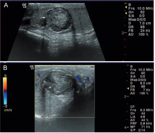
Figure 1 - a. Longitudinal US image of the right testis show a bilobulated mass in the upper medial aspect of the testis. The mass had sharp margins, composed of a central echogenic area and of alternating hyperechogenic and hypoechogenic rings in the periphery (onionskin sign), measuring 2.4 x 1.7 x 1.7 cm. b. Longitudinal Doppler-US image of the right testis demonstrates the absence of vascularization of the mass.
Ultrasound (US) revealed a solid mass in the upper pole of the right testis that was well circumscribed, avascular, and displayed the on-ionskin appearance suggestive of testicular epider-moid cyst (1,2) (Figure-1).
Magnetic resonance (MR) conirmed the well-circumscribed intra-testicular mass with a
A
535
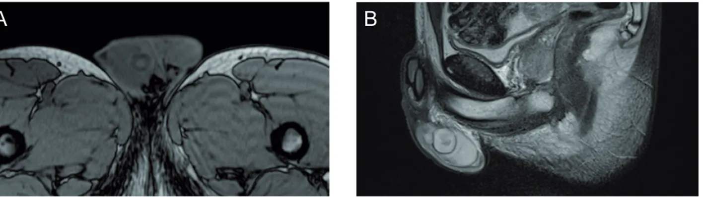
Radiology PageFigure 2 - a. Axial T2-weigthed image conirms the well-circumscribed mass in the upper medial pole of the right testis. The mass has a central slightly hyperintense center and an isointense periphery. b. Sagittal T2-weigthed image conirms the well-circumscribed mass in the upper medial pole of the right testis. The mass had a central slightly hypointense center and a hyperintense periphery.
Figure 3 - Photograph of the sectioned surgical specimen shows a bilobulated mass with a ibrous capsule that was composed of a laminated white-yellow paste like material, typical of keratin.
REFERENCES
1. Loya AG, Said JW, Grant EG: Epidermoid cyst of the testis: radiologic-pathologic correlation. Radio-graphics. 2004; 24(Suppl 1): S243-6.
2. Cho JH, Chang JC, Park BH, Lee JG, Son CH:
Sono-graphic and MR imaging indings of testicular epider -moid cysts. AJR Am J Roentgenol. 2002; 178: 743-8. 3. Heidenreich A, Engelmann UH, Vietsch HV,
Ders-chum W: Organ preserving surgery in testicular epi-dermoid cysts. J Urol. 1995; 153: 1147-50.
4. Dieckmann KP, Loy V: Epidermoid cyst of the tes-tis: a review of clinical and histogenetic consider-ations. Br J Urol. 1994; 73: 436-41.
_____________________
Correspondence address:
Dr. Lígia Pires-Gonçalves
Department of Imagiology, Hospital de Braga Largo Carlos Amarante, 2242
Braga, 4701-965, Portugal Fax: + 351 253 613-334
E-mail: [email protected] central area hyperintense on T1 and hypointense
on T2 and a peripheral area isointense on T1 and hyperintense on T2, displaying the target or bull’s eye appearance, typical of testicular epidermoid cyst (1,2) (Figure-2). The remaining testicular pa-renchyma was unremarkable.
Surgical enucleation of the right tes-ticular mass was performed (3). The pathologi-cal analysis of the surgipathologi-cal specimen confirmed the clinical and radiological proposed diagnosis
(1,2,4) (Figure-3). The postoperative follow-up was uneventful.
The case presented demonstrates the clini-cal, ultrasound, and MR indings diagnostic of tes -ticular epidermoid cyst with macroscopy correlation. The recognition of the typical imaging indings of testicular epidermoid cyst can suggest the pre-oper-ative diagnosis of this rare benign tumor thus allow-ing to considered organ-preservallow-ing surgery and its associated psychological and cosmetic beneits.