Latin American Consensus on the use of
transcranial Doppler in the diagnosis of brain death
INTRODUCTION
Transcranial Doppler (TCD) is a technique introduced by Aaslid in 1982,(1)
widely used at present to evaluate cerebral hemodynamics in patients with brain
injury.(2) his technique measures cerebral blood low velocity in the major
vessels of the base of the brain. In Latin America, transcranial Doppler has been used more frequently in recent years, including as an auxiliary technique in
the diagnosis of brain death (BD).(3) Because TCD is a non-invasive real-time
technique that is able to be performed at the bedside of the patient, it is ideal for serious, unstable, or diicult-to-transport patients. here are numerous studies that validate the usefulness of TCD in the diagnosis of cerebral circulatory
arrest, usually present in patients with BD.(4,5)
Consensus Group on Transcranial Doppler in the Diagnosis of Brain Death*
Transcranial Doppler evaluates cerebral hemodynamics in patients with brain injury and is a useful technical tool in diagnosing cerebral circulatory arrest, usually present in the brain-dead patient. his Latin American Consensus was formed by a group of 26 physicians experienced in the use of transcranial Doppler in the context of brain death. he purpose of this agreement was to make recommendations regarding the indications, technique, and interpretation of the study of transcranial ultrasonography in patients with a clinical diagnosis of brain death or in the patient whose clinical diagnosis presents diiculties; a working group was formed to enable further knowledge and to strengthen ties between Latin American physicians working on the same topic.
A review of the literature, concepts, and experiences were exchanged in two meetings and via the Internet. Questions about pathophysiology, equipment,
Conflicts of interest: None.
Submitted on November 26, 2013 Accepted on March 17, 2014
Corresponding author: Corina Puppo
Hospital de Clínicas - Departamento de Emergencia Avenida Italia s/n - Montevideo
Montevideo 11600 Uruguay
E-mail: [email protected]
Consenso Latinoamericano sobre el uso del Doppler transcraneal
en el diagnóstico de muerte encefálica
ABSTRACT
Keywords: Brain death/diagnosis; Brain death/ultrasonography; Ultrasonography, Doppler, transcranial; Consensus
techniques, indings, common problems, and the interpretation of transcranial Doppler in the context of brain death were answered. he basic consensus statements are the following: cerebral circulatory arrest is the inal stage in the evolution of progressive intracranial hypertension, which is visualized with transcranial Doppler as a “pattern of cerebral circulatory arrest”. he following are accepted as the standard of cerebral circulatory arrest: reverberant pattern, systolic spikes, and absence of previously demonstrated low. Ultrasonography should be used - in acceptable hemodynamic conditions - in the anterior circulation bilaterally (middle cerebral artery) and in the posterior (basilar artery) territory. If no ultrasonographic images are found in any or all of these vessels, their proximal arteries are acceptable to be studied to look for a a pattern of cerebral circulatory arrest.
he objective of this Latin American Consensus is to make recommendations regarding the indications, technique, and interpretation of the study of transcranial ultrasound in patients with a clinical diagnosis of BD and/or in patients whose clinical diagnoses present diiculties. However, protocols for the diagnosis of BD difer depending on the country or region of Latin America
(and the world)(6) with respect to the indications and
timing of certain conirmatory tests. herefore, we consider it useful to generate standardized recommendations regarding how to perform and analyze TCD to conirm the diagnosis of BD to improve the reproducibility and comparison between studies in a multicenter setting.
Another equally important goal was to form a working group that would further consolidate knowledge and constructive ties between Latin American doctors working on the same topic. Although the issue in pediatrics is similar, the consensus focused on adults.
METHODOLOGY
For the consensus process, we used a method based
on the “state of the art” Glaser approach.(7-10) A group
of doctors, predominantly neurointensivists and neurologists, with experience in the use of TCD in this clinical setting conducted a review of the literature. he group was heterogeneous, composed of physicians with more than 20 years of experience in TCD and with published research articles as well as young physicians with internship experience at referral medical centers.
A search was conducted in diferent electronic databases (SCOPUS, PubMed and LILACS). he terms used in the search strategy were as follows: brain death, cerebral circulatory arrest and transcranial Doppler. he search languages included Spanish, Portuguese, English, and French according to the knowledge of the participants.
In the irst step, 423 articles were found. Twenty-six doctors from eight Latin American countries with experience in this area reviewed these articles and categorized them according to their relevance. he bibliography was discussed and classiied via internet discussion.
Multiple tables and lists were shared via the internet, including the following: (a) A data table of the participants, including the physicians’ nationality, working hospital, specialty, previous experience in consensus, experience in literature searches, years of experience in TCD, and languages. (b) A list of bibliographic items, including a numbered list containing only the authors, titles, and sources (journal, year, and page number) of all the articles. According to the classiication of each participant, a
diferent color was given to each item according to their relevancy to the consensus, whether the abstract or the full article was available, and the language. In this manner, the bibliography was selected using (c) the chart features of each item; the original 423 articles were divided among participants into groups of 10 to 20 articles. he title, author, year, journal, and a summary of the article were included (classiied as revision, review, or clinical case. Additionally, the study design, number of patients studied, and relevance to the consensus were included).
he irst discussion meeting was conducted in Buenos Aires, Argentina in June 2012, in which the members of the consensus presented their indings and opinions. After the irst meeting, participants were divided into groups of 2-3 members; groups were formed based on diferent levels of experience. Each group focused on answering questions about relevant aspects of the topic, and the information was shared via the Internet. With the responses of each group and the results of the respective literature reviews, opinions, and suggestions, the consensus coordinator prepared an initial manuscript. he manuscript was discussed at a second meeting in Buenos Aires in June of 2013. Prior to this second meeting, a new search of the literature published in the period between the two meetings was performed, and 71 additional articles were reviewed.
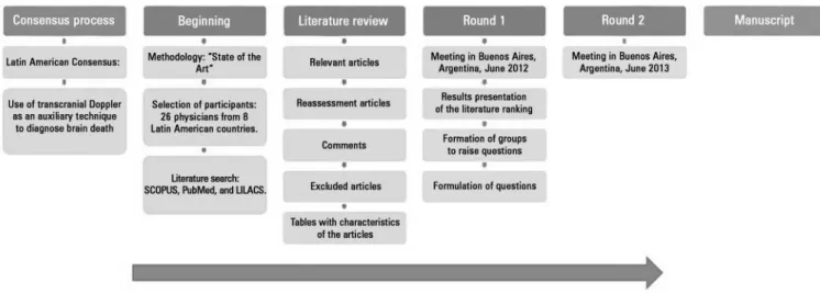
Figure 1 schematically depicts the evolution of the consensus. No other methodology was used, such as the Delphi method, because we wanted the consensus to focus on the Latin American reality and because another methodology would have involved the incorporation of the suggestions by experts outside of our region with diferent experiences. All consensus participants endorsed the recommendations presented in this paper. he following aspects were considered: pathophysiology, equipment, technique, indings, solutions to common problems, and interpretation.
RESULTS
1. Preliminary considerations: What is the alteration of cerebral blood flow in the brain-dead patient? Can cerebral blood flow be observed in a patient who is clinically brain-dead? Does the finding of preserved cerebral blood flow exclude the diagnosis of brain death? What is the pathophysiological significance of the pattern of cerebral circulatory arrest?
Figure 1 - Consensus process.
hypertension, decreased cerebral perfusion pressure, and global ischemia. However, it should be noted that there are cases of complete and irrecoverable absence of brain functions (i.e., brain dead) even in the presence of sustained cerebral blood low. A clear example is reperfusion in a patient who has sufered a heart attack and is resucitated too late. Under these circumstances, the patient’s brain has been irreversibly damaged by global ischemia. However, the low is restored, and for a time, persists. herefore, the discovery of low does not rule
out BD.(11) However, if cardiorespiratory function in this
patient is artiicially maintained, dead brain cells generate edema and intracranial hypertension, eventually leading to decreased cerebral perfusion pressure (CPP) and inally the absence of low. If the ultrasound study is repeated, eventually it will be possible see the pattern of cerebral circulatory arrest (Figure 2).
Cerebral circulatory arrest is the inal stage in the evolution of progressive untreated or refractory to treatment intracranial hypertension (ICH). he decrease in CPP is accompanied by characteristic changes. he normal sonographic pattern of the arteries of the brain stem is continuous low. he blood moves in a single direction within the vessel and is slowed during diastole and accelerated in systole without stopping (the low velocity never reaches zero) or reversing direction. If intracranial pressure begins to rise, resistance opposes CBF and slows its low. Flow initially slows more in diastole, which is the part of the cycle in which it moves with lower energy. he cardiogenic response is a stronger contraction,
Figure 2 - A 19-year-old patient with asthma who experienced a cardiac arrest during an asthma attack; she was resuscitated via CPR. Admission GCS was 4 with initial brainstem reflexes; over 12 days, the Doppler pattern deteriorated, with all clinical neurological activity disappearing. The initial sonogram revealed normal flow velocity, evolving to slowing velocity, and cerebral circulatory arrest. Tomographic image post-CPR demonstrates diffuse anterior ischemia, as noted by the contrast "white cerebellum".
which increases systolic velocity; coincidentally, this also increases the pulsatility index (PI). he formula is as follows: PI=(SV-DV)/MV, SV: systolic velocity; DV: diastolic velocity; MV: mean velocity. his phenomenon is gradually accentuated.
velocity and high pulsatility occupying the entire systole. When the ICP continues to rise and reaches the blood pressure (BP), diastolic low is reverberant, i.e., moving very slowly in the systolic phase and in reverse direction in the diastolic phase. When the ICP reaches the mean arterial pressure (MAP), there are systolic spikes. When the ICP reaches the systolic blood pressure, low is not
observed in the vessels.(12) Later, we will further address
the sonographic image of each pattern.
Some authors(13) disagree with the patterns of cerebral
circulatory arrest being called “low” patterns because they represent a ‘stagnant’ column, i.e., the blood does not “low”; the genesis of the reverberating patterns and systolic spikes should, according to this hypothesis, include the percussive efect of cardiac systole on a thrombosed blood column.
In a series of 40 patients, Poularas et al. conirmed 100% concordance between the cerebral angiography and
TCD in the diagnosis of cerebral circulatory arrest.(14)
he irst consensus on the usefulness of TCD in cerebral circulatory arrest, published in 1998, emphasized the value of this technique in cases where the existence of sedative drugs prevented the execution of an EEG for the
diagnosis of BD.(15) Petty published a study in 1990 with
suggestions on their value and performance as well as data
on sensitivity and speciicity.(16)
In 2000, an update was published deining the efectiveness of the technique in diferent disorders and included the diagnosis of cerebral circulatory arrest as
type B recommendation, class II evidence.(17) Since then,
there have been several publications, including the 2004 guidelines of the American Neurological Association (Sloan et al.), in which a type A Class II evidence recommendation
is made to conirm the diagnosis of BD by TCD.(3)
Regarding the speciicity and sensitivity of the method, there are techniques that compare TCD with other low surrogate methods that are related to the diagnosis of BD. he diagnosis of BD depends on which standard is used. In the case of Uruguay, where TCD is performed only if there is clinical diagnosis uncertainty (the most common use being the setting of depressant drugs, mainly thiopental), a CCA pattern was observed in 75% of 445
patients with clinical suspicion of BD.(18)
As will be discussed later in the text, a brain-dead patient may in certain circumstances exhibit sustained blood low. hus, the presence of cerebral blood low in a clinically BD patient does not correspond to a false negative. However, the opposite is not true; the absence of cerebral blood low for a given period inevitably accompanies the death of the brain and therefore the death of the individual.
One circumstance in which it is possible to observe normal low and clinical BD that would correspond to a false negative in the diagnosis of BD is the misidentiication of an extracranial vessel with normal low-as intracranial low. his is avoided with proper technique (topography of each window, window depth, and direction of the ultrasonic beam).
Consensus declaration
Cerebral circulatory arrest is the inal stage in the evolution of progressive intracranial hypertension.
2. Is it mandatory to confirm the diagnosis of brain death with an ancillary study? What ancillary studies are available to the physician to support the diagnosis of brain death? For which patients should we be incli-ned toward certain technical methods?
he diagnosis of brain death is clinical. Diagnostic criteria vary in diferent countries, and even in some countries such as the USA, the protocols used vary in diferent states. According to the protocol, diagnosis should a) always be supported by a technical method or b) only applied in doubtful cases.
Conirmatory studies of brain death can be classiied into the following:
a) hose that measure or evaluate cerebral blood low: TCD, 4-vessel cerebral arteriography, digital subtraction angiography, multi-slice CT angiography, and angioscintigraphy with radiotracers. here are also nuclear medicine studies to assess cerebral blood low, such as positron emission tomography or single photon emission tomography. In general, all have little practical applicability compared with TCD; they are limited by the high cost, the need to transport the patient, the inability of immediate assessment, and others.
Studies evaluating low are of particular interest in the following:
Cases in which brainstem relexes cannot be evaluated, which are primarily cases of maxillofacial injuries. he existence of toxic CNS depressants.
Cases in which the apnea test cannot be completed. Cases in which diagnosis cannot be delayed while waiting for the clearance of long-acting drugs.
of this by itself, independent of the etiology, allows for the diagnosis of brain death. When transcranial Doppler is used, evidence of the absence of useful low is termed cerebral circulatory arrest.
b) Electrophysiological methods: electroencephalogram (EEG) and evoked potentials to assess the absence of multimodal encephalic function.
he main limitation of the use of ECG in these intensive patients is the presence of depressants of the central nervous system. EEG becomes useless when the absence of a function is due to reversible extrinsic causes, such as the use of central nervous system depressants and hypothermia.
Consensus declaration
TCD is a low study. Studies evaluating low are of particular interest in the following: cases in which brainstem relexes cannot be evaluated, cases in which the apnea test cannot be completed, and cases involving CNS depressants.
3. What are the characteristics of transcranial Doppler equipment used for the diagnosis of cerebral circulatory arrest? Can transcranial color Doppler also be used?
Equipment specifications
TCD requires an equipment with a pulsed Doppler transducer emitter of 2MHz. Sample volume studied may be variable with a transmission potential (intensity
or amplitude) of 100mW/cm2 (the intensity of an
ultrasound beam is the energy per unit area expressed in
milliwatts per square centimeter, (mW/cm2); ultrasound
intensities lower than 100mW/cm2 may be used)(19) and
sound ilters. It should be possible to increase or decrease the transmission power of the ultrasound depending on the window used.
From a technical point of view, approximately 5-15% of patients do not exhibit adequate temporal bone windows; this prevents the evaluation of the middle cerebral arteries. he ability to evaluate the cerebral arteries also depends on operator experience.
Transcranial color Doppler (TCCD) or transcranial color-coded duplex ultrasonography (TCCS) can also
be used in these cases.(20) This technique combines the
B mode analysis of the frequency spectrum with that of the pulsed mode, which differs from transcranial Doppler because it adds the two-dimensional representation of the brain parenchyma and intracranial vascular structures in
real time. TCCS is more accurate in imaging the vascular anatomy of even small arterial and venous branches. TCCS enables the correction of the insonation angle between the ultrasound beam and the blood low direction, enabling a more accurate low velocity calculation. In the context of temporal hyperostosis, TCCS helps diferentiate between an absence of vascular signal and a lack of permeability of the window, as a poorly-permeable temporal window does not allow the visualization of the vessel and cannot visualize parenchymal structures in mode B.
TCCS exhibits high sensitivity for weak lows if the energy or power mode is used. A disadvantage of TCCS is that it takes longer to handle the keyboard during
insonation, and the transducer is somewhat heavier.(21,22)
he limitations of this method to pass through diicult
windows are similar to those of TCD.(23)
Consensus declaration
Both conventional TCD and TCCD can be used for the diagnosis of CCA.
4. What is the correct technique and what are the clinical conditions necessary to perform a Doppler study seeking to confirm or rule out cerebral circulatory arrest? How and where does one measure? How long is insonation performed for each window?
Clinical conditions
It is essential to know the patient’s blood pressure to avoid false positives in the presence of hypotension. TCD cannot validate cerebral circulatory arrest with systolic/ diastolic arterial blood pressures lower than 90/50mmHg or mean arterial blood pressures lower than 60 mmHg.
hree sectors should be studied: two anterior symmetrical sectors, both right and left middle cerebral arteries (MCAs), and the posterior basilar artery (BA) or
both vertebral arteries (VA).(24)
Useful access
Anterior sector
ultrasound. In fact, a percentage of individuals, between 5 and 15% in diferent studies, exhibit temporal windows
with no permeability to ultrasound.(1,25,26) Because of
this technical limitation, the absence of signals during the performance of TCD is not considered a diagnostic pattern of cerebral circulatory arrest unless a study was previously conducted demonstrating that the windows are permeable to ultrasound. he temporal window becomes impermeable to ultrasound as age increases. he group with the lowest ultrasound permeability of the temporal window consists of older women. he group of patients in brain death is predominately composed of young males whose temporal window is often permeable to ultrasound. he middle cerebral artery is found between 45 and 55mm depth. If the MCA is not found at these depths, the search can go as deep as 60-65mm.
he ultrasonic beam is directed slightly upward in all cases at an angle of approximately 10° to 20°. he beam is directed slightly backwards if the anterior temporal window is used, slightly forward if the posterior temporal window is used, and only slightly upward if the average temporal window is used.
he intensity emitted initially should be equal to
100mW/cm2 but can be raised up to 400mW/cm2 to
increase the equipment detection power for cases in which the signal becomes diicult to detect. he registration of each artery should be maintained for at least 30 seconds, registering the bilateral middle cerebral arteries and the basilar artery. If detection must be achieved through the orbital window to look at the internal carotid sinus (see
below), the power must be decreased to 10mW/cm2 to
avoid damage to the ocular structures.
Posterior sector
In the case of the posterior sector, the occipital or transforaminal window is used. his window is at the level of the foramen magnum, and the ultrasonic beam direction is from here to the base of the nose. Sonography is performed at a depth between 80 and 100mm when searching for the basilar artery. Ringelstein reported the union of the vertebral arteries to form the basilar artery and the bifurcation of the latter were found at depths of 84+/-8 and
108+/-8mm, respectively.(27) Other authors report somewhat
lower values, 58.8 and 66.6 mm in men and women for the start of the basilar artery and 90.0 and 80.4 at
the termination, respectively.(28) he position of these
patients can be varied to perform sonography through the occipital window. It is necessary to know which
cervical movements can be performed; in the absence of contraindications, lateral lexion or decubitus supine position with the head lateralized to the opposite side of the transducer should be used, with a small pillow or pad used to lex the head, leaving a space for the hand holding the transducer. Alternatively, forward lexion of the head in the decubitus supine position can be used.
It should be noted that a position change can trigger hemodynamic changes ranging from hypertension to irreversible cardiac arrest and neurological or spinal reflexes (opisthotonos, triple flexion, reflex of Lazarus).
Insonation can be performed on the dorsal decubitus position without moving the head; in this case, the insonation is performed laterally, down and into the mastoid. It is possible to perform insonation of both vertebral arteries instead of the basilar artery. If both arteries have reverberating low or spikes (and similar patterns also exist simultaneously anteriorly), cerebral circulatory arrest is diagnosed.
Time in each window
he registration of each artery should be maintained for at least 30 seconds, registering the bilateral middle cerebral arteries and the basilar artery or the vertebral arteries. he study should be repeated after 30 minutes.
Consensus declaration
Perform insonation of anterior and posterior circulation vessels under acceptable hemodynamic conditions.
he pattern of CCA should be maintained over time.
5. What patterns are accepted for the diagnosis of cerebral circulatory arrest?
Images
he pattern of cerebral circulatory arrest is given by any of the three indings listed below: a) Reverberant low, also called “to and fro” oscillating motion, is characterized by a spectrum of two-phase low velocities, with equivalent components of inward and outward low such that the net result is zero average velocity. his pattern was reported by
Hassler in 1988 and 1989 and by Powers in 1989.(24,29) he
top row and the two sides of the bottom row, and Figure 4, sonogram of the right) (Figures 3 and 4). b) Systolic spikes, very short sonograms with a duration lower than 200ms, appearing at the beginning of the systolic phase without a low signal in the remainder of the cycle, and a systolic velocity not higher than 100cm/s. (Figure 4, left sonogram). c) he third pattern is the disappearance of a previously detected low. In this case, no image can be obtained through a bone window previously demonstrated to be permeable to ultrasound, meaning a real low abscence. In cases of reverberating low and systolic spikes, low is minimal or oscillating and is not efective. hese indings were initially described only in the arteries of the anterior sector but quickly began to be included in descriptions of the posterior.(24)
hese features are considered conirmatory when they are described in the following three sectors: 1 and 2) left and right anterior sectors and 3) posterior sector (either
basilar artery or both vertebral arteries).(5,30,31)
he record for each artery should be maintained for at least 30 seconds. he study must be repeated after 30 minutes to check whether the pattern persists. his persistance rules out that the circulatory arrest could be caused by a transient wave of intracranial
hypertension.(32,33) In the event that the sonograms
are very small and unclear, the consensus group recommended examination without the envelope of the maximum velocities because it can produce confusing images in these cases.
Consensus declaration
he three following patterns are accepted as cerebral circulatory arrest: reverberant low, systolic spikes, disappearance of a previous registered low.
6. What solution can be sought when no sonogram can be found through a temporal window? What if no image is found in through the occipital window?
When there is an absence of a temporal window, increased transmission power ultrasound should be used. If an unclear image is obtained, the gain should be increased (such as by placing an ampliier).
If there is a lack of a sonographic image despite these changes, one must accept the following: a) the window has no permeability to ultrasound; or b) there is no signal. In all cases, the physician must ask if there was a preliminary examination demonstrating the permeability of the window
Figure 3 - Two repeated studies over time in a patient with clinical brain death. In the top row, the left central sonogram and the center belong to the first study; the other sonograms belong to the second study. MCA - middle cerebral artery; R - right; L - left. The right middle cerebral artery (left sonogram of the top row) shows normal velocity and pulsatility blood flow, and continuous flow during the arterial pulse. These findings rule out rules out the diagnosis of cerebral circulatory arrest. The left middle cerebral artery (central sonogram of the top row) demonstrates a reverberating flow pattern, but a unilateral finding does not constitute cerebral circulatory arrest of the three sectors needed to support a diagnosis of brain death. After repeating the test (the remaining 4 sonograms), the flow pattern of the right middle cerebral artery has worsened; a unidirectional peak occupies approximately half of the cycle (systolic peak), these sonograms are not systolic spikes, because their duration is longer. The reverberant pattern in the left middle cerebral artery persists, showing lower velocities. The basilar artery has a shimmering pattern. The right vertebral artery exhibits systolic peaks with some ipsilateral flow during diastole. Together, these sonograms do not constitute a pattern of cerebral circulatory arrest of the three territories; thus, the right middle cerebral artery visualization should be repeated. When corroborating reverberating flow in the basilar artery, the vertebral artery should not be included in the assessment because the distal vessel provides sufficient evidence of the hemodynamic status of each sector studied.
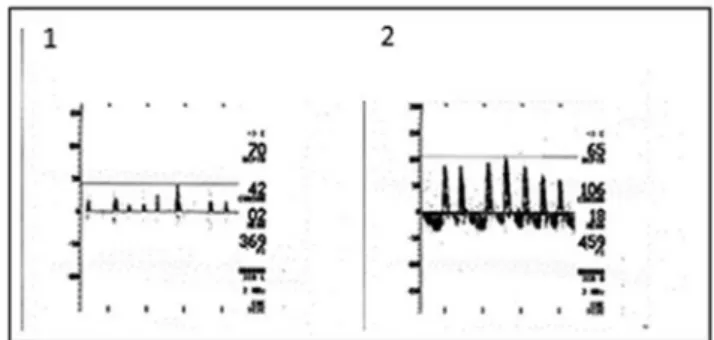
Figure 4 - A 65-year-old male patient with chronic atrial fibrillation, anticoagulation, cerebellar infarction with hemorrhagic transformation, hydrocephalus, external shunt, fever, venous sinus thrombosis, and multiple bilateral infarcts. GCS 3 without brainstem reflexes and without sedation for 24 hours. Mean arterial pressure 100mmHg. Transcranial Doppler does not demonstrate temporal window sonograms. Reverberating flow and systolic spikes at the right (1) and left (2) carotid syphon and basilar artery (not shown) are consistent with the diagnosis of brain death.
window is permeable to ultrasound, we cannot conirm nor rule out anything with respect to the low at that particular vessel. It is possible to solve this problem by studying the low in the proximal vessels. In the case of a cerebral circulatory arrest pattern in a proximal vessel, we can conclude that in the distal region, there will be no low. For the temporal window, if there is no image, a possible solution that is used frequently is looking for a carotid sonogram at the level of the
carotid sinus through the orbital window.(34-37) Sonographic
indings of cerebral circulatory arrest permit to conclude that the low in the MCA (intracranial vessel, terminal branch of the internal carotid artery (ICA)) is equal to or worse than the low observed in the carotid. However, the MCA could receive low through the contralateral collateral circulation
MCA or the posterior cerebral communicating arteries.(38)
his is not the case because the diagnosis of cerebral circulatory arrest requires a conirmation of a circulatory arrest pattern in all territories (bilateral anterior and posterior). A inding of reverberating low or spikes on a carotid sinus should be accompanied by the same indings in the contralateral anterior and in the posterior circuit.
Finally, we must insist that the study of the orbital window should only be performed when a sonogram
cannot be obtained through the temporal window.(38-40)
In the case of the occipital window, if there is no low in the basilar artery, vertebral arteries can be insonated through the same window, at a lower depth. If both vertebral arteries exhibit cerebral circulatory arrest patterns, it can be assumed that the low in the BA is equal or worse.
Because of the diiculty in changing the position of the head of some patients, the physician may choose to perform insonation on both vertebral arteries instead of the BA. In cases in which both arteries exhibit a pattern of systolic spikes or reverberating low in association with a similar pattern in both middle cerebral arteries, cerebral circulatory arrest is diagnosed.
It is possible to validate the pattern of cerebral circulatory arrest through an extracranial vessel only if it contains a pattern of reverberating low or spikes. If there is an absence of low or a diferent low pattern, the pattern cannot be validated, and the study results are inconclusive.
Figure 5 presents the diagnostic algorithm of cerebral circulatory arrest (CCA).
Consensus declaration
In the absence of any ultrasound image in either MCA or BA, its proximal vessels: (ipsilateral internal carotid
artery initially at the carotid siphon o secondarily at the neck, or both vertebral arteries, respectively) are acceptable for a diagnosis of CCA.
7. Which situations can confound the interpretation of transcranial Doppler records in cerebral circulatory arrest? In which cases can an ultrasound pattern of cerebral circulatory arrest be detected while the patient is not clinically brain dead? Are these considered false positives?
Clinical brain death and CCA are parallel phenomena that may not always coincide at the time of presentation, especially when the source of brain death is primarily infratentorial. his temporal asynchrony between the two phenomena leads to the concept of BD as an integrated and multi-phenomenological process. herefore, to avoid misinterpretation, it is desirable that both clinical and instrumental phenomena that determine BD are simultaneous so that it is possible to establish the diagnosis of cerebral circulatory arrest with certainty.
Infrequently, records can be found compatible with CCA while the patient continues to exhibit some clinical sign of neurological activity, including corneal relex, extensor reactivity, etc., which refutes the clinical diagnosis of BD
weak breathing.(41) his phenomenon has also been described
in pediatric patients with reverberating low. Another group analyzed 12 patients and described one patient who presented
with a pattern of CCA with breathing preserved.(42) A study
16 patients with cerebral circulatory arrest patterns. Several of these papers are from a irst period in which the study of the posterior portion was not required for the diagnosis of cerebral circulatory arrest.(43)
he meta-analysis by Monteiro described patients with cerebral circulatory arrest and weak breathing for
several hours that inevitably evolved to BD.(44) Since
1998, diferent consensus groups have noted that the pattern of cerebral circulatory arrest in both MCAs may precede the loss of function at the level of the brainstem. he importance of studying the posterior circulation is
emphasized.(15) A Chilean study evaluated 53 patients,
including 25 in cerebral circulatory arrest.(45) he study
described a patient who was breathing at the time of cerebral circulatory arrest. he patient completed the BD criteria at 24 hours. All patients described in this work inexorably developed BD at varying times within 24 hours. None were described as false positive. hese scenarios have been reported in the literature with a frequency of approximately 1/100. hey are interpreted as cases of imminent brain death. To our knowledge, there are no reports of patients who have survived in these conditions. hese situations reairm that the two conditions are not exactly simultaneous.
he situations in which we can observe patterns of circulatory arrest that do not imply or do not match clinical brain death include cases of hypotension, sudden and transient elevations in ICP (spontaneous A waves or sudden active bleeding, such as in the initial rupture
of a brain aneurysm(33)) and incomplete study, e.g., the
evaluation of the superior circuit only.
We must insist on the following: 1) good systemic hemodynamics; 2) an examination for a period of at least 30 minutes, because the plateau waves last 15 minutes or less; and 3) an evaluation of the three sectors.
In other cases, the clinical diagnosis is made, but Doppler patterns of cerebral circulatory arrest are not yet observed due to the lack of synchronization between the two processes. In such circumstances, the diagnostic accuracy of TCD to conirm BD decreases. However, a large percentage of these patients will eventually exhibit a pattern of cerebral circulatory arrest.
hese cases include the presence of ventriculostomy, decompressive craniectomy, opening of the calvarium, skull base fracture, traumatic arteriovenous istula, anoxic-ischemic encephalopathy, and the use of intra-aortic balloon counterpulsation. he latter preserves diastolic
low, therefore the balloon has to be put in stand by mode if possible to diagnose by means of TCD the real cerebral hemodinamic situation.
In an as-of-yet unpublished study conducted by members of this consensus (GH, CP, GF, LM), 36 patients with decompressive craniectomy with clinically suspected BD were described in whom a complete clinical diagnosis was not possible due to diferent factors (depressant drugs, inability to perform an apnea test, etc.). In the irst study, a CCA pattern was found in 43% of cases (14 patients). Of the 22 for which CCA was not initially veriied, the study was repeated in seven patients, and CCA was detected in three patients. Of the remaining four patients, the study was repeated in two, and one of them exhibited a CCA pattern in the third study, and the other in the fourth.
Finally, it was veriied that 19 patients exhibited CCA in the initial group of 34 patients, although there were 15 cases in which the study was never repeated. hus, despite the fact that decompressive craniectomy may sustain low, more than half of the patients with DC whose clinical status worsened to clinical diagnosis or high suspicion of brain death exhibited CCA.
Consensus declaration
here are situations (loss of hermeticity of the skull) that delay the onset of the CCA. However, a large percentage of these patients progress to CCA; therefore, the study involves no contraindications, but its repetition over time becomes peremptory for the diagnosis of such evolution.
8. What is the practical significance of circulatory arrest in both sectors separately? Is it ethically sound that either or both situations allow organ donation, thereby gaining time and optimal donor conditions? What degree of irreversibility is possible in both situations?
In patients with focal lesions, there may exist
interhemispheric pressure gradients(46) between supra
and infratentorial regions.(47) In the first years after the
of BD;(37) this was interpreted as a false positive for
the diagnosis of BD but was actually an incomplete study. Thereafter, different consensuses groups began requiring the study of posterior (basilar artery or both vertebral arteries) vessels in addition to the bilateral
anterior sections.(37)
In patients examined with TCD, it is common to observe that the pattern of CCA is not presented simultaneously in all territories. he usual behavior is to repeat the study until the pattern is displayed and appears in all three sections; this observation supports the diagnosis of BD.
In countries where the criteria of brainstem death are accepted as death of the individual, it would be consistent to accept a pattern of circulatory arrest in the basilar artery or both vertebral arteries as supporting the clinical diagnosis of BD. In these cases, the presence of irreversible posterior circulatory arrest (CA) could save time and allow for better donors. We did not ind any articles in the research literature that speciically focused on this topic. We have not found any work that attempts to validate the diagnosis of circulatory arrest BD through only the basilar artery in countries or regions that accept the criterion of brain stem death as the criterion of death. his type of study is very diicult to perform because often there is doubt in the clinical diagnosis of BD due to the presence of CNS depressant drugs involved in the therapeutics of intracranial hypertension. However, given the collateral circulation in the brain via the circle of Willis, the possibility exists that the distal portion of the basilar artery receives blood from the anterior sector; if this circulation is present, the brain stem can be perfused simultaneously to an absent circulation in the initial portion of the basilar artery. In our opinion, therefore, the pattern of CCA in just the basilar artery cannot be accepted as conclusive of brain stem absence of low.
his consensus has not considered ethical factors due to the large variability in the diagnostic criteria of death in diferent countries. Independent of the
diagnostic criteria of brain death, the presence of regional
circulatory arrest has a very poor neurological functional outcome, as observed in the hemispheric infarction by carotid occlusion, malignant infarction of the middle cerebral, or poor outcome criterion in the CA of the
MCA subsequent to thrombolysis (TIBI 1).(48)
Consensus declaration
Regardless of whether the accepted diagnostic criterion for BD is that of “whole-brain death” or “brainstem death”, TCD supports the diagnosis of brain death only when vessels exhibit anterior and posterior CA patterns.
9. What training and experience are required to perform and report a study of cerebral blood flow with transcranial Doppler in the context of ancillary examination in the diagnosis of brain death?
To ensure the diagnosis of cerebral circulatory arrest to support the legal diagnosis of BD, the physician must be properly trained, familiar with their performance and interpretation. Each country or region may certify these doctors diferently, but this consensus group recommends a minimum of 50 TCD studies conducted in neurocritical patients and 20 diagnoses of cerebral circulatory arrest.
All TCD studies must be documented and controlled by a an experienced professional, and the individual must pass a written examination on clinical pathophysiology, the diagnostic protocol of BD, and the physical principles of transcranial Doppler.
Consensus declaration
A minimum number of documented studies, approval by a tutor, and a theoretical test is recommended.
CONCLUSIONS
In an attempt to standardize the methodology of transcranial Doppler in Latin America, the present work presents consensus opinions of a group of experienced Latin American physicians intention to standardize when and how to use this technique to aid the diagnosis of the cerebral circulatory arrest that often accompanies brain death. A consensus statement by all members was issued for each point that was considered.
Cerebral circulatory arrest is the inal stage in the evolution of progressive intracranial hypertension, the most common etiology of brain death.
transcranial Doppler. To support the diagnosis of brain death, transcranial Doppler must show one of these three patterns of cerebral circulatory arrest maintained over time in the anterior arteries (bilateral middle cerebral arteries) and posterior (basilar artery). Insonation must be performed under acceptable hemodynamic conditions in the bilateral anterior and posterior sector. In cases in which ultrasonographic imaging of either one or both middle cerebral arteries or basilar artery is not possible, the vessels proximal to them (ipsilateral internal carotid siphon or both intra- and extracranial vertebral, respectively) are acceptable for the diagnosis of cerebral circulatory arrest.
here are situations (loss of hermeticity of the braincase) that delay the onset of cerebral circulatory arrest; however, a percentage of these patients progress to
cerebral circulatory arrest. herefore, repeated use of TCD is critical for the diagnosis of this evolution.
Both conventional transcranial Doppler and transcranial color Doppler ultrasound can be used for the diagnosis of cerebral circulatory arrest.
Regardless of whether the diagnostic criteria of brain death accepted is “death of the whole brain” or “death of the brainstem”, a diagnosis of brain death is supported when the vessels of the anterior (bilaterally) and posterior circulation exhibit a pattern of cerebral circulatory arrest.
A minimum number of documented studies are recommended that must be made by a physician, together with a theoretical test, to qualify for performing the diagnosis of cerebral circulatory arrest to support the legal diagnosis of brain death.
El Doppler transcraneal evalúa la hemodinámica cerebral en el paciente neurocrítico. Se destaca su aporte como técnica auxiliar en el diagnóstico del paro circulatorio cerebral, que habitualmente presenta el paciente en muerte encefálica. Este Consenso Latinoamericano se conformó por un grupo de 26 médicos con experiencia en el uso de Doppler transcraneal en el contexto de muerte encefálica. El propósito de este consenso es realizar recomendaciones en relación a las indicaciones, técnica e interpretación del estudio de la ultrasonografía transcraneal en el paciente con diagnóstico clínico de muerte encefálica o en aquel paciente cuyo diagnóstico clínico presenta diicultades; formar un grupo de trabajo que permita profundizar conocimientos y consolidar lazos entre médicos latinoamericanos trabajando en el mismo tema.
Se revisó la literatura, se intercambiaron conceptos y experiencias en dos encuentros presenciales y vía Internet. Se contestaron preguntas sobre isiopatología, equipo,
técnica, hallazgos, problemas frecuentes e interpretación del Doppler transcraneal en el contexto de muerte encefálica. Las declaraciones fundamentales del consenso son: El paro circulatorio cerebral es la última etapa en la evolución de la hipertensión intracraneana progresiva, donde se visualiza con el Doppler transcraneal un “patrón de paro circulatorio cerebral”. Se acepta como patrón de paro circulatorio cerebral: patrón reverberante, espigas sistólicas y ausencia de lujo previamente evidenciado. Se debe insonar - en condiciones hemodinámicas aceptables - sector anterior bilateralmente (arterias cerebrales medias) y sector posterior (arteria basilar). De no encontrarse ninguna imagen ultrasonográica en éstas, las arterias proximales (carótida interna ipsilateral en sifón o ambas vertebrales respectivamente) son aceptables para el diagnóstico de paro circulatorio cerebral.
RESUMEN
Descriptores: Muerte encefálica/diagnóstico; Muerte en-cefálica/ultrasonograia; Ultrasonografía Doppler transcraneal; Consenso
REFERENCES
1. Aaslid R, Markwalder TM, Nornes H. Noninvasive transcranial Doppler ultrasound recording of flow velocity in basal cerebral arteries. J Neurosurg. 1982;57(6):769-74.
2. Schatlo B, Pluta RM. Clinical applications of transcranial Doppler sonography. Rev Recent Clin Trials. 2007;2(1):49-57. Review.
3. Sloan MA, Alexandrov AV, Tegeler CH, Spencer MP, Caplan LR, Feldmann E, Wechsler LR, Newell DW, Gomez CR, Babikian VL, Lefkowitz D, Goldman RS, Armon C, Hsu CY, Goodin DS; Therapeutics and Technology Assessment Subcommittee of the American Academy of Neurology. Assessment: transcranial Doppler ultrasonography: report of the Therapeutics and Technology Assessment Subcommittee of the American Academy of Neurology. Neurology. 2004;62(9):1468-81.
4. Escudero D. [Brain death diagnosis]. Med Intensiva. 2009;33(4):185-95. Review. Spanish.
5. Segura T, Calleja S, Irimia P, Tembl JI, Spanish Society of Neurosonology. Recommendations for the use of transcranial Doppler ultrasonography to determine the existence of cerebral circulatory arrest as diagnostic support for brain death. Rev Neurosci. 2009;20(3-4):251-9.
6. Wijdicks E. The transatlantic divide over brain death determination and the debate. Brain. 2012;135(Pt 4):1321-31.
7. Glaser EM. Using behavioral science strategies for defining the state-of-the-art. J Appl Behav Sci. 1980;16(1):79-92.
9. Nedelmann M, Stolz E, Gerriets T, Baumgartner RW, Malferrari G, Seidel G, Kaps M; TCCS Consensus Group. Consensus recommendations for transcranial color-coded duplex sonography for the assessment of intracranial arteries in clinical trials on acute stroke. Stroke. 2009;40(10):3238-44. 10. Rotondi AJ, Kvetan V, Carlet J, Sibbald WJ. Consensus conferences in critical
care medicine. Methodologies and impact. Crit Care Clin. 1997;13(2):417-39. 11. Flowers WM Jr, Patel BR. Persistence of cerebral blood flow after brain
death. South Med J. 2000;93(4):364-70.
12. Marinoni M, Alari F, Mastronardi V, Peris A, Innocenti P. The relevance of early TCD monitoring in the intensive care units for the confirming of brain death diagnosis. Neurol Sci. 2011;32(1):73-7.
13. Topcuoglu MA, Arsava EM. How Doppler effect occurs in absence of intracranial blood flow in brain death? Med Hypotheses. 2013;80(1):103-4. 14. Poularas J, Karakitsos D, Kouraklis G, Kostakis A, De Groot E, Kalogeromitros
A, et al. Comparison between transcranial color Doppler ultrasonography and angiography in the confirmation of brain death. Transpl Proc. 2006;38(5):1213-7.
15. Ducrocq X, Hassler W, Moritake K, Newell DW, von Reutern GM, Shiogai T, et al. Consensus opinion on diagnosis of cerebral circulatory arrest using Doppler-sonography: Task Force Group on cerebral death of the Neurosonology Research Group of the World Federation of Neurology. J Neurol Sci. 1998;159(2):145-50.
16. Petty GW, Mohr JP, Pedley TA, Tatemichi TK, Lennihan L, Duterte DI, et al. The role of transcranial Doppler in confirming brain death: sensitivity, specificity, and suggestions for performance and interpretation. Neurology. 1990;40(2):300-3.
17. Babikian VL, Feldmann E, Wechsler LR, Newell DW, Gomez CR, Bogdahn U, et al. Transcranial Doppler ultrasonography: year 2000 update. J Neuroimaging. 2000;10(2):101-15.
18. Abstracts of the Neurocritical Care Society 10th Annual Meeting. October
4-7, 2012. Denver, Colorado, USA. Neurocrit Care. 2012;17 Suppl 2:S1-337. 19. Ortiz MA. Ultrasound physics [Internet]. Corrientes (AG): Northeas
National University School of Medicine. Continuous training program on general medicine: s.d. [Redacted by Sicari V]. [cited Mar 23 2014]. Disponível en: http://med.unne.edu.ar/posgrado/cursomedgral/clases/ La%20f%EDsica%20de%20los%20ultrasonidos.doc.
20. Stulin I, Solonskiy D, Sinkin M, Musin S, Mnushkin A, Kascheev A, et al. The role of color duplex sonography in the brain death diagnostics. Perspect Med. 2012; 1(1-12):. 362-5.
21. Abadal JM, Llompart-Pou JA, Homar J, Pérez-Bárcena J, Ibáñez J. [Applications of transcranial color-coded duplex sonography in monitoring neurocritical patients]. Med Intensiva. 2007;31(9):510-7. Spanish. 22. Egido JA, Simal P. Neurosonology [Internet]. In: Guia Neurologica 8:
Enfermedad Cerebrovascular. Bobotá: ACN; s.d. cap. 6, p. 88-121. [citado 2014 Mar 23]. Disponível en: http://www.acnweb.org/guia/g8cap6.pdf 23. Hoksbergen AW, Legemate DA, Ubbink DT, Jacobs MJ. Success rate
of transcranial color-coded duplex ultrasonography in visualizing the basal cerebral arteries in vascular patients over 60 years of age. Stroke. 1999;30(7):1450-5.
24. Hassler W, Steinmetz H, Pirschel J. Transcranial Doppler study of intracranial circulatory arrest. J Neurosurg. 1989;71(2):195-201. 25. Halsey JH. Effect of emitted power on waveform intensity in transcranial
Doppler. Stroke. 1990;21(11):1573-8.
26. Itoh T, Matsumoto M, Handa N, Maeda H, Hougaku H, Hashimoto H, et al. Rate of successful recording of blood flow signals in the middle cerebral artery using transcranial Doppler sonography. Stroke. 1993;24(8):1192-5. 27. Ringelstein EB, Kahlscheuer B, Niggemeyer E, Otis SM. Transcranial
Doppler sonography: anatomical landmarks and normal velocity values. Ultrasound Med Biol. 1990;16(8):745-61.
28. Volc D, Possnigg G, Grisold W, Neuhold A. Transcranial Dopplersonography of the vertebro-basilar system. Acta Neurochir (Wien). 1988;90(3-4):136-8.
29. Powers AD, Graeber MC, Smith RR. Transcranial Doppler ultrasonography in the determination of brain death. Neurosurgery. 1989;24(6):884-9. 30. Calleja S, Tembl JI, Segura T; Spanish Neurosonology Society.
[Recommendations in the use of transcranial Doppler to determine the existence of cerebral circulatory arrest as diagnostic support of brain death]. Neurologia. 2007;22(7):441-7. Spanish.
31. Lange MC, Zétola VH, Miranda-Alves M, Moro CH, Silvado CE, Rodrigues DL, Gregorio EG, Silva GS, Oliveira-Filho J, Perdatella MT, Pontes-Neto OM, Fábio SR, Avelar WM, Freitas GR; Task Force Group of the Neurosonology Department, Brazilian Academy of Neurology. Brazilian guidelines for the application of transcranial ultrasound as a diagnostic test for the confirmation of brain death. Arq Neuropsiquiatr. 2012;70(5):373-80. 32. Kirkham FJ, Levin SD, Padayachee TS, Kyme MC, Neville BG, Gosling RG.
Transcranial pulsed Doppler ultrasound findings in brain stem death. J Neurol Neurosurg Psychiatry. 1987;50(11):1504-13.
33. Steinmetz H, Hassler W. Reversible intracranial circulatory arrest in acute subarachnoid haemorrhage. J Neurol Neurosurg Psychiatry. 1988; 51(10):1355-6.
34. Dominguez-Roldan JM, Jimenez-Gonzalez PI, Garcia-Alfaro C, Rivera-Fernandez V, Hernandez-Hazañas F. Diagnosis of brain death by transcranial Doppler sonography: solutions for cases of difficult sonic windows. Transpl Proc. 2004;36(10):2896-7.
35. Soldatos T, Karakitsos D, Wachtel M, Boletis J, Chatzimichail K, Papathanasiou M, et al. The value of transcranial Doppler sonography with a transorbital approach in the confirmation of cerebral circulatory arrest. Transplant Proc. 2010;42(5):1502-6.
36. Lampl Y, Gilad R, Eschel Y, Boaz M, Rapoport A, Sadeh M. Diagnosing brain death using the transcranial Doppler with a transorbital approach. Arch Neurol. 2002;59(1):58-60.
37. Ducrocq X, Braun M, Debouverie M, Junges C, Hummer M, Vespignani H. Brain death and transcranial Doppler: experience in 130 cases of brain dead patients. J Neurol Sci. 1998;160(1):41-6.
38. de Freitas GR, André C, Bezerra M, Nunes RG, Vincent M. Persistence of isolated flow in the internal carotid artery in brain death. J Neurol Sci. 2003;210(1-2):31-4.
39. Jacobs BS, Carhuapoma JR, Castellanos M. Clarifying TCD criteria for brain death-are some arteries more equal than others? J Neurol Sci. 2003; 210(1-2):3-4.
40. de Freitas GR, Andre C. Routine insonation of the transorbital window for confirming brain death: a double-edged sword. Arch Neurol. 2003;60(8):1169. 41. Hadani M, Bruk B, Ram Z, Knoller N, Spiegelmann R, Segal E. Application
of transcranial doppler ultrasonography for the diagnosis of brain death. Intensive Care Med. 1999;25(8):822-8.
42. Newell DW, Grady MS, Sirotta P, Winn HR. Evaluation of brain death using transcranial Doppler. Neurosurgery. 1989;24(4):509-13.
43. Puppo C. Transcranial Doppler in brain death. In: III International Symposium on Coma and Death. Montevideo, Uruguay; 2000.
44. Monteiro LM, Bollen CW, van Huffelen AC, Ackerstaff RG, Jansen NJ, van Vught AJ. Transcranial Doppler ultrasonography to confirm brain death: a meta-analysis. Intensive Care Med. 2006;32(12):1937-44.
45. Brunser A, Hoppe A, Cárcamo DA, Lavados PM, Roldán A, Rivas R, et al. [Validation of transcranial Doppler in the diagnosis of brain death]. Rev Med Chil. 2010;138(4):406-12. Spanish.
46. Sahuquillo J, Poca MA, Arribas M, Garnacho A, Rubio E. Interhemispheric supratentorial intracranial pressure gradients in head-injured patients: are they clinically important? J Neurosurg. 1999;90(1):16-26.
47. Slavin KV, Misra M. Infratentorial intracranial pressure monitoring in neurosurgical intensive care unit. Neurol Res. 2003;25(8):880-4. 48. Demchuk AM, Burgin WS, Christou I, Felberg RA, Barber PA, Hill MD, et al.
* Consensus Group on Transcranial Doppler in the Diagnosis of Brain Death