Brazilian recommendations of mechanical ventilation
2013. Part 2
INTRODUCTION
Invasive or non-invasive mechanical ventilation (MV) must be performed in an adequate and safe manner to avoid the occurrence of ventilation-induced lung injury. Based on physiological principles, evidence collected in laboratory experiments, and randomized clinical or observational studies involving actual patients that were available in the literature, current MV recommendations indicate that ventilatory support should be performed at a tidal volume (Vt) of 6mL/kg predicted body weight, with a delta between plateau pressure and positive
end-expiratory pressure (PEEP) not greater than 15cmH2O, and end-expiratory
pressure levels suicient to avoid airway and alveolar collapse and ensure adequate gas exchange. Other recommendations include positioning the patient to guarantee adequate and harmless ventilation (such as prone positioning in cases of severe acute
Carmen Sílvia Valente Barbas, Alexandre Marini Ísola, Augusto Manoel de Carvalho Farias, Alexandre Biasi Cavalcanti, Ana Maria Casati Gama, Antonio Carlos Magalhães Duarte, Arthur Vianna, Ary Serpa Neto, Bruno de Arruda Bravim, Bruno do Valle Pinheiro, Bruno Franco Mazza, Carlos Roberto Ribeiro de Carvalho, Carlos Toufen Júnior, Cid Marcos Nascimento David, Corine Taniguchi, Débora Dutra da Silveira Mazza, Desanka Dragosavac, Diogo Oliveira Toledo, Eduardo Leite Costa, Eliana Bernadete Caser, Eliezer Silva, Fabio Ferreira Amorim, Felipe Saddy, Filomena Regina Barbosa Gomes Galas, Gisele Sampaio Silva, Gustavo Faissol Janot de Matos, João Claudio Emmerich, Jorge Luis dos Santos Valiatti, José Mario Meira Teles, Josué Almeida Victorino, Juliana Carvalho Ferreira, Luciana Passuello do Vale Prodomo, Ludhmila Abrahão Hajjar, Luiz Claudio Martins, Luis Marcelo Sá Malbouisson, Mara Ambrosina de Oliveira Vargas, Marco Antonio Soares Reis, Marcelo Brito Passos Amato, Marcelo Alcântara Holanda, Marcelo Park, Marcia Jacomelli, Marcos Tavares, Marta Cristina Paulette Damasceno, Murillo Santucci César Assunção, Moyzes Pinto Coelho Duarte Damasceno, Nazah Cherif Mohamed Youssef, Paulo José Zimmermann Teixeira, Pedro Caruso, Péricles Almeida Delfino Duarte, Octavio Messeder, Raquel Caserta Eid, Ricardo Goulart Rodrigues, Rodrigo Francisco de Jesus, Ronaldo Adib Kairalla, Sandra Justino, Sergio Nogueira Nemer, Simone Barbosa Romero, Verônica Moreira Amado
Perspectives on invasive and noninvasive ventilatory support for critically ill patients are evolving, as much evidence indicates that ventilation may have positive efects on patient survival and the quality of the care provided in intensive care units in Brazil. For those reasons, the Brazilian Association of Intensive Care Medicine (Associação de Medicina Intensiva Brasileira - AMIB) and the Brazilian horacic Society (Sociedade Brasileira de Pneumologia e Tisiologia - SBPT), represented by the Mechanical Ventilation Committee and the Commission of Intensive herapy, respectively, decided to review the literature and draft recommendations for mechanical ventilation with the goal of creating a document for bedside guidance as to the best practices on mechanical ventilation available to their members. he document was based
The present recommendations are a joint initiative of the Mechanical Ventilation Committee of the Brazilian Intensive Care Medicine Association (Associação de Medicina Intensiva Brasileira - AMIB) and the Commission of Intensive Therapy of the Brazilian Thoracic Society (Sociedade Brasileira de Pneumologia e Tisiologia - SBPT).
Completion of the drafting of the document:
October 20, 2013
Conflicts of interest: With the help of the Brazilian Thoracic Society, the AMIB Division of Scientific Issues procured financial support from industrial companies and laboratories, distributed as sponsorship quotas, to cover part of the event costs (participants’ air tickets, food and lodging). None of those companies participated in the drafting of the present document, nor had access to its content until it was disclosed (after its final format was approved) as brochures distributed at the Brazilian Congress of Intensive Care Medicine in Rio de Janeiro in 2013. The companies that collaborated with the present project are: Air Liquide, Covidien, GE, Intermed, Magnamed, Mindray and Philips.
Corresponding author:
Carmen Silvia Valente Barbas
Disicplina de Pneumologia, Hospital das Clínicas da Faculdade de Medicina da Universidade de São Paulo Avenida Dr. Eneas de Carvalho Aguiar, 44 Zip code - 05403-900 - São Paulo (SP), Brazil E-mail: [email protected]
Recomendações brasileiras de ventilação mecânica 2013. Parte 2
ABSTRACT on the available evidence regarding 29
subtopics selected as the most relevant for the subject of interest. he project was developed in several stages, during which the selected topics were distributed among experts recommended by both societies with recent publications on the subject of interest and/or signiicant teaching and research activity in the ield of mechanical ventilation in Brazil. he experts were divided into pairs that were charged with performing a thorough review of the international literature on each topic. All the experts met at the Forum on Mechanical Ventilation, which was held at the headquarters of AMIB in São Paulo on August 3 and 4, 2013, to collaboratively draft the inal text corresponding to each sub-topic, which was presented to, appraised, discussed and approved in a plenary session that included all 58 participants and aimed to create the inal document.
(such as extracorporeal carbon dioxide (CO2) removal) in cases of refractory ARDS. he development of increasingly more sophisticated ventilators allow for ine adjustment of sensitivity and include several trigger mechanisms, diferent inspiratory low speeds, acceleration, mechanisms for ending inspiratory time, and monitoring options, which enable adjustment of the patient-ventilator synchrony and MV as a function of the patient’s disease. In this regard, the possibility of providing diferential ventilatory support for restrictive and obstructive conditions stands out.
For that reason, joint analysis of the available evidence on ventilatory support by Brazilian experts who deal with mechanical ventilation like anesthesiologists, intensivists, pulmonologists, physical therapists, nurses, nutritionists and speech therapists was necessary. Such evidence, taken together with experience gathered by the various specialties, may provide guidance to health care professionals in Brazilian intensive care units (ICU) on how to provide safe and efective respiratory support for patients with respiratory failure, based on the best evidence available, in order to avoid the occurrence of ventilator-associated lung injury.
herefore, the aim of the present study was to review the available literature on 29 subtopics related to ventilatory support for individuals with respiratory failure, and following presentation, discussion, and approval at a plenary session including all 58 participating specialists, to present the results in the form of recommendations and suggestions.
METHODS
Literature available from MedLine (2003-2013) and the Cochrane Central Register of Controlled Trials (CENTRAL) was reviewed by specialists with a higher education (intensivists, anesthetists, pulmonary specialists, physical therapists, and nurses) who were distributed in pairs for review of each of the 29 selected subtopics related to non-invasive and invasive ventilatory support for patients with respiratory failure.
After reviewing the articles available in the literature, each pair answered the questions formulated by the organizing commission (composed by Carmen Silvia Valente Barbas, President of the Committee of Respiratory Failure and Mechanical Ventilation of AMIB, Alexandre Marini Isola, National Coordinator of the Course of MV in ICU - VENUTI, and Augusto Manoel de Carvalho Farias, Coordinator of the Department of Intensive Care of the SBPT) according to criteria previously suggested
by other authors.(1-4) hus, the term “recommendation”
was used when the level of evidence was high, i.e., derived from randomized studies conducted with more than
100 participants, meta-analyses, all-or-nothing efect, or patient safety. he term suggestion was used when the available evidence was weak, i.e., based on observational or case-control studies, case series, or on the experience of specialists to provide guidance for eicient and safe ventilatory support in Brazil. We therefore hoped that these evidence-based recommendations would help to avoid potential deleterious efects associated with inadequate ventilatory support in our patients.
he 58 participating specialists were requested to answer the proposed questions during an eight-hour session conducted at the AMIB on August 3, 2013. he answers were formulated based on the evidence available in the literature and on the experience of the specialists and were then presented at a plenary session that included all 58 participating specialists, which was held on August 4, 2013 at AMIB headquarters. During that session, the answers were discussed, modiied when needed, voted on, and approved in accordance with the suggestions and observations of the specialists who attended the meeting.
he reports made by all the pairs of specialists were gathered by the project organizing commission, which revised, formatted and drafted the inal document, following the authors’ revisions. he document was then printed in the form of a bedside manual of recommendations to be distributed to ICUs all across Brazil, and it was also sent for publication in the Brazilian Journal of Intensive Care (Revista
Brasileira de Terapia Intensiva - RBTI) and the Brazilian
Journal of Pulmonology (Jornal Brasileiro de Pneumologia).
MECHANICAL VENTILATION IN CHEST TRAUMA Noninvasive mechanical ventilation
Recommendation - Noninvasive ventilation (NIV) is contraindicated in patients with upper airway injury, in the presence of hemodynamic instability, and in severe craniocerebral trauma.(5-10)
Recommendation - In patients with isolated chest trauma, early application of NIV can improve gas exchange, prevent orotracheal intubation (OTI), and
reduce complications and ICU length of stay.(5-10)
oxygen saturation (SpO2), and reduction of the partial
pressure of carbon dioxide (PaCO2) without signiicant
abdominal distension. In unsuccessful cases, OTI and invasive MV should be performed immediately.
Invasive mechanical ventilation(11)
Recommendation - Patients with severe chest trauma, respiratory failure, and speciic contraindications to NIV should be promptly intubated and ventilated.
Recommendation - Initially, use an assist-control mode of ventilation, i.e., volume-cycled ventilation (VCV) or pressure-control ventilation (PCV), in chest trauma with severe respiratory failure.
Recommendation - Regardless of the mode selected (VCV or PCV), patients with chest trauma should be initially ventilated with a Vt of 6mL/kg predicted body weight, an f of 16-20 breaths/min, and a fraction of
inspired oxygen (FiO2) that is suicient to maintain
an SpO2>92% and a PEEP of 5-10cmH2O. In cases of
ARDS, follow the instructions in the related section of the present Recommendations (Part 1).
Recommendation - In cases of high output bronchopleural istula, use the PCV mode, which will compensate for the leak. Another option is the use of high frequency oscillatory ventilation, only in centers with this capability and specialized personnel. In cases that are more severe, asynchronous independent lung ventilation can be either used or not, and the lung with the istula is ventilated in the PCV mode with a distending pressure of
<15cmH2O and low PEEP levels (<10cmH2O).
Pain control
Suggestion - horacic epidural analgesia, as part of a multimodal strategy, is recommended. If epidural analgesia cannot be used or is contraindicated, patient-controlled i.v. analgesia or intercostal nerve blockade can be used.(11)
Suggestion - Administration of intermittent analgesia can be used in cases of less severe pain.
MECHANICAL VENTILATION DURING SURGICAL PROCEDURES
Comment - Postoperative pulmonary complications (PPC) contribute to a substantial proportion of the risks associated with surgery and anesthesia, being the major cause
of morbidity and mortality in the postoperative period.(12)
PPC, which include respiratory infections, respiratory failure, pleural efusion, atelectasis, pneumothorax,
bronchospasm, and aspiration pneumonitis, afect
approximately 5% of patients undergoing surgery.(12,13)
Specific care before intubation Preoperative risk stratification
Recommendation - All patients should be assessed for risk of PPC by using a speciic scale. he American Society of Anesthesiology (ASA) classiication is a scale that is subjective and imprecise. Among the scales suggested for stratifying patient risk for PPC are the European Surgical
Outcomes Study (EuSOS) scoring system and the
Assess Respiratory Risk in Surgical Patients in Catalonia
(ARISCAT).(13)
Pre-oxygenation during induction of anesthesia
Recommendation - Pre-oxygenation with an FiO2 of approximately 100% for all patients aims at increasing
reserve and reducing the risk of hypoxemia.(14)
Use of noninvasive ventilation during induction of anesthesia
Suggestion - Use NIV during induction of anesthesia for elective surgery in patients with decreased abdominal compliance and in those in whom it is necessary to use
an FiO2 of 100% for pre-oxygenation. he use of NIV
can prevent the development of atelectasis in patients with decreased abdominal compliance and in those undergoing
pre-oxygenation with an FiO2 of 100%.(14)
Use of positive end-expiratory pressure and alveolar recruitment maneuvers during induction of anesthesia
Suggestion - Use recruitment maneuvers and a PEEP
of approximately 5-8cmH2O after induction of anesthesia
in patients with decreased abdominal compliance or in
those who develop hypoxemia.(14)
Specific care during the intraoperative period Mode of ventilation
Suggestion - Use PCV to improve pulmonary mechanics in patients undergoing video-assisted laparoscopic surgery. Observe appropriate expired Vt values.(15,16) In the remaining scenarios, there is no beneit
Tidal volume
Recommendation - he use of MV with a Vt of 6mL/kg predicted body weight should be considered in all patients. Various studies in various scenarios (abdominal, thoracic, and cardiac surgery) have demonstrated the beneit of using low Vt during surgery.(17-19)
Positive end-expiratory pressure
Suggestion - he use of a PEEP of approximately
5-8cmH2O should be considered. All studies assessing
conventional MV strategies versus protective strategies in patients undergoing surgery considered using a low Vt and a high PEEP in the protective strategies. In general, the use of a high PEEP results in better oxygenation and a lower incidence of PPC.(17,20)
Alveolar recruitment maneuvers
Suggestion - Use alveolar recruitment maneuvers (RM) during the intraoperative period to reverse alveolar collapse and improve oxygenation. he use of RM, in association with maintenance of high PEEP levels, reduces the amount of collapsed lung and improves patient oxygenation
during surgery.(17,20) Among the RM most often cited in
the literature is maximum recruitment strategy (MRS), which seeks to maintain a high plateau pressure (Pplat) in
the airway (approximately 40-45cmH2O) for 60 seconds
(as described in part 1 of the present Recommendations).
Fraction of inspired oxygen
Suggestion - he lowest FiO2 level that can maintain
SpO2 at approximately 96-98% should be used. he
combination of an FiO2 of approximately 30-40% and
high PEEP levels can maintain adequate oxygenation and reduce lung atelectasis. he role of high levels of oxygen in preventing surgical wound infection remains controversial.(14)
Respiratory rate
Recommendation - Use f to maintain PaCO2 at
38-43mmHg (end-tidal CO2 [CO2 at end-expiration] of
approximately 35-40mmHg). he trend to use a low Vt during surgery requires the adoption of a higher f.(17,19)
Discontinuation of mechanical ventilation
Recommendation - Discontinue MV in the postoperative period as early and as quickly as possible, as soon as the patient is hemodynamically stable, is under
has regained a suicient level of consciousness to ensure maintenance of ventilatory drive and airway protection. Extubation can be performed in the operating room, in the post-anesthesia care unit, or in the ICU.(21)
Noninvasive ventilation after extubation
Suggestion - he use of NIV should be considered in patients undergoing cardiac, thoracic, bariatric, or upper abdominal surgery, because it is associated with better oxygenation and a lower incidence of atelectasis. It should be performed with low pressure levels in upper abdominal and esophageal surgery. NIV should not delay reintubation.
Recommendation - NIV should not be used following the onset of an acute respiratory failure episode after extubation.(22)
MECHANICAL VENTILATION IN OBESE PATIENTS
Comment - Patients with a body mass index
(BMI) ≥30kg/m2 are considered obese. his condition
is characterized by a series of physiological changes, such as decreased lung compliance, which is caused by the direct mechanical efect of fat distribution and an abnormal position of the diaphragm, because of increased intra-abdominal pressure (IAP), reduced functional residual capacity (FRC), and reduced total lung capacity (TLC), and by the increased work of breathing, because of increased airway resistance (Raw) and increased chest wall resistance, as well as a need for high minute volumes, leading to an increase in PaCO2.(23,24)
Recommendation - Consider every obese patient as potentially diicult airway. In obese patients with a Malampatti score ≥3 and a Cormack score of 3-4, as well as increased neck circumference, consider diicult airway and prepare the necessary infrastructure to manage this condition.(25)
Suggestion - Use the reverse Trendelenburg position
during ventilation.(26) he goal is to improve PaO
2, static
respiratory system compliance (Crs) and cardiac output, as well as reducing the development of atelectasis.
Suggestion - Avoid the supine position because of reduced FRC, cardiac output, and increased work of breathing. If it is possible to use it, the “beach chair position” is suggested.(27)
Suggestion - Use NIV in cases of hypercapnic respiratory failure, taking the necessary precautions. Care should be exercised in the use of NIV in patients with a
Suggestion - In invasive MV, no mode is superior to another mode. It is suggested that the assist-control mode
(AC) as either PCV or VCV be initially used.(28)
Suggestion - Monitor respiratory mechanics. Monitoring of IAP should be considered in cases of increased PaCO2 levels and/or increased airway pressures that cannot be explained by pulmonary causes.
Recommendation - Use a Vt of 6mL/kg predicted body weight;(26-29) for FiO
2, it is suggested that the lowest
level that can maintain oxygen saturation (SatO2) ≥92%
be used.
Suggestion - To perform PEEP/recruitment
maneuvers,(30,31) aiming to increase FRC, prevent the
development of atelectasis, and reduce the risk of ventilation-induced lung injury. In addition, it is suggested that MRS be performed in cases of hypoxemia, decreased Vt, or increased PaCO2 levels.
Suggestion - Use PEEP levels ≥10cmH2O.
Recommendation - Maintain Pplat at ≤35cmH2O.(32)
Suggestion - In cases of moderate and severe ARDS, a
Pplat of up to 40cmH2O may be tolerable, provided that
the distending pressure is maintained at ≤15cmH2O.
Recommendation - Extubate patients as soon as their clinical status allows, and NIV can be used to facilitate it.
MECHANICAL VENTILATION IN PATIENTS WITH CENTRAL NERVOUS SYSTEM INVOLVEMENT
Gas exchange - oxygen
Recommendation - Avoid hypoxemia in patients with acute neurological injury, because it leads to increased morbidity and mortality.(33,34)
Suggestion - Avoid hyperoxia in cases of anoxic-ischemic encephalopathy.(35)
Gas exchange - carbon dioxide
Recommendation - Do not use prophylactic or
prolonged hyperventilation, and maintain PaCO2 between
35 and 40mmHg during the acute phase of injury.(36-38)
Recommendation - Acute hyperventilation is indicated as rescue therapy in cases of cerebral herniation.(36-38)
Recommendation - Monitor CO2 by capnography.
When this tool is unavailable, assess PaCO2 levels through
blood gas sampling, more frequently during the acute phase.
Suggestion - In patients with acute ischemic stroke,
avoid PaCO2<35mmHg because of risk of ischemia in the
penumbra region.
Acute respiratory distress syndrome
Recommendation - Use a protective ventilation strategy for the treatment of ARDS in patients with neurological injury, accompanied by intracranial pressure
(ICP) and cerebral perfusion pressure monitoring.(29,39)
More details are provided in the related section of the present Recommendations (Part 1).
Suggestion - In cases of severe ARDS, the use of a high PEEP should be individualized and ICP should be monitored, because increased ICP can occur when there is
a concomitant decrease in lung and brain compliance.(40,41)
Modes of ventilation
Suggestion - Use the VCV mode for patients with
severe neurological injury during the acute phase,(42) to
avoid oscillations in Vt.
Recommendation - Patients with severe neurological injury, with intracranial hypertension (ICH) during the acute phase, should not be maintained on a spontaneous mode of ventilation.(43)
Airway approach
Recommendation - Intubate patients with a Glasgow coma scale score ≤8 and in whom the cough relex is absent.(44)
Suggestion - Use rapid sequence intubation in patients with ICH or with suspected ICH, minimizing the risk of secondary brain injury due to increased intracranial pressure. his technique uses a combination of sedatives that have minimal cardiodepressant efects, such as ketamine (1-2mg/kg i.v.) or etomidate (0.3mg/kg i.v.), particularly in patients with hypotension or at risk of hypotension, and short-acting neuromuscular blocking agents (succinylcholine, 1.5mg/kg i.v.) so that, within 45 to 60 seconds, the goal of paralysis and sedation is achieved.
Unconventional ventilation strategies
Suggestion - In patients with severe pulmonary involvement, individualize the use of new ventilation strategies such as recruitment maneuvers, prone
positioning, arteriovenous extracorporeal membrane CO2
removal (AV-ECCO2R), and extracorporeal membrane
oxygenation (ECMO), assessing risk versus beneit on a case-by-case basis.(45-47)
Head of the bed between 30º and 45º
MECHANICAL VENTILATION IN PATIENTS WITH NEUROMUSCULAR DISEASES
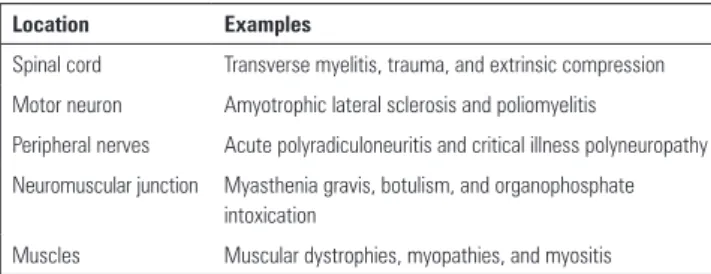
Comment - In respiratory failure from neuromuscular disease, ventilatory support depends on the location of the injuries (from spinal cord injuries to direct muscle involvement) (Table 1).
Myasthenic-speciic treatment with immunoglobulin or plasmapheresis should be initiated early.(53-55)
Suggestion - Consider using NIV (bilevel positive airway pressure [BiPAP]) in patients with myasthenic crisis
in an attempt to prevent OTI, with PCO2 >50mmHg
being a predictor of BiPAP failure; the use of NIV can be considered in persistent or recurrent weakness following extubation.(53-55)
Suggestion - Periodically assess patients with myasthenic crisis by measuring PImax, PEmax, and VC.
Patients with VC <20mL/kg, PImax <-30cmH2O, and
PEmax >40cmH2O can undergo a trial treatment with
NIV (BiPAP) and, if there is failure, they should be
electively intubated to prevent emergency OTI.(53-55)
Suggestion - Conduct an intensive respiratory program, including sighs, use of PEEP, frequent aspiration of the bronchial tree, respiratory therapy, body position change, and administration of antibiotic therapy (in cases of documented infection), in patients placed on MV for a myasthenic crisis.(53-55)
Duchenne muscular dystrophy
Comment - Duchenne muscular dystrophy (DMD) is a recessive X-linked disease that afects 1:3,000 male births. It is characterized by progressive loss of muscle strength, with VC decreasing progressively until the onset of respiratory failure, usually between 18 and 20 years of age, with a consequent need for ventilatory support. Most patients develop cardiomyopathy. Forced VC (FVC) <1L and the onset of nocturnal hypoventilation are signs of poor prognosis, and NIV can be used to improve survival and quality of life outcomes.(56-58)
Suggestion - Use NIV in cases of nocturnal hypoventilation and/or decreased VC (<1L).
Suggestion - Invasive ventilation via elective tracheostomy is indicated for patients who do not tolerate
NIV or who have been intubated for an acute event.(56-58)
Amyotrophic lateral sclerosis
Comment - Amyotrophic lateral sclerosis (ALS) is a degenerative motor neuron disease, during the course of which respiratory failure due to muscle failure may occur. Chronic aspiration due to bulbar muscle dysfunction and inefective cough are additional complications. Most patients die from respiratory
complications that vary in course.(59,60)
Table 1 - Location of the neuromuscular injuries and examples
Location Examples
Spinal cord Transverse myelitis, trauma, and extrinsic compression Motor neuron Amyotrophic lateral sclerosis and poliomyelitis
Peripheral nerves Acute polyradiculoneuritis and critical illness polyneuropathy Neuromuscular junction Myasthenia gravis, botulism, and organophosphate
intoxication
Muscles Muscular dystrophies, myopathies, and myositis
Acute polyradiculoneuritis (Guillain-Barré syndrome)
Comment - One third of patients with acute polyradiculoneuritis (Guillain-Barré syndrome) require MV during the course of the disease. Generalized weakness, rapid progression, and bulbar involvement are associated with the need for MV in such patients.(49-52)
Suggestion - Patients with acute polyradiculoneuritis should be periodically assessed by measuring maximum inspiratory pressure (PImax) from residual volume, maximum expiratory pressure (PEmax) from TLC, and
vital capacity (VC). Patients with PImax <-30cmH2O,
PEmax <40cmH2O, and VC <20mL/kg or a reduction of
more than 30% in VC should be electively intubated to prevent emergency OTI.(49-52)
Recommendation - NIV should be used with care because of the instability of acute polyradiculoneuritis. herefore, OTI and invasive MV should not be delayed when there is deterioration of lung function.(49-52)
Suggestion - he decision to tracheostomize patients with Guillain-Barré syndrome may be postponed for 2 weeks. If, after 2 weeks, lung function test results do no improve signiicantly, tracheostomy should be considered. If lung function test results are improving, tracheostomy
may be postponed until weaning is achieved.(49-52)
Myasthenia gravis
Recommendation - Use NIV in patients with ALS, except in the subgroup of patients with severe bulbar dysfunction.
Recommendation - Invasive MV via tracheostomy is indicated in patients with impaired airway protection and severe bulbar dysfunction, and it should only be instituted after its complications, as well as its social and logistical implications, are discussed in detail with the patients and their families.
Recommendation - Indications for ventilatory support
include VC <50% of predicted, PImax <-30cmH2O or
<60% of predicted, peak expiratory low (PEF) <270L/min, PCO2 >4 mmHg, and nocturnal hypoventilation.(59,60)
Suggestion - he parameters for invasive and NIV are as follows. An oral or nasal mask can be used provided that it is properly itted. BiPAP should be used. Invasive MV via tracheostomy is usually carried out in the ventilation mode that is most suitable for the type of demand, if there is associated lung disease. Patients should be monitored for episodes of atelectasis, accumulation of secretions, and
pneumonias.(61)
MECHANICAL VENTILATION IN PATIENTS WITH CARDIOVASCULAR DISEASE
Comment - he goal of MV in patients with cardiovascular disease is to adjust oxygenation and ventilation and to ensure cardiac output.
Recommendation - Achieve an SpO2 ≥94% by using
the lowest possible FiO2.
Recommendation - Noninvasive MV with either continuous positive airway pressure (CPAP) or BiPAP is safe, and both modes have similar efects and are efective in preventing OTI. hey should be applied as a form of
ventilatory support during acute pulmonary edema.(62-68)
Recommendation - Apply the protective strategy to MV patients with cardiovascular disease.(69,70)
Recommendation - Applying recruitment maneuvers is safe in patients with cardiovascular disease provided that there is proper monitoring and appropriate care.(69,70)
Suggestion - Monitoring of cardiac output and measurement of extravascular lung water are suggested in MV patients with cardiovascular disease and ARDS, with the aim of volume adjustment and hemodynamic optimization.(71)
Suggestion - Monitoring of cardiac output, in patients with cardiovascular disease, can be performed with a pulmonary artery catheter (PAC) or by means of pulse contour analysis.(69,70)
Suggestion - Transthoracic echocardiography can be performed in MV patients with cardiovascular disease
who are hemodynamically unstable.(70)
Suggestion - Perform transthoracic echocardiography in patients who exhibit hemodynamic instability after undergoing RM, in order to assess them for volume status and right ventricular (RV) dysfunction.(69,70)
Recommendation - Discontinuation of MV in patients with cardiovascular disease follows the recommendations for patients with no cardiovascular disease. he use of NIV should be prioritized to facilitate the process of discontinuation of MV, and NIV should be applied immediately after extubation.(62-70)
Suggestion - Increased brain natriuretic peptide (BNP) levels during weaning from ventilation in patients with cardiovascular disease show accuracy in predicting weaning failure.(70,72)
Recommendation - A positive luid balance should be avoided in MV patients with cardiovascular disease who
are hemodynamically stable.(70)
Recommendation - Inhaled nitric oxide is an efective strategy in MV patients with cardiovascular disease, RV
dysfunction, and pulmonary hypertension.(70)
Recommendation - No mode of ventilation is recommended as preferable in patients with cardiovascular disease.(70)
Suggestion - In patients on inotropic support, this
support may be continued until after extubation.(70)
Mechanical ventilation in patients with cardiovascular disease undergoing surgery
Tidal volume
Recommendation - he use of a Vt of 6mL/kg predicted body weight, in the volume-control mode or suicient delta of inspiratory pression to maintain this
same volume in PCV.(70)
Positive end-expiratory pressure
Recommendation - Apply PEEP during general anesthesia, because it is associated with improved oxygenation and prevention of atelectasis.(70)
Alveolar recruitment maneuvers
Fraction of inspired oxygen
Recommendation - At induction of anesthesia, use
an FiO2 of 100% to ensure adequate oxygenation for
intubation. Fractions of oxygen needed to maintain SpO2
>94% are recommended.(70)
Discontinuation of mechanical ventilation
Recommendation - Discontinuation of MV should be gradual, and pressure support ventilation (PSV) can be used. NIV is an important resource, which should be used immediately after extubation.(62-70)
Postoperative analgesia
Recommendation - Achieving adequate postoperative analgesia is associated with optimization of postoperative pulmonary function.
MECHANICAL VENTILATION IN INTERSTITIAL LUNG DISEASES
Comment - Interstitial lung diseases (ILDs) are a heterogeneous group of diseases that predominantly afect the lung interstitium, with varying degrees of
inlammation and ibrosis,(73,74) and that can progress
with varying degrees of hypoxemia and gradual reduction in lung volumes. Patients with ILD may require MV because of a number of factors, including the anesthesia process in surgical procedures, such as open lung biopsy and other elective or emergency operations, respiratory infections leading to respiratory failure, and acute exacerbations (AEs - noninfectious) of the underlying interstitial disease.(75,76)
Indications for mechanical ventilation
Comment - Respiratory failure in patients with ILD should be divided into two groups: progression of the underlying disease and AEs. AEs are characterized by acute, unexplained worsening of clinical symptoms of ILD, especially dyspnea and cough, usually in the last 30 days, accompanied by radiological worsening, often in the form of ground-glass changes superimposed on previous changes. AEs were initially described in patients with idiopathic pulmonary ibrosis (IPF), but they can
occur in other forms of ILD.(76-79) he onset of AEs
seems to occur at some point in the course of IPF in 5 to 10% of patients, and mortality in those requiring MV is nearly 100%.
Recommendation - Before diagnosing an AE, it is necessary to exclude infections, pulmonary thromboembolism, cardiac dysfunction, drug-induced pulmonary toxicity, etc.(76-80)
Acute complication
Suggestion - In AEs of ILD, the patient’s previous status should be assessed. Invasive MV is indicated if the cause of acute respiratory failure is diagnosed as not being due to progression of the underlying disease.
Progression of the underlying disease
Recommendation - ICU admission and invasive MV should be avoided, and any decision in this regard should be discussed with the patients or their families.
Noninvasive mechanical ventilation
Suggestion - NIV can be used as initial ventilatory support in patients with ILD who develop acute respiratory failure or as palliative ventilatory support in patients who have previously expressed the desire not to be intubated. Either CPAP or bilevel NIV can be used and should be applied early.
Recommendation - he use of NIV should be monitored at the bedside by a healthcare professional within thirty minutes to two hours. For NIV to be considered successful, the following criteria should be met: reduction of f, increase in Vt, improvement of the level of consciousness, reduction or cessation of the use
of accessory muscles, increase in PaO2 and/or SpO2,
and reduction of PaCO2 without signiicant abdominal
distension. In unsuccessful cases, OTI and invasive MV should be performed immediately. Success is expected in 50% of this population.(80)
Noninvasive mechanical ventilation
Comment - Since the histological inding in AEs is difuse alveolar damage (DAD), similar to that observed in patients with ARDS, and given the lack of prospective studies, some authors have suggested that strategies used for ARDS could be extrapolated to patients with AEs of ILD. herefore, some experts advocate the use of protective ventilation, with a low Vt, set at approximately
6mL/kg ideal body weight, and a Pplat ≤30cmH2O.(80-84)
of patients with ILD who underwent MV: Suh et al. reported low mortality in a group of patients with acute interstitial pneumonia who received an early intervention strategy, which involved a series of measures, including MV with a low Vt and a moderate PEEP, with a median
of 11cmH2O.(79) In contrast, in a more heterogeneous
group of patients with ILD who underwent MV,
Fernandez-Perez et al. observed that a PEEP >10cmH2O
on the irst day of MV was associated with high mortality, but they themselves comment that a high PEEP may be a marker of greater respiratory failure severity.(76)
Suggestion - Patients with ILD who require MV should be ventilated with a low Vt, set at approximately
6mL/kg ideal body weight, and a Pplat <30cmH2O; a
high f (>30 breaths/min) and a short inspiratory time can be used to prevent hypercapnia. Use a PEEP between 5
and 10cmH2O.
Suggestion - he use of a high PEEP can be attempted with caution and should be individualized for each patient. Rescue therapies for refractory hypoxemia, such as prone positioning, RM, and nitric oxide, can be used in referral centers with expertise in these therapies.
DISCONTINUATION OF MECHANICAL VENTILATION Identifying patients who are ready to wean
Recommendation - Discontinue patients from invasive MV as soon as it is clinically possible.(85,86)
Recommendation - he deinitions of terms related to discontinuation of MV must be made clear in ICU guidelines. he concept of “successful weaning” refers to a patient successfully completing the spontaneous breathing trial (SBT) while still connected to the ventilator. “Successful extubation” refers to a patient having the endolaryngeal tube removed (extubation) after passing the SBT and not being reintubated in the following 48 hours. In tracheostomized patients, successful extubation means tolerating being disconnected from the ventilator after a well succeeded SBT and not needing to be reconnected to the ventilator in the following 48 hours.(87-89)
Recommendation - Assess and identify patients daily (active surveillance by means of internal guidelines established by a multidisciplinary team) with a view to the possibility of discontinuing ventilation, in order to reduce duration of MV and decrease costs.(90-94)
Sedation
Recommendation - Sedation should be interrupted daily to assess the patient’s ability to maintain spontaneous ventilation(95) (see the “Sedation and Analgesia” section in
part 1 of the present Recommendations).
Criteria required for assessment of readiness to wean
Recommendation - Perform active surveillance, including the topics in table 2.(88,90-96)
Table 2 - Topics that are to be routinely assessed during active surveillance in mechanically ventilated patients
PaO2≥60mmHg on FIO2≤0.4 and PEEP≤5 to 8cmH2O
Hemodynamic stability, with good tissue perfusion, with or without low doses of vasopressors, and absence of decompensated coronary artery disease or arrhythmias with hemodynamic effects
Resolution or control of the cause of respiratory failure Capability to initiate an inspiratory effort
Zero or negative fluid balance in the last 24 hours Normal acid-base and electrolyte balance
Delay extubation when patients are scheduled to be taken to tests or surgery requiring general anesthesia in the next 24 hours
PaO2 - partial pressure of oxygen; FIO2 - fraction of inspired oxygen; PEEP - positive
end-expiratory pressure.
Predictive indices
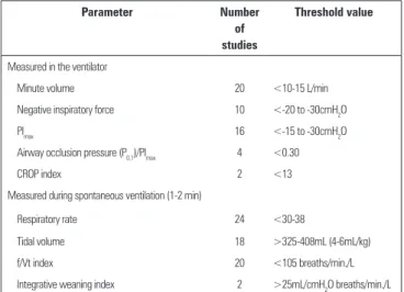
Recommendation - he most accurate weaning-predictive indices are the rapid shallow breathing index (RSBI), i.e., f divided by Vt (f/Vt), and the integrative weaning index (IWI) (Table 3). hey should only be calculated in situation in which the decision is diicult and must NOT be used as the sole tool in the decision-making process regarding the SBT.(97-99)
Autonomous breathing trial (spontaneous breathing trial)
Recommendation - In the SBT, patients should be
placed on T-tube or ventilated with PSV of 5-7cmH2O
for 30-120 minutes. During the SBT, patients should be
monitored for signs of failure.(85-94) Successful SBTs are
deined as those in which patients maintain an adequate breathing pattern, as well as adequate gas exchange, hemodynamic stability, and comfort (Table 4).(85-94,100-102)
Table 3 - Significant parameters for predicting successful weaning
Parameter Number
of studies
Threshold value
Measured in the ventilator
Minute volume 20 <10-15 L/min Negative inspiratory force 10 <-20 to -30cmH2O
PImax 16 <-15 to -30cmH2O
Airway occlusion pressure (P0.1)/PImax 4 <0.30
CROP index 2 <13 Measured during spontaneous ventilation (1-2 min)
Respiratory rate 24 <30-38
Tidal volume 18 >325-408mL (4-6mL/kg) f/Vt index 20 <105 breaths/min./L Integrative weaning index 2 >25mL/cmH2O breaths/min./L CROP - compliance, respiratory rate, oxygenation, and pressure; PImax - maximum inspiratory
pressure; f - respiratory rate; Vt - tidal volume.
Table 4 - Signs of intolerance to the spontaneous breathing trial
Respiratory rate >35 breaths/min Arterial de O2 saturation <90% Heart rate >140 bpm
Systolic blood pressure >180mmHg or <90mmHg
Signs and symptoms of agitation, sweating, altered level of consciousness
How to determine the timing of extubation Assessment of airway protection
Recommendation - Determine whether the patient has the required level of consciousness (a Glasgow coma scale score >8), an efective cough (a positive white card test result and a peak low >60 lpm), and a small amount of secretions (with no need for suctioning every 1 or 2 hours).(103)
Assessment of airway patency
Suggestion - Test airway patency in patients at higher risk for laryngeal stridor and airway obstruction (prolonged ventilation and trauma); to that end, a qualitative or quantitative approach can be used. Perform thorough suctioning of the mouth and larynx before delating the tube cuf for the test, in order to prevent unwanted material from entering the lower airways iatrogenically (Table 5).(104)
Use of corticosteroids
Recommendation - In patients at high risk for laryngeal stridor and laryngeal edema, as identiied by the
Table 5 - How to perform the endotracheal tube cuff-leak test in mechanically ventilated patients
1. Before performing the cuff-leak test, perform suctioning of tracheal and oral secretions and set the ventilator to assist-control mode (VCV).
2. With the cuff inflated, record Vti and Vte and observe whether they are similar.
3. Deflate the cuff.
4. Record Vte during six consecutive breaths; observe that Vte will reach a plateau after the first breaths.
5. With the cuff deflated, Vte is expected to be <90% of (the preset) Vti, situation in which the test is considered adequate.
VCV - volume-cycled ventilation; Vte - expired tidal volume; Vti - inspired tidal volume.
cuf leak test, the preventive use of corticosteroids may be beneicial. he prescribed doses range from 20 to 40mg of i.v. methylprednisolone every 4 to 6 hours, started at least 4 hours, more commonly 12 to 24 hours, before extubation.(105)
Use of noninvasive ventilation in the discontinuation of mechanical ventilation
Use of noninvasive ventilation to facilitate
discontinuation of mechanical ventilation - early weaning (noninvasive ventilation as a facilitation technique)
Recommendation - It is recommended that NIV be used to facilitate early discontinuation of MV in patients with chronic obstructive pulmonary disease (COPD), even in those who fail the SBT, provided that their clinical status is adequate. he patient should be managed in
centers with experience in the use of NIV (Figure 1).(106)
Use of noninvasive ventilation to prevent extubation failure (noninvasive ventilation as a preventive technique)
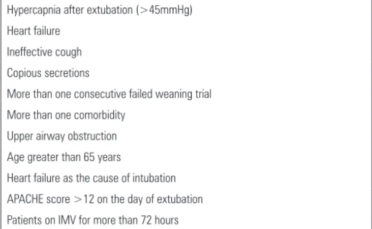
Recommendation - NIV should be used immediately after extubation, in a preventive manner, in patients identiied as high risk, especially in hypercapnic patients (Figure 1 and Table 6).(107-111)
Use of noninvasive ventilation in respiratory failure after extubation (noninvasive ventilation as a curative technique)
Figure 1 - Use of noninvasive ventilation for discontinuation of mechanical ventilation. SBT - spontaneous breathing trial; NIV - noninvasive ventilation; COPD - chronic obstructive pulmonary disease; RF - acute or exacerbated respiratory failure.
Table 6 - Noninvasive ventilation as a preventive technique: risk factors for respiratory failure
Hypercapnia after extubation (>45mmHg) Heart failure
Ineffective cough Copious secretions
More than one consecutive failed weaning trial More than one comorbidity
Upper airway obstruction Age greater than 65 years
Heart failure as the cause of intubation APACHE score >12 on the day of extubation Patients on IMV for more than 72 hours
APACHE - Acute Physiology and Chronic Health Evaluation; IMV - invasive mechanical ventilation.
How to manage patients with weaning failure (patients who fail their first spontaneous breathing trial)
Recommendation - Reinstate ventilatory support that provides patients with comfort and adequate gas exchange, maintaining it for 24 hours, so that the SBT can be repeated. Try to identify the causes of failure(86) (for diicult-to-wean
patients and those requiring long-term weaning, see the related section of the present Recommendations).
Gradual weaning methods
Recommendation - Avoid the use of synchronized inspiratory mandatory ventilation, because it can increase
the time to discontinuation of invasive MV.(113)
How to manage patients with extubation failure
Recommendation - Reintubate patients as soon as possible; identify and treat the causes of failure; and, as soon as possible, restart the discontinuation process (exception: noninvasive ventilation as a curative technique may be tried in surgical patients).
PATIENTS WITH PROLONGED WEANING
Suggestion - Use classiication deinitions for the duration of the weaning process to categorize your
patients as undergoing simple weaning(114) (when patients
successfully complete their irst SBT), diicult weaning (when patients fail their irst SBT and require up to three SBTs or up to seven days of MV following the irst SBT), and, inally, prolonged weaning (when patients fail more than three consecutive SBTs or require >7 days of MV following the irst SBT).
Suggestion - Use the concept of prolonged MV deined as MV that is needed for ≥21 consecutive days for 6 hours daily.
Recommendation - Identify causes of failure to discontinue MV (Table 7).(88,114-130)
Table 7 - Causes of failure to discontinue mechanical ventilation(89,115-131)
Age ≥65 years
Decreased diaphragmatic function Presence of comorbidities
Presence de delirium, depression, anxiety Persistent infection/inflammatory states
Decompensated cardiac, respiratory, neurological, and psychiatric diseases
Muscle disorders
Suggestion - Assess the possibility of critical illness polyneuropathy and of phosphorus, magnesium, calcium, and potassium disorders.
Endocrine and metabolic diseases
Recommendation - Adequately control diabetes,
hypothyroidism, and adrenal failure.
Comment - Obesity can be an additional factor for prolonged weaning, because it is characterized by an
increase in O2 consumption (VO2) and CO2 production
(VCO2), a decrease in static compliance, VC, and TLC,
Electrolyte and acid-base disturbances
Recommendation - Monitor, diagnose, and treat states of hyperhydration, which are associated with increased morbidity, mortality, and ICU length of stay. Identify and treat cases of metabolic alkalosis, the most common causes of which are chronic respiratory acidosis and use of diuretics. Metabolic alkalosis is associated with increased
mortality, decreased respiratory drive, decreased O2
delivery (DO2), shift of the oxyhemoglobin curve to the
left, ventilation/perfusion (V/Q) mismatch, and systemic vasoconstriction. Provide an adequate means of nutrition (see the related section of the present Recommendations) to prevent malnutrition, increased protein catabolism, reduced body muscle mass and decreased efectiveness of the thoracic pump mechanism, with reduced strength and
endurance and increased VO2, and perpetual dependence
on the ventilator. Decreased albumin is associated with
prolonged weaning and should be monitored.(114-130)
Rehabilitation strategies and strategies to facilitate discontinuation from mechanical ventilation
Reassessment of the underlying disease and comorbidities
Recommendation - Treat cardiac, pulmonary, psychiatric, and infectious underlying diseases as much as possible, and maintain clinically adequate nutrition.
Specific care for discontinuation of mechanical ventilation
Suggestion - Transfer patients undergoing prolonged weaning to a unit specialized in discontinuation of MV, if available.
Recommendation - Tracheostomy is indicated in patients who repeatedly fail SBTs, from the tenth day of MV onward, as part of a discontinuation protocol and in accordance with speciications in the related section of the present Recommendations.
Suggestion - Perform SBTs daily, using a tracheostomy collar or T-piece. In cases of tolerance (f<35 breaths/minute or over 35 breaths/minute for less than 5 consecutive minutes; SaO2 >90%; HR <140 beats/minute or a sustained change of 20% in any direction; 90mmHg> blood pressure <180mmHg with no anxiety or diaphoresis), gradually increase the duration of T-piece use and rest the patient overnight on assist-control ventilation. In cases of failure, return the patient to an assist-control mode for rest, in
Suggestion - Long-term MV is characterized when there is failure of the entire discontinuation process, especially in patients with spinal trauma, end-stage COPD, advanced dementia, pulmonary ibrosis, and irreversible neuromuscular disease. In these situations, explain the concept of futile treatment and palliative care to patients and families in order to arrive at a joint decision regarding the best approach.(114)
Recommendation - Early physical therapy and passive mobilization should be performed during MV and also during the discontinuation process. hese activities are considered safe and are associated with better functional results and shorter duration of MV(121,130) (see the related
section of the present Recommendations).
Suggestion - Inspiratory muscle training can be considered in patients who fail to wean, in order to increase PImax and facilitate discontinuation of ventilatory support.(130,131)
HEMODYNAMIC CHANGES AND HEMODYNAMIC CARE IN PATIENTS ON INVASIVE MECHANCIAL VENTILATION
Comment - he cardiovascular efects of positive pressure MV are described in Table 8.
Table 8 - Cardiovascular effects of positive pressure mechanical ventilation
Hemodynamic effect of positive pressure ventilation
Likely effect on cardiac output
Preload dependent Afterload dependent RV Decreases preload
↓ ↓
Increases afterload LV Decreases preload
↓ ↑
Decreases afterload
RV - right ventricle; LV - left ventricle.
Hemodynamic care in mechanically ventilated patients
Recommendation - Resuscitation aimed at restoration of tissue perfusion begins in the early acute phase, in which resuscitation should be performed as soon as possible
to achieve mixed venous O2 saturation (SvO2) >65% or
central venous saturation (SvcO2) >70%, blood lactate
concentration <2mmol/L (18mg/dL), and adequacy
of DO2/VO2.(132-135) he process should continue in the
post-resuscitation phase (when adequate tissue perfusion is achieved), and luid restriction should be performed to maintain a zero or negative luid balance.(136)
Suggestion - In ARDS patients receiving PEEP
≥15cmH2O and <20cmH2O, perform echocardiography at
ARDS patients receiving PEEP ≥20cmH2O or experiencing hemodynamic instability, perform monitoring with serial echocardiography and/or with a volumetric PAC, if available.(137,138)
Suggestion - During MRS with decremental PEEP titration, use invasive blood pressure monitoring; perform echocardiography after 6 hours, or earlier if the patient exhibits hemodynamic instability, to assess RV dysfunction.(137,138)
Suggestion - In moderate/severe ARDS, consider extravascular lung water monitoring (if available).(139,140)
Suggestion - Avoid systemic systemic vasodilators in refractory hypoxemia (they inhibit hypoxic vasoconstriction).(141)
Suggestion - Interpret SvO2 by considering PaO2. High PaO2 values can increase SvO2.(142)
Mechanical ventilation in patients with left ventricular failure
Recommendation - For the diagnosis of left ventricular (LV) failure, use Doppler echocardiography, which should show ejection fraction, velocity-time integral, as well as assess diastolic function [E/A, E/E’, global
end-diastolic volume].(143) If you use a PAC, a pulmonary
artery occlusion pressure >18mmHg and a cardiac index <2.2L/min/m2 characterize LV failure.(143)
Recommendation - Use inotropes, vasopressors if necessary, diuretics, and vasodilators when possible. In selected cases, use mechanical circulatory support.(143)
Suggestion - Favor the use of high PEEP (because of decreased LV preload and afterload). If there is concomitant RV failure, increase PEEP carefully (monitor
the RV and RV low).(144) Prevent severe hypercapnia
(pH <7.15 or PaCO2 >80mmHg).(145) Consider kidney
ultrailtration for achieving a negative luid balance in situations of refractoriness to diuretics.(146)
Mechanical ventilation in patients with right ventricular failure
Recommendation - Regarding diagnosis, use Doppler echocardiography (RV diastolic diameter >3.5cm, RV/LV ratio >1, intraventricular septal lattening or paradoxical interventricular septal motion, pulmonary artery systolic pressure >35mmHg, tricuspid annular plane systolic
excursion <1.8cm)(147) or a PAC (a volumetric PAC, if
available): central venous pressure >pulmonary artery
Table 9 - Suggestions for monitoring, treatment, and specific care in patients with right ventricular failure(146,148-152)
Perform monitoring with a PAC (a volumetric PAC, if available)(148)
Prevent hypervolemia (reverse Bernheim effect), promote a negative fluid balance(149)
Favor the use of a low PEEP (<10cmH2O) and a Vt of 6mL/kg predicted weight or lower(149)
Prevent hypoxemia (it increases pulmonary vascular resistance caused by hypoxic vasoconstriction)(149)
Prevent severe hypercapnia (it increases RV afterload)(150)
Consider using dobutamine at low doses (to prevent tachycardia) or milrinone(149)
Use nitric oxide/sildenafil testing associated with monitoring with a PAC or transthoracic echocardiography. In centers where nitric oxide is unavailable, a therapeutic trial of sildenafil can be used(151)
Avoid abrupt withdrawal of inhaled nitric oxide(152)
Consider kidney ultrafiltration for achieving a negative fluid balance in situations of refractoriness to diuretics(146)
PAC - pulmonary artery catheter; PEEP - positive end-expiratory pressure; Vt - tidal volume; RV - right ventricular.
occlusion pressure; mean pulmonary artery pressure
>25mmHg; systolic index <30mL.min-1.m-2; RV
end-diastolic volume index >140mL.m-2.(137)
Suggestion - Details regarding monitoring, treatment, and speciic care are shown in table 9 and igure 2.
Resources available for hemodynamic monitoring in mechanically ventilated patients
Suggestion - Suggested methods of hemodynamic monitoring in MV patients include predicting the response to luid loading (a >15% increase in the cardiac index) in patients with the following characteristics:
PEEP <10cmH2O; Vt of 8-10mL.kg-1 ideal body weight;
f <30 min-1; respiratory compliance >30mL.cmH2O; no
arrhythmias; no respiratory efort; no cor pulmonale; delta pulse pressure >13%; systolic volume variation >10%; delta velocity-time integral >15%; superior vena cava collapsibility >36%; and inferior vena cava distensibility >18%.(153,154) Also perform the expiratory
port occlusion maneuver - in spontaneously breathing
patients receiving PEEP ≤10cmH2O,(155) with a delta
central venous pressure >1mmHg (1.36cmH2O).(156) In
patients receiving high PEEP and/or low Vt, passive leg
raising maneuver,(157) luid challenge with small aliquots
Recommendation - Regarding the timing for speech-language pathology evaluations after extubation, it is
recommended that patients be evaluated 24(166) to 48 hours
after extubation and that those who have dysphagia and are at risk for aspiration be started on speech therapy.(167-171)
Suggestion - Do NOT perform speech-language therapy in orotracheally intubated patients, because there is a lack of clinical evidence regarding its beneits to the swallowing function; however, early identiication of patients who, even during OTI, have several associated risk factors that may compromise swallowing dynamics (patients with cardiovascular disease, patients with Parkinson’s disease, post-stroke patients, and patients with dementia) is indicated.
Suggestion - Perform clinical speech-language evaluations (structural and functional) at the bedside(163,172)
and determine the need for instrumental examination of swallowing (functional nasal iberoptic endoscopy and
modiied barium swallow test).(173)
Suggestion - he speech-language pathologist should deine the type of food consistency and the need for the use of thickeners for the administration of luids, in collaboration with the nutrition service, when the patient is ready to initiate oral intake.
Suggestion - Fit a speaking valve into the mechanical ventilator circuit or directly into the tracheostomy, with the aid of a physical therapist and/or a physician, provided that cuf delation is feasible and patient tolerance is assessed.(174,175)
Suggestion - In tracheostomized patients, use a blue food dye in the food delivered orally and/or in the saliva swallowing test, to evaluate the occurrence of discharge of blue saliva and/or secretions through the tracheostomy, characterizing an aspiration event.(176-178)
Suggestion - Assess for signs and symptoms of dysphagia during the delivery of oral nutrition, especially those that may be associated with bronchial aspiration, such as choking, cough, and wet voice.(179-181)
Suggestion - Discuss with the medical team the use of xerostomic medications in patients on MV and/or with tracheostomy who do not tolerate cuf delation or who experience signiicant aspiration of saliva.(182)
Suggestion - Perform direct and indirect swallowing therapy in patients who have dysphagia and/or are at risk for aspiration.(183)
NURSING CARE IN PATIENTS RECEIVING INVASIVE OR NONINVASIVE VENTILATORY SUPPORT
Comment - he nursing team, as part of the multidisciplinary ICU team, actively participates in the
Figure 2 - Hemodynamic management algorithm suggested in mechanically ventilated patients. LV - left ventricular; RV - right ventricular; MV - mechanical ventilation; PEEP - positive end-expiratory pressure.
SPEECH-LANGUAGE PATHOLOGY CARE IN THE REHABILITATION OF PATIENTS AFTER MECHANICAL VENTILATION
Comment - he role of speech-language pathologists in the ICU is endorsed by Resolution-RDC 07/2010 of
the Brazilian Agência Nacional de Vigilância Sanitária
(ANVISA, National Health Oversight Agency). In dysphagia, the role of speech-language pathologists in the ICU is regulated by Resolution 356 of the Brazilian Federal Speech-Language Pathology Council, passed
on December 6, 2008, and published in the Diário
Oficial da União (Oicial Federal Government Gazette)
on December 9, 2008. In a multidisciplinary team, the speech-language pathologist evaluates the safety of oral feeding/swallowing and manages swallowing diiculties, thereby contributing to the prevention of aspiration pneumonia and to the tracheostomy weaning process.
Suggestion - Request a speech-language pathology
evaluation(159-165) for all patients who required OTI ≥48
administrative and healthcare activities involving invasive and noninvasive support in MV patients.
Care of circuits, filters, and humidifiers
Recommendation - Maintain the lower airways warm and moist during MV.
Recommendation - Replace (hygroscopic and hydrophobic) heat and moisture exchangers every 7 days, provided that appropriate height and positioning of the devices relative to the endotracheal tube are maintained (the devices should be connected vertically to the tube and the circuit so that they are not looded with droplets and dirt). If there is dirt, condensation, or damage, the ilter should be replaced.(184)
Recommendation - Do not replace the mechanical ventilator circuit routinely; replace it only when there is dirt visible to the naked eye, when there is damaged, or in cases of prolonged ventilation (>30 days).(185,186)
Cleaning and maintenance of the equipment
Recommendation - Mechanical ventilator circuits require high-level disinfection (0,5% sodium hypochlorite and a contact time of 60 minutes) or sterilization.(187)
Precautions during bed bath and patient repositioning
Recommendation - Assess the vital signs, analyze and record the MV parameters (ventilation mode, peak
pressure, PEEP, f, Vt, and FIO2), and check the alarms
and clinical parameters before a bed bath and before patient repositioning. Continue cardiac monitoring and
SatO2 monitoring during the bed bath and during patient
repositioning. Allow a 5- to 10-minute equilibrium period before determining hemodynamic intolerance/instability due to patient repositioning and/or a bed bath.(188,189)
Recommendation - Work with the multidisciplinary team to determine the most appropriate time to perform a bed bath in critically ill, clinically unstable patients. he nurse should assess the patient before allowing the bath, postponing it in cases of severity that can compromise patient safety.
Recommendation - Perform patient repositioning every 2 hours, with a lift sheet and at least two nursing professionals.(190)
Suggestion - Perform continuous lateral rotation therapy with the use of a bed for kinetic therapy, when available.(191)
Recommendation - Maintain the head of the bed between 30º and 45º in MV patients. he evidence is conlicting as to aspiration of gastric content (45º) and pressure ulcers (30º). Positioning at 30° is preferred as long as it does not pose risks to or cause conlict with
medical and nursing procedures.(192)
Suggestion - Use the beach chair position 2 to 4 times/day; it requires less personnel than do other interventions and therefore allows early mobility of ICU
patients and improvement in pulmonary function.(193)
Recommendation - Maintain the cuf pressure of the endotracheal tube between 18 and 22mmHg or between
25 and 30cmH2O (cuf meter) in order to prevent air leaks
without excessive compression of the tracheal mucosa.
Avoid cuf pressures >22mmHg or 30cmH2O. Check the
cuf pressure at least 4 times/day and before performing oral hygiene.
Recommendation - Maintain the endotracheal tube secured and centralized by using an adhesive device or a shoelace so that the cuf pressure is homogeneously distributed in the trachea. Pay attention to lesions in the oral cavity, in the corners of the lips, and on the face.(194)
Recommendation - he precautions to be followed during patient repositioning and while tilting the patient laterally in a bed bath are described in table 10.(195)
Table 10 - Precautions during patient repositioning and while tilting the patient laterally in a bed bath
Visualize all of the stretchers and equipment that are connected to the patient Be careful not to pull the mechanical ventilator circuit during head-of-bed elevation, lateral tilting for patient repositioning and/or a bed bath, in order to prevent accidental extubations. Check whether the ventilatory device is fixed; release the ventilator circuit from the rack
Maintain the head of the bed at 30º
Pull the patient up in bed, monitoring continuously the ventilator Tilt the patient laterally with his/her head supported by the headrest
Perform hygiene of the patient's back and buttocks, most of it already with the patient placed in the lateral decubitus position, facing the side where the ventilator is
Elevate the head of the bed and fix the circuit to the ventilator rack loosely so that, if the patient is moved in bed, the ventilator circuit is not pulled
and nipples should be protected. Patient rotation should be performed in two steps, with total attention being paid to the invasive devices. he dorsal electrocardiogram should be monitored, and the reverse Trendelenburg position may be used to decrease facial edema.(194,195)
Recommendation - Use a closed suctioning system to perform tracheal suctioning in hemodynamically unstable patients, in order to prevent desaturation in at-risk patients (i.e., patients with cardiovascular disease) and to maintain alveolar recruitment and prevent atelectasis in
ARDS patients receiving PEEP ≥10cmH2O. he system
should be changed every 7 days. Closed suctioning systems have not been shown to reduce the occurrence of ventilator-associated pneumonia, mortality, or ICU
length of stay, when compared with open systems.(196)
Specific instructions for oral hygiene, oral feeding, and enteral feeding
Recommendation - It is recommended that oral hygiene with brushing be performed every 12 hours, with an aqueous solution containing 0.12% chlorhexidine gluconate. In the irst part of these recommendations, the theme “Mechanical ventilation in PAV” recommended the use of 2% based on evidence of best result at this concentration. Nevertheless, it is not yet available in the Brazilian market. In the intervals, oral hygiene should be performed with distilled or iltered water and/or
alcohol-free mouthwash four times/day.(196-198)
Recommendation - Check the cuf pressure of the endotracheal tube or tracheostomy before performing oral hygiene.(199)
Recommendation - he gastric and post-pyloric routes can be used for enteral feeding in MV patients, with post-pyloric tube placement being reserved for patients with gastric intolerance and/or contraindication.(200)
Recommendation - Secure the nasogastric tube with a securing device (a commercially available nasal bridle or an adhesive device) in order to reduce the rate of
unintentional tube dislodgement.(201)
Suggestion - Monitor the diference between prescribed and delivered enteral nutrition as a marker of dietary compliance.(202)
PHYSICAL THERAPY CARE IN PATIENTS ON VENTILATORY SUPPORT
Comments - ICU patients may experience respiratory and muscle dysfunction and, over time, develop neuromuscular weakness and complications of immobility,
which can make discontinuation of MV diicult. Prolonged immobility leads to loss of motor function and loss of quality of life, both of which can be minimized with the institution of early mobilization and respiratory care. he incidence of ICU-acquired muscle weakness (neuromuscular weakness) in patients requiring prolonged MV ranges from
25 to 60%,(203) which contributes to increasing ICU and
hospital length of stay. Physical therapy works to maintain and/or restore the functionality of the patient by preventing musculoskeletal changes and respiratory complications.
Recommendation - A physical therapy diagnosis should precede any intervention.(204)
Recommendation - Physical therapy in MV patients in the ICU should be delivered 24 hours/day, having beneits in reducing duration of MV, ICU and hospital length of stay, hospital costs, and mortality.(200,205)
Physical therapy maneuvers and approaches in mechanically ventilated patients
Recommendation - Bronchial hygiene therapy (positioning, manual inlation, vibration, and chest compression): indicated in patients with increased Raw due to the presence of secretions, causing asynchrony of MV and/or reduced oxygenation; and mandatory in lobar atelectasis.(206)
Suggestion - Lung expansion techniques can be used in the presence of lung collapse, with reduced compliance and oxygenation.(207)
Recommendation - Perform inspiratory muscle training in patients with inspiratory muscle weakness who are undergoing prolonged MV, in order to improve muscle strength. he role of inspiratory muscle training in reducing duration of MV and successfully discontinuing MV has yet to be established.(208)
Early mobilization in noninvasive and invasive mechanical ventilation
Recommendation - Early mobilization should be initiated less than 72 hours following the initiation of mechanical ventilation, because it is feasible, safe, and results in signiicant functional beneits.(206)
Suggestion - Neuromuscular electrical stimulation and a cycle ergometer can be considered as a complement to the early mobilization program.(208)