LEAL GN ETAL.
490 REV ASSOC MED BRAS 2016; 62(6):490-493
AT THE BEDSIDE
What are the benefits of two-dimensional speckle tracking
echocardiography for diagnosis and treatment follow-up of
childhood-onset systemic lupus erythematosus myocarditis?
GABRIELA NUNES LEAL1*, MARIADE FÁTIMA DINIZ1, JULIANA BRUNELLI2, ALESSANDRO C. LIANZA1, ADRIANA M. E. SALLUM2,CLOVIS A. SILVA2
1Echocardiography Service, Instituto da Criança, Hospital das Clínicas, Faculdade de Medicina, Universidade de São Paulo (HC-FMUSP), São Paulo, SP, Brazil 2Pediatric Rheumatology Unit, Instituto da Criança, HC-FMUSP, São Paulo, SP, Brazil
Study conducted at the Echocardiography Service and at Pediatric Rheumatology Unit, Instituto da Criança, Hospital das Clínicas, Faculdade de Medicina, Universidade de São Paulo (HC-FMUSP), São Paulo, SP, Brazil
Article received: 6/29/2016 Accepted for publication: 7/26/2016
*Correspondence:
Address: Av. Dr. Enéas de Carvalho Aguiar, 647 São Paulo, SP – Brazil
Postal code: 05403-000 [email protected]
http://dx.doi.org/10.1590/1806-9282.62.06.490
Financial support: Supported by grants from Conselho Nacional de Desenvolvimento Cientíico e Tecnológico
(CNPq 472155/2012-1 and 303422/2015-7 - 1A to CAS), Federico Foundation to CAS and by Núcleo de Apoio à Pesquisa “Saúde da Criança e do Adolescente” / USP (NAP-CriAd) to CAS.
C
ASE REPORTA 17-year-old white female had a previous diagnosis of childhood-onset systemic lupus erythematosus(c-SLE) based on American College of Rheumatology classiication criteria: arthritis, pericarditis, proteinuria > 0.5 g/day, and the presence of antinuclear (ANA), double-stranded DNA (ds-DNA) and antiphospholipid autoantibodies.1 She pre-sented to the emergency room with a one-day history of fever (38oC), shortness of breath, chest pain on inspiration, and orthopnea. Regarding classic cardiovascular risk fac-tors, there was no history of arterial hypertension or chron-ic renal failure. Her lipid proile was considered normal for age and sex and her body mass index was 27.5 kg/m2 (be-tween 90th and 95th percentile). Previously to emergency room admission, she had never shown signs or symptoms of heart failure, and left ventricle (LV) ejection fraction (EF) documented by a routine echocardiogram one month ear-lier was 66% (normal ≥ 55%, by Teichholz method). There was no chamber enlargement, ventricular hypertrophy or signs of diastolic dysfunction. Despite that, a concomitant deformation analysis of LV by two-dimensional speckle trackingechocardiography (2DST) identiied reduced lon-gitudinal peak systolic strain in the apical 4-chamber view: -15.2%; normal range -22.18%±3.06 (My Lab 60 – Esaote,
Florence, Italy).2 Of note, LV circumferential peak systolic strain in the mid cavity was still preserved: -25.7% (normal
WHATARETHEBENEFITSOFTWO-DIMENSIONALSPECKLETRACKINGECHOCARDIOGRAPHYFORDIAGNOSISANDTREATMENTFOLLOW-UPOFCHILDHOOD-ONSETSYSTEMICLUPUSERYTHEMATOSUSMYOCARDITIS?
REV ASSOC MED BRAS 2016; 62(6):490-493 491
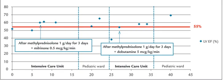
initiated. However, it was only after receiving 1 g of meth-ylprednisolone for three consecutive days that heart fail-ure symptoms disappeared and a normal LV ejection frac-tion was achieved (60%). She was discharged from the pediatric ICU on day 17 and admitted to the pediatric ward with a prescription of hydroxychloroquine 400 mg/day, prednisone 40 mg/day, and furosemide 20 mg/day. Anoth-er echocardiogram with 2DST was obtained on day 23, which revealed normal LV ejection fraction (65%) but still a striking reduced longitudinal peak systolic strain in api-cal 4 chamber view (-9%). Circumferential peak systolic strain in mid cavity was also below acceptable values (-16.8%). On day 24, she presented tachypnea, basal crept, and hepatomegaly. Glasgow score was reduced (11) and endotracheal intubation was necessary. The patient was sent back to pediatric ICU and inotropic support was re-introduced. A bedside echocardiogram showed LV ejection fraction of 38% and a small pericardial effusion. By that time, SLEDAI-2K was 30. Again, it was only after a three-day pulse of methylprednisolone that a normal LV ejection fraction (58%) was achieved. Computed tomography scan ruled out the hypothesis of pulmonary embolism or brain hemorrhage. Cerebral luid analysis showed aseptic men-ingitis, suggesting neuropsychiatric lupus manifestation. She was progressively weaned from mechanical ventilation and inotropes, and was sent back to the pediatric ward on day 36. A inal echocardiogram with 2DST on day 40 showed that, although LV ejection fraction was 58% and she had no more signs or symptoms of heart failure, lon-gitudinal peak systolic strain in apical 4-chamber view was still depressed (-15.4%). Circumferential peak systolic strain in mid cavity was once more within normal limits (-19.84%). Intravenous cyclophosphamide infusion (1 g/m2) was
pre-scribed and she was inally sent home after 42 days of hos-pitalization, with a prescription of captopril 100 mg/day, furosemide 20 mg/day, spironolactone 50 mg/day, pred-nisone 40 mg/day, and carvedilol 75 mg/day. LV ejection fraction throughout hospitalization is shown in Figure 1 and 2DST strain analysis in Figure 2. The Ethics Commit-tee of our University Hospital approved this study.
D
ISCUSSIONWe reported here the irst case of c-SLE myocarditis, in which 2DST deformation analysis was prospectively able to detect LV systolic function impairment even before the development of congestive heart failure or decreased ejec-tion fracejec-tion. Addiejec-tionally, this novel technique was clear-ly helpful in decision making throughout treatment.
Myocarditis is a well-known life threatening compli-cation of c-SLE and may present itself as an acute illness or have a chronic course, due to myocardial inlamma-tion, small vessel vasculitis and interstitial ibrosis.4 Di-agnosis is usually based on clinical indings of congestive heart failure, elevated cardiac enzymes, and decreased LV ejection fraction on echocardiogram. Other comorbidi-ties also contribute to myocardial dysfunction in c-SLE and should always be ruled out, such as systemic hyper-tension, dyslipidemia, obesity, and chronic renal failure.5 Magnetic resonance imaging may support the diagnosis of myocarditis, although it is limited by poor availabili-ty on emergency basis. Endomyocardial biopsy is reserved for doubtful cases, owing to its invasive nature and pro-cedure related risks.
Of note, 2DST is an angle-independent method for echocardiographic assessment of myocardial deforma-tion in longitudinal, radial and circumferential direcdeforma-tions,
FIGURE 1 Left ventricle (LV) ejection fraction (EF) vs. days from admission. After methylprednisolone 1 g/day for 3 days
+ milrinone 0.5 mcg/kg/min After methylprednisolone 1 g/day for 3 days + dobutamine 5 mcg/kg/min
Intensive Care Unit Intensive Care Unit
55%
Pediatric ward Pediatric ward
80
70
60
50
40
30
20
10
0
0 5 10 15 20 25 30 35 40 45
LEAL GN ETAL.
492 REV ASSOC MED BRAS 2016; 62(6):490-493
and was recently considered useful in two cross-section-al studies as a tool to investigate c-SLE patients.6,72DST deformation analysis has also been proven as a valuable echocardiographic tool for detection of subclinical myo-cardial compromise in a wide range of diseases, with un-doubted prognostic and therapeutic implications.8 Al-though negative correlation between SLEDAI-2K and LV longitudinal peak systolic strain has already been docu-mented by transversal studies in c-SLE patients with no signs or symptoms of heart failure,7 no prospective study has ever documented LV myocardial deformation pattern of patients throughout as well as in-between lares.
Longitudinal mechanics, which relies on subendocar-dial ibers, is the most vulnerable and sensitive to the pres-ence of myocardial aggression, thus being affected irst. Circumferential mechanics, resultant of mid-myocardial layers deformation, are the last to be affected in the pro-gression of diseases and usually maintains LV pump and
guarantees a preserved ejection fraction in a subclinical stage.9 This explains why the routine echocardiogram per-formed one month before emergency room admission of our patient showed reduced longitudinal strain, albeit pre-served circumferential strain and ejection fraction. Indeed, as inlammation status progressed, a huge decrease of LV longitudinal and circumferential strain preceded ejection fraction reduction and her second intensive care unit ad-mission. In face of such troublesome evolution, although she eventually experienced recovery of LV ejection fraction and was completely asymptomatic at the time of discharge, the still impaired longitudinal strain motivated clinicians to prescribe additional intravenous cyclophosphamide, as well as to maintain heart failure therapy at home, adding carvedilol to the therapeutic arsenal.
SLE is a systemic disease with simultaneous involve-ment of various organs and systems.10 Treatment options for severe lupus manifestations, including myocarditis,
FIGURE 2 Longitudinal and circumferential peak systolic strain.
1 month before hospitalization
Longitudinal peak sy
stolic str
ain
Circumf
er
ential peak sy
stolic str
ain
Day 23 Day 40
-15.2%
-25.71%
-9.6%
-16.81%
-15.44%
WHATARETHEBENEFITSOFTWO-DIMENSIONALSPECKLETRACKINGECHOCARDIOGRAPHYFORDIAGNOSISANDTREATMENTFOLLOW-UPOFCHILDHOOD-ONSETSYSTEMICLUPUSERYTHEMATOSUSMYOCARDITIS?
REV ASSOC MED BRAS 2016; 62(6):490-493 493
may include high-dose glucocorticoids, intravenous cy-clophosphamide, and intravenous immunoglobulin,11 even though there is no consensus on the most appropri-ate way to manage those patients. We wonder if the in-troduction of carvedilol by the time longitudinal strain compromise was irst detected by a routine echocardio-gram could have altered the clinical course in this partic-ular case. In fact, carvedilol antioxidant properties were associated with inlammatory cytokines suppression in experimental autoimmune myocarditis.12
Prospective multicenter studies using 2DST myocar-dial deformation analysis should be conducted in order to establish the ideal moment to address speciic treat-ment for lupus myocarditis, given that myocardial com-promise contributes considerably for morbidity and mor-tality among c-SLE patients. We hypothesize that better cardiovascular outcome in future life may be achieved with earlier myocarditis diagnosis and management in c-SLE patients.
R
EFERENCES1. Hochberg MC. Updating the American college of rheumatology revised criteria for the classification of systemic lupus erythematosus. Arthritis Rheum. 1997; 40(9):1725.
2. Bussadori C, Moreo A, Di Donato M, De Chiara B, Negura D, Dall’Aglio E, et al. A new 2D-based method for myocardial velocity strain and strain rate quantification in a normal adult and paediatric population: assessment of reference values. Cardiovasc Ultrasound. 2009; 7:8.
3. Gladman DD, Ibañez D, Urowitz MB. Systemic lupus erythematosus disease activity index 2000. J Rheumatol. 2002; 29(2):288-91.
4. Apte M, McGwin G Jr, Vilá LM, Kaslow RA, Alarcón GS, Reveille JD; LUMINA Study Group. Associated factors and impact of myocarditis in patients with SLE from LUMINA, a multiethnic US cohort. Rheumatology (Oxford) 2008; 47(3):362-7.
5. Zawadowski GM, Klarich KW, Moder KG, Edwards WD, Cooper LT Jr. A contemporary case series of lupus myocarditis. Lupus. 2012; 21(13):1378-84. 6. Leal GN, Silva KF, França CM, Lianza AC, Andrade JL, Campos LM, et al.
Subclinical right ventricle systolic dysfunction in childhood-onset systemic lupus erythematosus: insights from two-dimensional speckle-tracking echocardiography. Lupus. 2015; 24(6):613-20.
7. Leal GN, Silva KF, Lianza AC, Giacomin MF, Andrade JL, Kozu K, et al. Subclinical left ventricular dysfunction in childhood-onset systemic lupus erythematosus: a two-dimensional speckle-tracking echocardiographic study. Scand J Rheumatol. 2016; 45(3):202-9.
8. Russo C, Jin Z, Elkind MS, Rundek T, Homma S, Sacco RL, et al. Prevalence and prognostic value of subclinical left ventricular systolic dysfunction by global longitudinal strain in a community based cohort. Eur J Heart Fail. 2014; 16(12):1301-9.
9. Mor-Avi V, Lang RM, Badano LP, Belohlavek M, Cardim NM, Derumeaux G, et al. Current and evolving echocardiographic techniques for the quantitative evaluation of cardiac mechanics: ASE/ EAE consensus statement on methodology and indications endorsed by the Japanese Society of Echocardiography. J Am Soc Echocardiogr. 2011; 24(3):277-313. 10. Gomes RC, Silva MF, Kozu K, Bonfá E, Pereira RM, Terreri MT, et al. Features
of 847 childhood-onset systemic lupus erythematousus patients in three age groups at diagnosis: a Brazilian multicenter study. Arthritis Care Res (Hoboken). 2016 (in press).
11. Silva CA, Aikawa NE, Pereira RM, Campos LM. Management considerations for childhood-onset systemic lupus erythematosus patients and implications on therapy. Expert Rev Clin Immunol. 2016; 12(13):301-13.