VI nerve palsy (abducens palsy)
Paralisia do VI nervo (abducente)
Renato Luiz Nahoum Curi¹, Ian Curi Bonotto de Oliveira Costa², Tábatta Graciolli Moreira Barroso
31Fluminense Federal University – UFF – Rio de Janeiro (RJ), Brazil. Niterói Institute of Ophthalmology – ION – Rio de Janeiro (RJ), Brazil. 2Medical Student; Intern at the Ophthalmology Service, Antônio Pedro University Hospital (HUAP), Fluminense Federal University — UFF – Rio de
Janeiro (RJ), Brazil.
Study conducted at the Ophthalmology Department, Fluminense Federal University Medical School – UFF - Rio de Janeiro (RJ), and the ION — Niterói Institute of Ophthalmology - Rio de Janeiro (RJ), Brazil
A
BSTRACTThe authors review the basic aspects, etiology, clinical signs, diagnosis and treatment of the VI nerve palsy. Review the possible causes of abducent paralysis and location of determinant lesions. The clinical signs and clinical follow up are also observed in order to guide the etiology and therapeutic. The authors describe the clinical, pharmacological and surgical treatment. The authors emphasizes their proposal of VI nerve palsy correction using the isolated Carlson-Jampolsky transposition.
Keywords: Abducens nerve; Abducens nerve diseases/etiology; Abducens nerve diseases/surgery; Palsy/surgery; Paresis; Ophthalmologic surgery procedures/methods
R
ESUMONeste trabalho foi realizada uma revisão da literatura com o objetivo de integrar e compilar artigos disponíveis sobre a paralisia do VI nervo (abducente) para rever suas características clínicas, etiologias possíveis e os procedimentos clínicos, farmacológicos e cirúrgicos para seu tratamento. Primeiramente, descreve-se sua ação, localização, trajeto e possíveis lesões, depois seus principais fatores etiológicos para em seguida abordar-se o diagnóstico e o tratamento. Proposta de transposição de Carlson-Jampolsky isolada no tratamento cirúrgico da paralisia do VI nervo é também apresentada.
Descritores: Nervo abducente; Doenças do nervo abducente/etiologia; Doenças donervo abducente/cirurgia; Paralisia/cirur-gia; Paresia; Transposições musculares; Procedimentos cirúrgicos oftalmológicos/métodos
The authors declare no conflicts of interest
Figure 1: Left VI nerve (abducens) paresis or paralysis. Left esotropia with major limitation of abduction, increasing on left gaze
Figure 2: Bilateral Duane syndrome, pre- and postoperatively INTRODUCTION
Cranial nerve VI (abducens) innervates only one extraocular muscle, the ipsilateral lateral rectus (LR), whose basic action is abduction of the eye. Impairment leads to a limitation in abduction of varying intensity and an imbalance of horizontal forces, with a predominance of the intact medial rectus (MR), resulting in esotropia and homonymous diplopia (Figure 1). Small esotropias can be masked or compensated by torticollis with the head turned toward the action of the impaired muscle, as well as increased horizontal fusional amplitude. With near fixation, equilibrium in convergence can occur without diplopia, thus improving symptoms. Greater and older deviations, with secondary muscle disorders, are not generally compensated and permanent diplopia occurs both on distant or near fixation(1-5).
Older statistics cite VI nerve palsy as the most frequent extraocular muscle palsy. With the increasing frequency of traumatic brain injury, largely due to traffic accidents among other factors, IV nerve (trochlear) paresis became the most frequent palsy in all statistical surveys, followed by VI nerve palsy.
Isolated VI nerve paresis or paralysis is easy to observe clinically, but aetiological diagnosis is often difficult and always requires a complete neurological and medical examination(1,3,5,6).
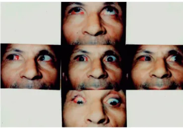
It is important to rule out congenital or acquired abduction deficiencies not related to abducens palsy, such as Möbius syndrome, Duane syndrome, and mechanical restrictions as in Graves’ disease and in fractures of the medial wall of the orbit(1,3,4,6) (Figures 2 and 3).
Aetiology
The length of the VI nerve, which starts in the brain stem, runs along the base of the skull, rises through the petrous part of the temporal bone to the cavernous sinus, crossing it before entering the orbit and then innervating the LR, makes it prone to the action of several aetiologic factors in peripheral lesions(1,3,5-7).
Central nuclear lesions produce gaze palsy to the affected side. Fascicular (intrapontine) lesions are associated with other lesions to cranial nerves V (trigeminal), VII (facial), and the motor pathways of the limbs, causing contralateral hemiparesis and pontine syndromes. In the clivus region nerve VI can be affected by pre-pontine tumours, compressions caused by brain
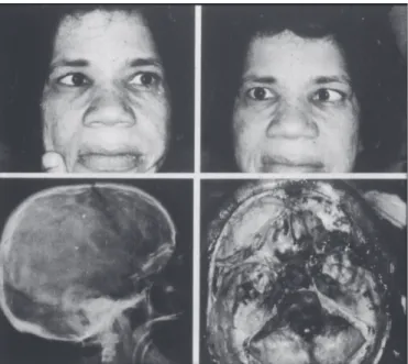
displacements due to tumour masses in the middle and posterior fossae, and meningitis, among other conditions. Intracranial hypertension often causes bilateral VI nerve palsy due to compression(5-7,9) (Figure 4).
Before entering the cavernous sinus, the VI nerve ascends along the petrous portion of the temporal bone. Infection of the middle ear with bone involvement, often with osteomyelitis of the petrous portion of the temporal (petrositis) can lead to paralysis of nerves VI and VII, with intense pain due to trigeminal involvement. The general condition depends on the severity of the infection and can lead to meningitis, which constitutes Gradenigo’s Syndrome(5-7,9).
Non-Localised Localised
Intracranial hypertension Pontine syndromes (vascular, demyelination, tumour)
Trauma Injury to the cerebellar-pontine angle (acoustic neuroma, meningioma) Lumbar puncture or spinal anaesthesia Lesions of the clivus (carcinoma, chordoma)
Hypertension Lesions of the middle fossa (tumour, petrositis)
Diabetes Lesions of the cavernous sinus (tumour, inflammation, aneurysm, fistula) Parainfectious causes (post-viral, middle
ear infections in children) Basal meningitis
Table 1
VI nerve paralysis and paresis: Aetiology
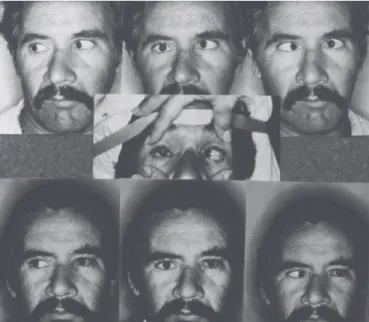
Figure 3 – A patient with high myopia and large esotropia, without eye movement either for abduction, elevation or depression in both eyes. Fixed strabismus due to bilateral VI nerve palsy with small movements of convergence. On attempted corrective surgery, none of the 12 extraocular muscles were found, but only very thin fibrous adhesions replacing the medial recti, hence the static position
Figure 4. Bilateral VI nerve paresis caused by intracranial hypertension due to a pituitary tumour. Note the enlarged sella and the tumour’s appearance on necropsy
Inside the cavernous sinus, nerve VI along with nerves III, IV, the ophthalmic division of nerve V, and sympathetic fibres can be affected by thrombosis, thrombophlebitis, aneurysm, and carotid-cavernous fistula, leading to characteristic syndromes. Total ophthalmoplegia, trigeminal pain in the affected territory, often Horner’s syndrome, and vascular signs due to venous stasis or fistula (proptosis, redness, chemosis, vascular dilatation [caput medusae], murmurs, and secondary glaucoma) are characteristic of lesions to the cavernous sinus(3,5-7). Multiple previous episodes
of oculomotor paresis with proptosis and pain suggest inflammation or a tumour of the orbital apex or the superior orbital fissure. Painful ophthalmoplegia due to inflammation of the superior orbital fissure is known as the Tolosa-Hunt syndrome(5-7). Expansive processes in the orbital apex may lead
to a similar presentation, either with normal visual acuity (Rochon-Duvignaud syndrome)(5-7) or with compression of the
optic nerve and visual loss (Rollet syndrome)(5-7).
Glaser and Bachynski(5) summarise the causes of VI nerve
palsies in Table 1.
The associated clinical pictures require in-depth neurological and clinical examination in order to find the causative factor. However, many cases are due to VI nerve palsy alone without any associated conditions. In these cases a medical and neurological examination should be performed, but the aetiology often remains obscure. Neuroimaging rarely shows any results and should only be performed in worsening cases or when other cranial nerves are affected.(3,5-8)
Clinical presentation
The intensity of LR impairment by the causative agent determines the presentation. Mild or early impairment can lead to homonymous diplopia and/or nystagmus toward the affected muscle, with esotropia proportional to the loss of function and no deviations in other positions of the gaze(1). The tonic imbalance
of horizontal muscles of the affected eye causes hyperactivity of the MR, which tends to increase the esotropia in the primary position, with diplopia and compensating torticollis, with the head turned toward the action of the affected muscle, maintaining bifixation on contralateral gaze, where there is no deviation and binocular vision is preserved(1). MR contracture is also
proportional to the intensity of LR impairment, creating a mechanical factor that restricts the already weak abduction. At this stage the angle of deviation increases in the primary position and esotropia also occurs on contralateral gaze, causing diplopia in all eye positions(1,5, 9,10).
Spontaneous recovery of the LR occurs in a large number of cases, depending on the aetiology and the effectiveness of
treatment. Functional recovery of the LR can lead to complete cure(5,7,9). Established MR contracture prevents clinical
improvement despite the improved function of the LR; in these cases, deviation remains predominantly due to mechanical restriction.(1,5,9,10). However, even though the onset of contracture
Figure 5 - Left VI nerve paresis in a child with varicella
Figure 6 – The same child after complete spontaneous cure
recover without eventual contracture. LR paralysis in the fixating eye can lead to hyperactivity and consequent contracture of the contralateral MR due to permanent nerve stimulation, manifested by greater deviation and esotropia which may become progressively alternating with bilateral motor changes, including mild LR deficits resulting from initial nerve inhibition and mechanical restriction due to MR contracture. Bilateral paralysis is not uncommon, with large angles, permanent diplopia and torticollis with the head turned toward the fixating eye. In this case, the torticollis is not to avoid diplopia, but due to the static position of the eye in esotropia(1,5,9-12).
Diagnosis
The presence of diplopia and oculomotor signs, with esotropia and unilateral or bilateral impairment of abduction easily lead to the diagnosis of VI nerve palsy. Clinical and neurological evaluation is essential for aetiologic diagnosis and appropriate treatment. The ophthalmic evaluation aims to ob-serve motor changes, their consequences on the oculomotor system and their progression, in order to determine the appropriate ophthalmic management to minimise symptoms and avoid sequels which, once established, can be treated independently of the aetiological treatment. It is essential to examine ocular deviation in different horizontal positions of the gaze to evaluate the function of the LR and the presence of MR contracture(1,3-5,7). Sequential measurements of the deviation at
each position of the gaze, the degree of incomitance, the presence of deviation in the primary position and with the gaze toward the opposite direction of the affected muscle help determine whether the case is improving or worsening after the onset of neurological treatment. An increased angle of deviation toward the affected muscle can point to a greater or lesser degree of LR deficiency as well as greater or lesser restriction caused by ipsilateral MR contracture, which is also manifested by esotropia of greater or lesser angle with the gaze toward the opposite direction(1,5,9). As regards the function of the LR, Scott’s force
generation test is a simple and reliable procedure, depending on the experience of the examiner. After topical anaesthesia, the temporal limbus of the affected eye is grasped with a forceps; the eye is taken to adduction and the patient is asked to perform a rapid movement toward the LR under study, and the muscle’s strength is felt at the forceps. Often the movement is so strong that damages the conjunctiva. A dynamometric forceps can be used, however this is not essential in clinical practice. It is important to note that major MR contractures that keep the eye fixed in adduction can lead to false results in Scott’s test due to the small range of motion toward abduction. This could lead to a
false diagnosis of severe paralysis or paresis, because even though the LR is effective, its action is not visible. Observing LR function using the force generation test in patients with a detached LR under topical anaesthesia can demonstrate the error(1,5,9,10,13).
The assessment of MR contracture is done with the forced duction test, in which the medial limbus is grasped with a forceps and the eye is forced toward abduction. Greater or lesser mechanical restriction will be noticed according to the strength needed to move the eye. Major contractures may prevent any passive movement of the eye. Restrictions induced by conjunctival changes can also be demonstrated, especially with the formation of conjunctival folds toward the forceps(1,3,5,9,10).
Electrooculography or electromyography can yield the same results, but these tests are not commonly used in clinical practice. The improvement or worsening of LR function can be assessed with sequential electrooculographies(9,10,13).
Diagnosing the condition of each individual muscle helps plan the correction of deviation(1,3).
Treatment
Spontaneous cure can occur, especially in cases of viral or vascular origin and those without aetiological diagnosis.
Medical and neurological treatment should be instituted, and ophthalmic therapy should target the clinical manifestations, improving the function of extrinsic muscles and motor and positional sequelae in the affected eye. In vascular diseases such as diabetes and arteriosclerosis gradual recovery tends to occur, with the patient noticing a progressive alignment of diplopic images. General treatment for disease control is effective. Similarly, infectious diseases, particularly viral infections, tend to improve, with remission of LR paresis depending on whether MR contracture has occurred(1,3,6,8,10,12) (Figures 5 and 6) .
Medical Treatment
order to fixate it in abduction, thus over-stimulating the LR and inducing increased neural inhibition of the MR in the same eye. This constant relaxation of the MR prevents contracture while allowing the LR to be activated. In more severe cases, with severely impaired abduction and torticollis due to MR contracture, this approach can be difficult. Occlusion of the healthy eye can lead to maintenance of torticollis depending on the position of static equilibrium of the eye, leading to difficulties in spatial orientation and rejection by the patient despite the absence of diplopia, but without any prospect of influencing muscle contraction in itself(1,5,9,10).
In cases with larger angles, longer progression or torticollis with large deviations of the head, in which a more important imbalance occurs in the PPG, or even with permanent diplopia in all horizontal positions of the gaze and consequent MR contracture, occlusion of the affected eye is satisfactory, as it prevents diplopia and the spatial disorientation induced by the static position of the affected eye in adduction, which may be the norm in all unilateral cases(1,3,10,12).
Alternating occlusion can be considered in cases where, due to muscle rebalancing, bilateral motor abnormalities occur leading to paralytic alternating esotropia, which is not uncommon and suggests a longer progression or bilateral VI nerve impairment. It is important to note that in alternating cases, both eyes can fixate in the PPG, thus improving the acceptance of occlusion(1,5,10).
Prism therapy: Temporal prisms can be used in cases with smaller angles or stable, residual esotropia after surgical treatment. Satisfactory correction of diplopia is achieved in the PPG, though it should be present in other positions of the gaze. Torticollis also improves, usually with good patient satisfaction
(1,3,8,10).
At greater angles acceptance is more difficult, either with unilateral therapy, due to the reduced vision and colour dissociation produced by the prism, or with bilateral therapy, due to the spatial disorientation caused by the prism in front of the fixating eye.
Assessment of prism therapy by the physician and the patient is critical, and the prism can be assembled in a test frame to allow the patient to get used to it.
Prism therapy can be associated with occlusion to control diplopia while acting on secondary effects (Guibor prism therapy)(3,9,10). The affected eye is occluded and the largest
possible prism is used in the healthy eye. Fixation of this eye is done in convergence, with great stimulation of the MR in this eye and transmission to the other eye, which is occluded. In case of LR paresis, overactivation will lead to improved performance; in case of paralysis there will be no contractile response, but in both cases there will be great inhibition of the MR in the affected eye, thus avoiding contracture. Despite being theoretically sound, the method is difficult to implement because of the need of a prism of great magnitude in front of the fixating eye with occlusion of the opposite eye, which leads to reduced vision, coloured halos, and often spatial disorientation(10).
Pharmacological Treatment
Botulinum toxin: Botulinum toxin can assist in the treatment of VI nerve paresis and it is very important depending on the clinical presentation. In most cases, progressive recovery starts early regardless of treatment. During the first month after the onset of symptoms, no procedure should be implemented if the patient is improving. However, occlusion can be used to alleviate the symptoms of diplopia. After this period, in case of continuous
spontaneous improvement, the patient should be monitored clinically, and botulinum toxin can be injected into the MR of the affected eye. Many physicians start treatment with botulinum toxin even earlier. The goal is to impair the MR, seeking to ba-lance out LR paresis and improve its contractile function due to the lack of antagonism, thus facilitating functional recovery. Ca-ses of vascular or diabetic origin are often cured with this method. When effective, botulinum toxin produces its greater effect in the first week, with exotropia induced by functional paralysis of the injected MR. Possible complications are vertical deviations and ptosis; both are reversed after the drug’s effect vanishes. In chronic cases with residual LR function, botulinum toxin can improve MR contracture and its consequences. Repeated injections are used if progressive improvement is seen. Traumatic brain injury and and sequelae of brain tumours can progress in this manner. Permanent paralysis with total impairment of the LR should not be treated with botulinum toxin only; the drug can be used to supplement surgery. The association of botulinum toxin in the MR with LR resection or muscle transposition has been proposed in order to weaken the muscle, thus preserving vascularisation(3,10,14).
Surgical Treatment
Approximately 6 months after the onset of VI nerve palsy, the clinical picture can be considered definitive if no angle and motor improvement or worsening occurs. Botulinum toxin should be considered, and enough time should be given to allow functional recovery before indicating surgery. Scott suggests that if no clinical improvement is seen in 3 months with repeated follow-up, surgery should be indicated(3,10,14,15).
It is necessary to evaluate the angle of deviation, the action of the affected LR, and the presence ipsilateral and contralateral MR contracture. Angular measurement, the force generation test or electrooculography, and the forced duction test can be used to evaluate muscle function. Despite the debate on the effectiveness of these tests, most authors indicate surgery based on their results(1,3,9,10,13,15). Once the intensity of MR contracture
and LR deficit (paresis or paralysis) is determined, surgery is planned. Surgery aims to correct the deviation and therefore diplopia and torticollis, increasing abduction and aiming for best binocular vision in the largest possible area of the visual field. These goals should also be considered for near fixation, as there is often an improvement of distant vision and a worsening of near vision, with patient dissatisfaction.
The surgical strategy varies depending on the presence of LR paresis or paralysis. Paresis can respond to resection proportional to the residual action of the LR. Paralysis does not respond to this procedure; in these cases, muscle transposition should be performed. Large LR resection does not improve contractility or muscular function, but can lead to severe mechanical restrictions of adduction.
Small-angle deviations with good residual LR function are usually associated with comitance without major MR contracture. Classical recession and resection surgery can be uesd with good results (Figures 7 and 8). Some authors recommend isolated resection of the LR in these cases(13), but the result is temporary,
with frequent relapse. Large MR recession in the affected eye has also been suggested when the LR shows some residual function, aiming for balance between the two muscles. This leads to good results for distant vision, but the loss of adduction can cause exotropia for near fixation, with diplopia while reading and worsening of symptoms.
Figure 7 – Right VI nerve paresis after peribulbar anaesthesia with bupivacaine for cataract surgery. Esotropia progressing to comitance (preoperative)
Figure 8 – The patient in Figure 6 after surgery, corrected with MR recession and LR resection in the right eye
and LR function is very good, with almost normal abduction despite the deviation in the primary position. On the other hand, a decreasing angle can indicate spontaneous recovery; in these cases, surgery should be postponed(1,5,10).
In similar cases with larger angles around 40-50 PD, recession/resection in the affected eye can be complemented with contralateral MR recession, improving nerve induction of the paretic LR and improving its function as proposed by Horta-Barbosa(11,12). This author also recommends a large recession of
the MR with the same goal (innervational surgery) (11).
In cases where LR function is absent or nearly absent, resection has no effect and muscle transposition is preferred. Still, Murray(16) and several other authors suggest a large (12 mm)
recession of the MR associated with exaggerated resection of the LR (12 to 14 mm) in these cases. This approach leads to good results in the primary position but causes other complications. The first is relapse due to failure of LR elasticity, which initially acts as a mechanical restriction, holding the eye in the PPG, but subsides with time. Also, while the mechanical action of the resected LR persists, there is great impairment of adduction since the MR was also largely recessed. This also impairs near fixation permanently, because even without the restrictive action of the LR, the recessed MR loses its adduction function. Therefore little eye movement remains, the binocular visual field is very small and, as the other eye moves freely, diplopia occurs almost constantly, with great variability and discomfort as it is homonymous, with esotropia with the gaze toward the affected LR and exotropia in the opposite direction.
Transposition of the vertical recti to the temporal side using any of the known techniques is the ideal approach in these cases, as it produces elastic and innervational actions that antagonise the action of the MR, avoiding deviation. Elastic factors due to vertical recti distension or transposition to the temporal side support LR action and may be responsible for the slight abduction achieved, since attempted abduction causes relaxation of the MR and vertical recti (adductors), facilitating the performance of these elastic components. Innervational factors produce a clinical picture similar to Duane Syndrome, i.e., during attempted adduction stimulation of the MR is also transferred to the verti-cal recti (adductors) which antagonise the MR and, once contracted, prevent adduction of the eye. This co-contraction not only stabilises eye movement but also acts in conjunction with elastic factors to prevent MR contracture.
RM recession associated with transposition of the vertical recti has become a more frequent procedure, regardless of the technique used for transposition, showing good results regarding parallelism in the primary position and more stability, with fewer relapses. Techniques of total transposition of the vertical recti tend to increase the risk of anterior segment ischemia.
Due to these vascular effects, we prefer to use the Carlson-Jampolsky(17) procedure associated with MR recession as it
pre-serves the medial arteries of the vertical recti, reducing the risk of ischemia. Nevertheless, cases of anterior segment ischemia still occur, even with successful surgery(1,18,19) (Figure 9).
From the mechanical point of view, a great loss of adduction occurs when MR recession is associated with muscle transposition. This is because of the excessive loss of function in the contractured muscle after large recessions due to mechanical and innervational restrictive factors introduced by the transposition (Figure 10). Inversion of diplopia when looking toward the healthy eye and exotropia on near fixation have been observed, as well as overcorrection, with secondary exotropia difficult to correct surgically (Figures 11 and 12). The Hummelshein(20), Jensen(21), Carlson-Jamposky(17), and Foster
(augmented transposition)(22) techniques lead to the same
problems when associated with MR recession. Large MR recession associated with large LR resection, a procedure advocated by Murray(16), usually shows very poor results from
the motor point of view. Not only does it result in a great loss of function of the MR, which would be problematic enough, but the MR is also affected by mechanical factors resulting from extensive LR resection, restricting adduction. Also, this surgical procedure is usually insufficient for a good result even in the primary position.
Due to such complications, and after reviewing the results of the Hummelshein(20), Jensen(21), and O’Connor(22) techniques
and the suggestion by Rosenbaum(14,24) of using botulinum toxin
instead of MR recession to prevent anterior segment ischemia, we observed that all these procedures led to complications(11,16,20).
In 1996 we started to use a modified, isolated Carlson-Jampolsky transposition(17) sparing the MR. We do not use
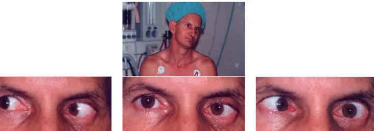
Figure 9 - Patient submitted to bilateral surgery for correction of bilateral VI nerve paresis after traumatic brain injury. The same procedure was performed in both eyes, with recession of the medial recti and Carlson-Jampolsky transposition. Surgery resulted in parallelism in the primary position with good abduction in both eyes, especially the left eye. The patient developed anterior segment ischemia with mydriasis, iris atrophy and cataract in the right eye, and mydriasis and cataract in the left eye
Figure 10 - Post-traumatic VI nerve paralysis, corrected with MR recession and Carlson-Jampolsky transposition. Note the loss of adduction in the operated eye and inverted deviation on right gaze (exotropia) and left gaze (esotropia)
of the vertical recti to slide better toward the LR, which also minimises vertical deviations. We seek to minimise the vascular deficit and the loss of adduction based on the notion that the mechanical and innervational factors produced by the transposition would be enough to balance the MR contracture, in addition to providing more stable results over time. On the one hand, the proposed transposition is a procedure in which the mechanical factor is key, whether through changing the functional position of the transposed parts of the vertical recti toward the LR or through the elasticity of these muscle fibres in these positions. However, the innervational factor should also be considered. During adduction, the innervational stimulus to the MR is also directed to the vertical recti, which also produce adduction. The increased tone of these muscles due to increased innervation also increases the tone of the transposed parts, which co-contract with the MR, creating an innervational situation si-milar to the Duane syndrome, leading to a better gaze position in the PPG as well as antagonising the action of the MR and preventing contracture.
Improvement in deviation occurred initially in all of the patients we operated (average residual angle lower than 12 PD in over 80% of cases), correcting torticollis, increasing abduction by 5-15 degrees (mean, 10 degrees), and increasing the field of binocular vision without loss of MR action and its consequences. MR contracture decreased progressively due to the opposing forces. No relapses were seen with this procedure. In some ca-ses, even with residual LR action, we believe it is possible to obtain good results regarding the angle of deviation in the primary position, as the residual LR action should lead to progressively better abduction. (Figures 13-17).
The patients in Figures 13-20 were some of the first to be submitted to this procedure in 1996.
We followed up 24 patients for 2 years, of which 16 were followed up for 5 years and 8 are still undergoing periodic control for over 10 years (Figure 21). Also, more patients have been operated with the same positive results, but these are being included in new sample to be assessed sequentially.
Among the initial 24 patients, all with unilateral VI nerve paresis, the average deviation was 29.79 prism diopters (angles of 20-45 diopters). One month after surgery, the mean deviation was 9.16 prism diopters, and after 2 years the mean deviation was 9.58 prism diopters.
We consider a postoperative angle in the primary position
of 10 prism diopters or less as ideal, which happened in 70.83% of patients in the immediate postoperative period and also in 70.83% after 2 years, despite individual variations, demonstrating the stability of the clinical condition. This shows that the technique is not associated with frequent relapse as seen with other surgical techniques, perhaps due to the mechanical restriction of abduction resulting from the transposition.
Other important factors to be considered in this first group of patients is that there was no over-correction with exotropia and no ischemic changes in the anterior segment, which were key factors in choosing the surgical procedure.
Figures 11 and 12 - Patient with right VI nerve palsy after traumatic brain injury, with central right facial paralysis and left hemiparesis. After recession of the right MR and Carlson-Jampolsky transposition in the same eye, the patient developed secondary exotropia with loss of action of the right MR, difficult to correct, with a poor functional outcome in terms of muscle function, aesthetics and control of diplopia
suture; the halves of the superior and inferior recti were tied to each other, a technique that theoretically prevents hypertropia.
R
EFERENCES1. Almeida HC, Curi R. Paresia e paralisia do VI nervo (abducente). In: Almeida HC, Curi R. Manual de estrabismo. Rio de Janeiro: [s.n.]; 1997. p. 133-40.
2. Curi RL. Enfermedad de Graves: oftalmopatia infiltrativa. In: Arroyo Y, llanes ME, editor. Actualidades del estrabismo latino-americano. México: Lithoimpresora Portales; 1998. p.169-82.
3. Curi RL. Paralisias oculares. In: Dantas AM, coordenador. Essencial em oftalmologia. Rio de Janeiro: Guanabara Koogan; 2011. p. 1112-39.
4. Dantas AM. Paralisias oculares. In: Dantas AM, Monteiro MLR. Neuro-oftalmologia. 2a ed. Rio de Janeiro: Cultura Médica; 2010. p. 503-55. 5. Glaser JS, Bachynsky B. Infranuclear disorders of eye movements. In:
Glaser JS, editor. Neuro-ophthalmology. 2nd ed. Philadelphia: Lippincott; c1990. p. 361-418.
6. Dantas AM. Paralisias oculares. In: Dantas AM, Monteiro MLR. Neuro-oftalmologia. Rio de Janeiro: Cultura Médica; 1999. p. 468-524. 7. Morterá-Rodrigues MP, Dantas JM, Dantas AM. Anatomia e
embriologia. In: Dantas AM, coordenador. Essencial em oftalmologia. Rio de Janeiro: Guanabara Koogan; 2011. p. 3-61.
8. Walsh T, Cohen K, Helveston EM, Stewart W. Diplopia. In: Walsh TJ, editor. Neuro-ophthalmology: clinical signs and symptoms. 4th ed. Baltimore: Williams & Wilkins; 1997. Cap. 6. p. 114-80.
9. Prieto-Diaz J, Souza-Dias C. Paresias y parálisis oculomotoras: parte III: paresias y parálisis del VI nervio. In: Prieto-Diaz J, Souza-Dias C. Estrabismo. 5a ed. Buenos Aires: Ediciones Cientificas Argentinas; 2005. p. 354-65.
10. Dias CR, Goldchmit M. Os estrabismos paralíticos. In: Souza-Dias CR, Goldchmit M. Os estrabismos: teoria e casos comentados. Rio de Janeiro: Cultura Médica / Guanabara Koogan; 2011. p. 243-351.
11. Horta-Barbosa P. VI nervo. In: Horta-Barbosa P. Estrabismo. Rio de Janeiro: Cultura Médica; 1997. p. 163-8.
12. Barbosa P. Aplicações clínicas das leis inervacionais. In: Horta-Barbosa P. Estrabismo. Rio de Janeiro: Cultura Médica; 1997. p. 169-70.
13. Santiago AP, Rosenbaum AL. Sixth cranial nerve palsy. In: Rosenbaum AL, Santiago AP, editors. Clinical strabismus management: principles and surgical techniques. Philadelphia: W.B. Saunders; 1999. p. 259-71. 14. Rosembaum AL, Kushner BJ, Kirschen D. Vertical rectus muscle trans-position and botulinum toxin (Oculinum) to medial rectus for abducens palsy. Arch Ophthalmol. 1989;107(6):820-3. Comment in Arch Ophthalmol. 1991;109(10):1345-6.
Figure 13 - Patient with traumatic left VI nerve palsy and significant limitation of abduction, not reaching the midline. The left LR is active, and the patient has severe torticollis. Good visual acuity in both eyes, with preferential fixation with the left eye (preoperatively)
Figure 14 – Postoperative period, 15 days after the previous figure, with orthotropia in the primary position, 10-degree abduction in the left eye, and marked improvement of torticollis
Figure 17 - Patient with traumatic right VI nerve palsy. The accident also caused a penetrating injury of the cornea and lens of the left eye, with loss of vision due to secondary glaucoma and intense corneal leukoma. Due to the fixed deviation of the right eye in esotropia in all positions of the gaze, the patient had torticollis with the head turned sharply to the right in order to preserve forward vision (top). The bottom row shows the result of the Carlson-Jampolsky transposition in the right eye, with orthotropia in the primary position and exotropia of the blind eye on right gaze, possibly due to innervational hyperactivity, with total correction of torticollis
Figure 18 - Patient with left VI nerve paresis of vascular origin, with parallelism on right gaze, esotropia of 30 prism diopters in the primary position and higher esotropia on left gaze, with the eye just reaching the midline. Intense left torticollis as the patient maintains preferential fixation with the left eye due to severe cataract in the right eye
Figure 19 – The same patient, 2 years after the Carlson-Jampolsky procedure and cataract surgery in the right eye. Note the improvement of torticollis
Figure 16 – Patient with a similar clinical picture to the previous figure, with left VI nerve paresis after infectious meningitis. Top: Preoperatively. Middle: Post-operative period, 15 days after isolated Carlson-Jampolsky transposition in the left eye. Orthotropia on right gaze, esotropia of 10 PD in the primary position and 25 DP on left gaze. Bottom: Improved primary position 1 year after surgery (6 PD)
16. Murray ADN. Supermaximal surgical treatment of total sixth nerve paralysis. In: Khoo CY, Ang BC. New frontiers in ophthalmology: pro-ceedings of the 26th International Congress of Ophthalmology, held in Singapore, 18-24 March 1990. Amsterdan: Elsevier; 1991. p. 970 17. Carlson MR, Jampolsky A. An adjustable transposition procedure for
abduction deficiences. Am J Ophthalmol. 1979;87(3):382-7. 18. Keech RV, Morris RJ, Ruben JB, Scott WE. Anterior segment ischemia
following vertical muscle transposition and botulinum toxin injection.
Arch Ophthalmol. 1990;108(2):176. Comment in Arch Ophthalmol. 1991;109(2):174.
19. Lee JP, Olver JM. Anterior segment ischemia following vertical muscle transposition and botulinum toxin injection. Arch. Ophthalmol. 1991;109(2):174. Comment on Arch Ophthalmol. 1990;108(2):176. 20. Hummelshein E. Über Sehnentransplantation am Auge. Ophthal
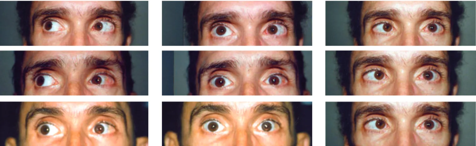
Figure 20 - Patient with traumatic bilateral VI nerve paralysis, fixation with the right eye, deviation angle over 100 prism diopters; moderate contracture of the right medial rectus and severe contracture of the left medial rectus. During the force generation test, both lateral recti showed a moderate force of contraction (top row). Isolated Carlson-Jampolsky transposition was performed in both eyes with excellent results (bottom row), maintained for over 2 years. This patient was not included in the sample due to bilateral involvement and a large deviation angle. However, this demonstrates the effectiveness of the procedure
Figure 21 – The patient from Figure 15 after 12 years of follow-up, showing the stability of surgical results.
Corresponding author:
Instituto de Oftalmologia de Niterói
Av. Ari Parreiras, nº 438 - Jardim Icaraí
CEP 24230-322 - Niterói (RJ), Brasil
Tel: 55 (21) 2610-1452
E-mail: [email protected]
[email protected]
[email protected]
21. Jensen CD. Rectus muscle union: a new operation for paralysis of the rectus muscles. Trans Pac Coast Otoophthalmol Soc Annu Meet. 1964;45:359-87.
22. Foster RS. Vertical muscle transposition augmented with lateral fixa-tion. J AAPOS. 1997;1(1):20-30.
23. O’Connor R. Transplantation of ocular muscles. Am J Ophthalmol. 1921;4:838-45.