Comparative study of the effects
of submucosal cauterization of
the inferior turbinate with or
without outfracture
Summary
Antonio Celso Nunes Nassif Filho1, Carlos Roberto
Ballin2, Carlos Augusto Seiji Maeda3, Gustavo
Fabiano Nogueira4, Matheus Moschetta5, Danielle
Salvatti de Campos6
1 Head of the Otolaryngology Department of the Santa Casa de Misericórdia, Curitiba - Brazil.
2 Otolaryngologist and Craniofacial surgeon of the Department of Otolaryngology of the Santa Casa de Misericórdia. Curitiba - Brazil. 3 2nd year otolaryngology resident of the Santa Casa de Misericórdia, Curitiba - Brazil.
4 2nd year otolaryngology resident of the Santa Casa de Misericórdia, Curitiba - Brazil. 5 3rd year otolaryngology resident of the Santa Casa de Misericórdia, Curitiba - Brazil. 6 1st year otolaryngology resident of the Santa Casa de Misericórdia, Curitiba - Brazil. Serviço de Otorrinolaringologia da Santa Casa de Misericórdia de Curitiba-PR e PUC-PR – Aliança Saúde.
Mail Address: Carlos A. S. Maeda – Av. Silva Jardim, 2014, ap. 801 Bairro Batel Curitiba PR 80250-200. E-mail: [email protected]
Paper sent to the ABORL-CCF SGP (Management Publications System) on August 12, 2004 and accepted on September 19, 2005.
A
im: The objective of this study was to compare the effects of submucosal cauterization of the inferior turbinate with or without outfracture. Study Design: clinical prospective. Method: Twenty patients with inferior turbinate hypertrophy were randomized and divided into two groups. The first one was submitted to submucosal cauterization associated with outfracture, and the second one without fracture. Five items were assessed to compare both methods: pain, nasal bleeding, scarring - analyzed through anterior rhinoscopy, observing edema, hyperemia and seropurulent secretion; crust formation (seen through anterior rhinoscopy); and nasal airway patency. Follow-up was performed on days 7, 14, 30. Results: In both groups crusting formation was similar. Scarring showed better results in the outfracture group in the first two weeks postoperative. The analysis of nasal airway patency showed good results in 80% of the patients submitted to submucosal cauterization with outfratcture on day 30 postoperatively. Conclusions: We concluded that submucosal cauterization of inferior turbinate with outfracture is better than the procedure without outfracture.Key words: submucosal cauterization, inferior turbinate hyper-trophy, turbinate outfracture
ORIGINAL ARTICLE Rev Bras Otorrinolaringol
INTRODUCTION
Nasal turbinates are arch-like bony structures that lay antero-posteriorly in the nasal cavities, having a border attached to the lateral nasal wall and a free medial ledge. There may be three or four nasal turbinates and, despite presenting similar framework, they bear distinct origins. The inferior turbinate, being larger and the one more eas-ily seen, has its own bone, called inferior nasal conchae, anchored to the maxilla, lacrimal, ethmoid and palatine bones. This turbinate tail has a posterior boneless protuber-ance that bulges into the choana, and it is formed almost exclusively by vascular erectile tissue, which is usually hypertrophic in nasal diseases. The middle, superior and supreme turbinates (the latter not always present), bear a bony structure formed by projections of the ethmoid bone. There is a modest build-up of vascular erectile tissue on the middle turbinate tail, lesser however than that of the inferior turbinate.
The nasal lateral wall, formed by the turbinates and the meatus, has a rather important role in nasal physiol-ogy as far as balancing temperature, moisture and also filtering of suspended particles present in inhaled air are concerned. Diseases that cause chronic nasal obstruc-tion – being alergic rhinitis the most prevalent – basi-cally involve the lateral wall of the nasal cavity, causing changes to both the mucosa and the submucosa of the nasal turbinates.
Most rhinopathies cause nasal obstruction, of which the initial treatment is usually clinical. The initial control of environmental factors maybe followed by anti-hista-mines, topic or systemic steroids and immunotherapy. Surgery is indicated for non-responders. The reduction of nasal turbinates is a surgical approach that has been well discussed in the scientific literature, bearing varied indications and techniques. Among described approaches are the injection of sclerogenic substances and corticoster-oids, cryosurgery, total resection of the inferior turbinate, laser ablation surgery, lateral fracture of the turbinate and submucosal ablation by electrocautery. The latter was initially described by Neres in 1907 and by Horn in 1908. In 1930, Beck reported the use of unipolar cautery; and in 1931, Hurd used bipolar ablation for the first time, a technique still broadly used2. In 1989, Woodhead et al. reported on the changes to the inferior turbinate caused by submucosal electrocautery ablation. It introduces mu-cosal changes that lead to fibrosis, apart from reducing the turbinate enlargement potential. The advantage of such procedure is that it can be performed under local anesthesia and it is technically simple to be performed. The major disadvantage is the short duration of the results, ranging from months to years. Meredith et al. reported that 31% of the ablated patients had relapsing symptoms within 33 months3. Differently from turbinectomy – which
is irreversible – this procedure can be performed numer-ous times. Electrocautery ablation usually causes edema and crust formation within 3 to 6 weeks after surgery1. About 20 to 30% of the cases may present adherence. Besides, bone necrosis of the inferior conchae may also occur if the bone is ablated during the procedure, which may then produce bone sequester, edema and erythema of long stand. Electrocautery ablation usually produces good postoperative hemostasis; notwithstanding, 6% of patients may present with late epistaxis3. The procedure should be very carefully performed so as to avoid burn-ing of the nasal ala, the columellae, the nasal septum and other structures.
Submucosal ablation is usually performed together with the lateral fracture of the inferior turbinate. However, the turbinate tends to return to its original position after an undetermined amount of time3. Thus, the real need for using the lateral fracture coupled with different surgical techniques of the inferior turbinate is yet to be ascertained. From these grounds, the author decided to make a clinical assessment of the patients who underwent submucosal electrocautery ablation with and without lateral fracture of the inferior turbinate, in the treatment of chronic hyper-trophic rhinitis.
OBJECTIVES
Comparative analysis of patients submitted to sub-mucosal electrocautery ablation with and without lateral fracture of the inferior turbinate.
MATERIAL AND METHODS
of the inferior turbinate coupled with lateral fracture, and group 2 underwent submucosal electrocautery ablation alone, without lateral fracture. The patients were assessed on the 1st, 7th, 14th and 30th day of post-op according to the protocol (attached). Breathing was assessed through the Likert Visual Analog Scale (0 to 10).
All patients underwent the following surgical pro-cedure:
1. Topic anesthesia with 10% xylocaine spray; 2. Sedation with midazolam hydrochloride and fentanyl citrate, or general anesthesia, depending on the case;
3. Local anesthesia with 2% xylocaine and epine-phrine, without vasoconstrictor, in a 1:100,000 concentra-tion.
4. Infiltration of the previous solution along the inferior turbinate;
5. Submucosal electrocautery ablation along the head, tail and body of the inferior turbinate by Wavetronic bipolar electrocautery (groups 1 and 2);
6. Medial fracture of the inferior turbinate (group 1);
7. Lateral refracture of the turbinate (group 1); 8. Anterior nasal packing with terramycin ointment and bismuth subgallate;
9. Identical procedure performed on the contralat-eral turbinate.
RESULTS
Demographics
The demographics of the trial groups did not show any significant difference as far as age and gender were concerned; in other words, age and gender distribution was practically the same in the assessed groups. Their ages ranged between 13 and 45 as shown in Table 1.
Day 1 post-op assessment
On the 1st day of post-op assessment, the groups did not show significant difference as far as nasal bleed-ing and pain were concerned. Regardless of the surgical technique (with or without lateral fracture), none of the patients presented nasal bleeding that would require nasal packing. Moreover, most of the patients did not report pain that called for potent analgesia (opioid derivatives) (Table 2).
Day 7 post-op assessment
On the 7th day post-op assessment (Table 3), the trial groups did not show any significant difference in terms of crust and nasal permeability. As for healing, the fracture group presented good healing (60.0%), while the group without fracture presented moderate healing (70.0%) (p=0.041) (Figure 1).
Day 14 post-op assessment
On the 14th day of post-op assessment, the trial groups did not show significant differences as far as crusts and nasal permeability are concerned. As for healing, no significant difference was seen. However, we must high-light the fact that the lateral fracture group showed good
Chart 1. Healing after 7 days of surgery of the trial groups.
Chart 2. Healing after 14 days post-op of the trial groups.
Table 1. Demographics of the trial groups.
Assessment with (n=10) without (n=10) total (n=20) p value
N % Nº % 1º %
Age (years) 0,733(1)
Average ± standard deviation 22,0 ± 10,2 21,9 ± 7,9 22,0 ± 8,9
Minimum and maximum 13,0 e 45,0 13,0 e 32,0 13,0 e 45,0
Median 45,0 32,0
Gender 0,655(2)
Male 04 40,0 06 60,0 10 50,0
Female 06 60,0 04 40,0 10 50,0
(1) Mann-Whitney. (1) Chi-squared.
Table 2. Day 1 post-op assessment of the trial groups.
Assessment with (n=10) without (n=10) Total (n=20) p value(1)
Nº % Nº % Nº %
Nasal Bleeding 0,500
None 06 60,0 07 70,0 13 65,0
Mild 04 40,0 01 10,0 05 25,0
Moderate - - 02 20,0 02 10,0
Pain 0,237
None - - 02 20,0 02 10,0
Mild 08 80,0 07 70,0 15 75,0
Moderate 02 20,0 01 10,0 03 15,0
(1) Fisher’s Exact.
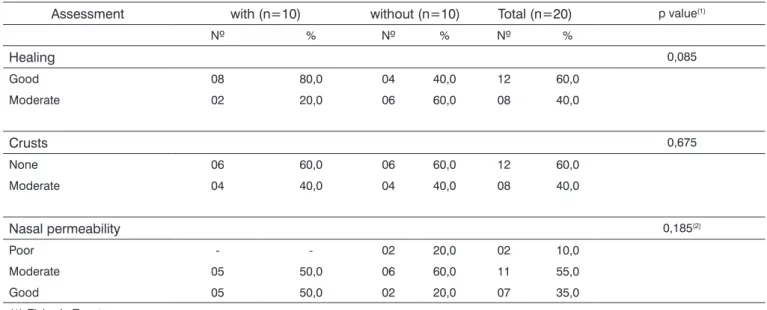
healing (80.0%) while the group without fracture presented moderate healing (60.0%) (p=0.085) (threshold likelihood indicating a trend) (Figure 2).
Day 30 post-op assessment
On the 30th day post-op assessment, no significant difference was seen on the trial groups as far as healing and crusts were concerned. Regarding nasal permeability, no significant difference was seen. However we must highlight the fact that the group with lateral fracture presented good nasal permeability (80.0%) while the non-fractured group had moderate permeability (50.0%) (p=0.062) (threshold likelihood indicating a trend) (Figure 3).
There were very few complications seen in the present study. Only one patient after 30 days developed posterior nasal synechiae in the right nasal cavity, which meant 5% of the cases considered. No patient complained of intense pain or late epistaxis on the follow-up. We used descriptive data analysis through tables, charts and figures. Non-parametric tests - “Mann-Whitney” (through
the “Primer of Biostatistics” software), “Chi-squared” and “Fisher’s Exact” (through the “Epi-Info” software) were uti-lized in order to corroborate the goal stated in this paper. The significance level (significance probability) used was lower than 5% (p<0.05).
DISCUSSION
Table 3. Day 7 post-op assessment of the trial groups.
Assessment with (n=10) without (n=10) Total (n=20) p value(1)
Nº % Nº % Nº %
Healing 0,041
Good 06 60,0 01 10,0 07 35,0
Moderate 04 40,0 07 70,0 11 55,0
Poor - - 02 20,0 02 10,0
Crusts 0,325(2)
None 05 50,0 07 70,0 12 60,0
Moderate 05 50,0 03 30,0 08 40,0
Nasal permeability 0,171
Poor - - 03 30,0 03 15,0
Moderate 07 70,0 05 50,0 12 60,0
Good 03 30,0 02 20,0 05 25,0
(1) Chi-squared. (2) Fisher’s Exact.
Table 4. Day 14 post-op assessment of the trial groups.
Assessment with (n=10) without (n=10) Total (n=20) p value(1)
Nº % Nº % Nº %
Healing 0,085
Good 08 80,0 04 40,0 12 60,0
Moderate 02 20,0 06 60,0 08 40,0
Crusts 0,675
None 06 60,0 06 60,0 12 60,0
Moderate 04 40,0 04 40,0 08 40,0
Nasal permeability 0,185(2)
Poor - - 02 20,0 02 10,0
Moderate 05 50,0 06 60,0 11 55,0
Good 05 50,0 02 20,0 07 35,0
(1) Fisher’s Exact. (2) Chi-squared.
brings about the least morbidity to the patients. Among the many methods presented in the literature, the submucosal electrocautery ablation is one of the most widely used.
The literature shows distinct opinions about the inferior turbinate submucosal electrocautery ablation. To begin with, the amount of coagulated tissue is difficult to be measured. According to Wengraf et al. (1986), there are no precise parameters to be used in the calculation of the amount of tissue to be ablated5. Likewise, in our trial we noticed that the amount ablated varied according to our experience. However, Williams et al. state that the
Table 5. Day 30 post-op assessment of the trial groups.
ASSESSMENT with (n=10) without (n=10) Total (n=20) p value(1)
Nº % Nº % Nº %
Healing 0,763
Good 09 90,0 09 90,0 18 90,0
Moderate 01 10,0 01 10,0 02 10,0
Crusts 0,500
None 02 20,0 03 30,0 05 25,0
Moderate 08 80,0 07 70,0 15 75,0
Nasal permeability 0,062(2)
Poor - - 02 20,0 02 10,0
Moderate 02 20,0 05 50,0 07 35,0
Good 08 80,0 03 30,0 11 55,0
(1) Fisher’s Exact. (2) Chi-squared.
Name: Age: Gender:
1. Assessment on the 1st day post-op: a. nasal bleeding:
[ ]none [ ]mild [ ]moderate [ ]severe. b. pain:
[ ]none [ ]mild [ ]moderate [ ]severe.
2. Assessment on the 7th day post-op:
a. healing (macro view by anterior rhinoscopy), evaluating edema, hyperemia and seropurulent secretion: [ ]good [ ]moderate [ ]poor.
b. crust formation in the nasal cavities: [ ]none [ ]moderate [ ]severe. c. nasal permeability:
[ ]poor [ ]moderate [ ]good.
3. Assessment on the 14th day post-op:
a. healing (macro view by anterior rhinoscopy), evaluating edema, hyperemia and seropurulent secretion: [ ]good [ ]moderate [ ]poor.
b. crust formation in the nasal cavities: [ ]none [ ]moderate [ ]severe. c. nasal permeability:
[ ]poor [ ]moderate [ ]good.
4. Assessment after 30 days of surgery:
a. healing (macro view by anterior rhinoscopy), evaluating edema, hyperemia and seropurulent secretion: [ ]good [ ]moderate [ ]poor.
b. crust formation in the nasal cavities: [ ]none [ ]moderate [ ]severe. c. nasal permeability:
[ ]poor [ ]moderate [ ]good.
the group of patients who underwent electrocautery abla-tion without lateral fracture. Despite such disadvantages, submucosal electrocautery ablation remains the procedure of choice for a large number of otolaryngologists, as it is a technically simple method to be performed and yields relatively few complications3.
Another method described in the literature is the lateral fracture of the inferior turbinate. This method was first introduced in 1904, by Killian, against the adverse ef-fects of turbinectomies2. In this case, the turbinate suffers a medial fracture and is repositioned laterally with the long nasal speculum. It is a simple procedure that does not cause major complications or bear great risks5. How-ever, it is not a very effective procedure when performed alone, not combined to other techniques1. Thus, lateral fracture is carried out during turbinectomy, submucosal electrocautery ablation, and septoplasty, to name a few. In agreement with this author, after 30 days we found good nasal permeability in 80% of the patients who underwent submucosal electrocautery ablation coupled with lateral fracture. On the other hand, in the patients who underwent only electrocautery ablation without lateral fracture, we found good nasal permeability in 30% and moderate per-meability in 50% (p=0.062) (threshold likelihood indicating a trend). Such findings show that submucosal electrocau-tery ablation coupled with inferior turbinate lateral fracture is more efficient than electrocautery ablation alone.
In relation to mucosal healing where edema, hy-peremia and seropurulent secretion were present, some differences between the groups were noticed. On Day 7 post-op the fractured group had good healing (60%) and the group without fracture showed moderate healing (70%) (p=0.041). On Day 14, the fractured group presented good healing (80%) and the group without fracture showed moderate healing (60%) (p=0.085). Day 30, all patients presented satisfactory healing. Jackson et al. observed long standing mucosal edema and hyperemia on patients in whom the ablation reached the turbinate bone1. In our study a better healing was noticed on the group that underwent lateral fracture of the turbinate. We can not ascertain the real cause of such difference. One might speculate that the group that did not suffer the lateral fracture may have had more ablated tissue, thus leading to less efficient healing.
CONCLUSIONS
From this comparative analysis of patients sub-mitted to submucosal electrocautery ablation with and without lateral fracture of the inferior turbinate, after 30 days of follow-up, we concluded that the lateral fracture has yielded better nasal permeability when compared to submucosal electrocautery ablation alone. We must stress that the patients who did not undergo lateral fracture of
the turbinate had unsatisfactory healing in the first two weeks, and this fact requires further clarification.
REFERENCES
1. Jackson L, Kock J. Controversies in the Management of Inferior Tur-binate Hypertrophy: A Comprehensive Review. Plast Reconstr Surg 1999;103:300-12.
2. Holm KS, Huizing EH. Treatment of inferior turbinate pathology: a review and critical evaluation of the different techniques. Rhinology 2000;38:157-66.
3. Fradis M, Goldz A. Inferior Turbinectomy versus Submucosal Diathermy for Inferior Turbinate Hypertrophy. Ann Otol Rhinol Laryngol 2000; 109:1040-5.
4. Patrocínio JA, Patrocínio LG, Paro JS, Alvarenga HA, Amaral PM, Reinhart RJY. Turbinectomia Parcial Inferior versus Cauterização Submucosa para Hipertrofia da Concha Nasal Inferior. Arq Fund Otorrinolaringol 2003;7 (3):225-30.
5. Wengraf CL, Gleeson MJ, Siodlak MZ. The stuff nose: a compara-tive study of two common methods of treatment. Clin Otolaryngol 11:61-8.
6. Williams HOL, Fisher EW, Golding-Wood DG. Two stage turbinec-tomy: sequestration of the inferior turbinate following submucosal diathermy. J Laryngol Otol 1991;105:14-16.
7. Butler J. The work of breathing through the nose. Clin Sci 1960; 19:55-62.
8. Elwany S, Harrison R. Inferior turbinectomy: comparison of four techniques. J Laryngol Otol 1990; 104:206-9.
9. Epi-Info – Centers for Disease Control & Prevention (CDC) USA World Heath Organization Geneva Switzerland Epi-Info Version 604b – January 1997 – A Word Processing Database and Statistics Program for Public Health.
10. Goode R. Surgery of the turbinates. J Otolaryngol 7: 262-8. 11. Holmes CR. Hypertrophy of the turbinated bodies. NY Med J
1900;72:529-34.
12. Inouye T, Nakanoboh M, Tanabe T, Ogura M. Laser surgery for al-lergic and hypertrophic rhinitis. Ann Otol Rhinol Laryngol 1999;108 (suppl.):1-19.
13. Jarvis WMC. Removal of hypertrophied turbinated tissue by écrase-ment with the cold wire. Arch Laryngo 1882;13:105-11.
14. Jones M. Turbinal hypertrophy. Lancet 1895;2:895.
15. Likert R. A technique for the measurement of attitudes. Arch Psychol-ogy 1932;22:5-55.
16. Mygind N. Measurement of nasal airway resistance – is it only for article writers? Clin Otolaryngo 1980;15:161-3.
17. Neres FE. Voltaic turbinal puncture for the relief of intumescent and hypertrophic rhinitis. J Am Med Ass 1907;49:1435¬8.
18. Ophir D, Schindel D, Halperin D, Marshak G. Long-term follow-up of the effectiveness and safety of inferior turbinectomy. Plast Reconstr Surg 1992;90:980-7.
19. Rakover Y, Rosen G. A comparison of partial inferior turbinectomy and cryosurgery for hypertrophic inferior turbinates. J Laryngol Otol 1996;110:732-5.
20. Smith TL, Correa AJ, Kuo T, Reinisch L. Radiofrequency tissue ablation of the inferior turbinates using a thermocouple feedback electrode. Laryngoscope 1999;109 (11):1760-5.
21. Stanton A, Glantz. Primer of Biostatistics – Version 40. Fourth Edition. New York: McGraw Hill; 1997 (Manual com 473 páginas).
22. Utley DS, Goode RL, Hakim I. Radiofrequency energy tissue abla-tion for the treatment of nasal obstrucabla-tion secondary to turbinate hypertrophy. Laryngoscope 1999;109(5):683-6.