Endoscopic cyclophotocoagulation in
refractory glaucomas: a long term study
Ciclofotocoagulação endoscópica em glaucomas refratários
com longo tempo de seguimento
Francisco Eduardo Lima1,2,3, José Beniz Neto1,2, Daniela Toscano3, Durval Moraes de Carvalho2,3, Marcos Pereira de Ávila1,2,3
A
BSTRACTPurpose: To evaluate the safety and efficacy of endoscopic cyclophotocoagulation (ECP) in the treatment of refractory glaucomas. Methods: The preoperative and postoperative courses of 539 eyes of 539 patients with refractory glaucoma who underwent ECP and had a minimum 5 years follow-up were retrospectively reviewed. All procedures were performed by a single surgeon (FEL). Eyes that were included had undergone at least one incisional glaucoma surgery and had intraocular pressure (IOP) equal to or above 35 mmHg on maximum tolerated medical therapy, and a visual acuity better than light perception. Success was defined as IOP greater than 6 mmHg and lower than 22 mmHg after 5 years postoperatively, with or without topical anti-hypertensive therapy. ECP included 210° of the ciliary body, corresponding to 2 to 9 hours in the right eye and from 3 to 10 hours in the left eye, including the anterior third of the pars plana. Results: The mean follow-up was 88.9 + 10.3 months (range 60 to 108 months). Mean preoperative IOP was 38.1 ± 6.5 mmHg, and postoperative IOP was 12.1 ± 3.4 mmHg, with or without topical antiglaucomatous medication (p<0.05). The mean number of glaucoma medications used by each patient was reduced from 3.9 + 1.2 preoperatively to 1.9 + 1.4 postoperatively (p< 0.05). Best corrected visual acuity was stable or improved postoperatively in 407 eyes (95.3%) while 20 eyes (4.7%) lost 2 or more lines of Snellen acuity. The success rate at 12 and 60 months were, 92.5% and 79% respectively. Kaplan-Meier survival analysis showed an overall probability of success at 60 months of 71.5%. Complications included fibrin exudation in 116 eyes (21.5%), hyphema in 58 eyes (10.7%), choroidal detachment in 31 eyes (5.7%), phthisis in 13 eyes (2.4%), retinal detachment in 8 eyes (1.4%) and hypotony in 7 eyes (1.2%).Conclusion: These results suggest that ECP is a safe and effective therapeutic modality for refractory glaucomas after a long-term follow-up.
Keywords: Glaucoma/surgery; Laser coagulation/methods; Fotocoagulation/methods; Visual acuity; Intraocular pressure
1MD, Universidade Federal de Goiás - UFG - Goiânia (GO), Brazil; 2MD, Centro Brasileiro de Cirurgia de Olhos - Goiânia (GO), Brazil; 3MD, Centro Brasileiro da Visão - Brasília (DF), Brazil.
I
NTRODUCTIONG
laucoma is the second leading cause ofblindness worldwide(1). Cyclodestructive procedureshave been used to treat glaucoma in patients for whom conventional filtration surgery has failed or has a poor predicted outcome(2,3).
Ablation of the ciliary body lowers intraocular pressure by decreasing the secretion of aqueous humor(4-6).
Most cyclodestructive procedures are performed by means of the transscleral route, either by freezing the ciliary body or by coagulating it with laser energy. However, this approach is limited by significant complications, treatment failure, and hypotony. These difficulties arise from the inability to visualize the ciliary process during the treatment, resulting in over – or under treatments as well as incorporation of non-aqueous producing tissue in the treatment zone.(4,5)
Endoscopic cyclophotocoagulation (ECP) has the unique ability of simultaneous visualization and treatment of the ciliary body through a pars plana or anterior chamber approach; it offers the possibility of selectively treating the ciliary body epithelium. On the other hand the disadvantage is the learning curve, the risk of lens damage in a phakic eye, zonular damage, and the risk inherent in any intraocular procedure.(8)
Uram described the technique of diode laser cyclophotocoagulation through an endoscopic system in 1992, reporting a series of 10 patients with neovascular glaucoma(7).
Trevisani et al.(10) had carried out histological
comparison between eyes treated with ECP and transscleral cyclophotocoagulation, and had concluded that the endoscopic route caused focal ablation of the ciliary epithelium, without destruction of the ciliary muscle while the transscleral route provoked coagulative alterations in stroma and in the ciliary muscle.
Due to the lack of studies evaluating long term follow up, the purpose of our study is to evaluate the safety and efficacy of ECP in the treatment of refractory glaucoma.
M
ETHODSThis was a retrospective, non-comparative study. The office charts off all patients who underwent ECP at Centro Brasileiro de Cirurgia de Olhos and Federal University of Goiás, Brazil, between 1995 and 2001, and had minimum 5 years follow-up were restrospectively reviewed. A signed informed consent and Ethics
Committee approval from the institutions were obtained before any patient enrollment. Patients were informed of the invasive nature of ECP and a written consent was obtained.
Eyes included had at least one intraocular glaucoma surgery and an intraocular pressure (IOP) greater than or equal to 35 mmHg on maximum tolerated therapy or eyes with advanced glaucomatous damage and IOP greater than target pressure, and visual acuity better than light perception. Exclusion criteria included eyes that had a previous cyclodestructive procedure, eyes that did not perceive light, eyes that had a retinal or choroidal detachment or eyes with failed corneal graft. All surgeries were performed by a single experienced surgeon (FEL). Success was defined as an IOP greater than 6 mmHg and bellow to 22 mmHg at last follow-up visit with or without maximum tolerated topical antiglaucomatous therapy. Failure treatment was defined as an IOP greater or equal to 22 mmHg during 3 consecutive postoperative visits, eyes that went to phthisis bulbi, and eyes that had to undergo another surgical intervention to control IOP. Patients who reached the failure endpoint were censored from further analysis. However, their IOP was included in the average IOP calculation at this time.
All patients underwent a thorough ophthalmic evaluation including a LogMar visual acuity , slitlamp biomicroscopy and dilated retinal exam. An ultrasound exam was performed when the media was not clear. Demographic data such as age and sex were collected. Three different physicians measured the IOP with the Goldmann tonometer in the morning, always around 10 o’clock. The IOP was considered as a single measured by one of the observers.
or duration were decreased if tissue explosion was observed. ECP was applied to 210° of the ciliary body, typically from 2:00 to 9:00 in the right eye and from 3:00 to 10:00 in the left eye, including the anterior third of the pars plana. Additionally, external scleral depression over the ciliary body was performed to reach the entire ciliary process and the valleys between the crests. Subconjunctival injection of tobramycin and dexamethasone was performed after procedure. Additionally, 0.1ml of dexamethasone was injected in the anterior chamber. Topical antibiotics, corticosteroids and atropine were prescribed postoperatively and tapered as the intraocular inflammation decreased. The antiglaucomatous medications were kept when necessary.
Statistical software SPSS was used to analyze the results. Preoperative and postoperative values were compared using Student t test. A Kaplan-Meier survival curve was created for success of IOP over time. Microsoft Excel was used to generate graphs and tables.
R
ESULTSFive hundred thirty nine patients were treated with endoscopic cyclophotocoagulation (ECP). Two hundred eighty eight (53.4%) female and 251 (46.6%) male patients ranging from 4 to 80 years of age (mean age 62 ± 14 years). Their underlying diagnoses were summarized in Table 1. All patients had undergone at least one previous glaucoma surgery (mean 2 ± 1 surgeries).
One hundred twelve (20.8%) eyes did not have 5 years follow-up because of treatment failure and were counted as failed; of which 2 eyes had aphakic glaucoma, 15 eyes had congenital glaucoma, 14 had neovascular glaucoma, 18 eyes had glaucoma associated with penetrating keratoplasty, 19 eyes had undergone combined surgery of cataract and glaucoma, 36 eyes had pseudophakic glaucoma, 2 eyes had glaucoma associated to uveitis and 6 eyes had glaucoma associated to vitreo-retinal disorders. For the 427 of the 539 eyes considered success and included in this analysis, follow-up ranged
Glaucoma classifications Frequency %
Aphakic 15 2.8
Congenital 20 3.7
Neovascular 93 17.3
Associated with penetrating keratoplasty 65 12.1
POAG or PCAG and cataract 112 20.8
Pseudophakic 172 31.9
Associated to uveitis 26 4.8
Associated to vitreo-retinal disorders 36 6.7
Total 539 100.0
Complications Eyes(n) % Total of eye
Fibrin exudates 116 21.5 539
Hyphema 58 10.7 539
Choroidal detachment 31 5.7 539
Failure of corneal graft 13 20 65
Phthisis 13 2.4 539
Retinal detachment 8 1.4 539
Hypotony 7 1.2 539
POAG = primary open-angle glaucoma; PCAG = primary closed angle glaucoma
Table 2
Postoperative complications of ECP in 539 eyes Table 1
from 60 to 108 months, with a mean of 88.9 + 10.3 months. Of the 427 eyes the mean preoperatively IOP was 38.1 ± 6.5 mmHg, at 1 month mean postoperative IOP was 12.4 ± 3.4 mmHg and at the last visit the mean postoperative IOP was 12.1 ± 3.4 mmHg with or without topical anti-glaucomatous medication (Figure 1). The difference between preoperative and postoperative IOP was statistically significant in all visits (p<0.05). Only 8.7% of the 539 eyes had IOP higher than 22 mmHg at the first postoperative day.
The mean number of glaucoma medications used by each patient, including oral acetazolamide, was reduced from 3.9 + 1.2 preoperatively to 1.9 + 1.4 at the last postoperatively visit (p< 0.05). The number of medications was decreased in 423 eyes (99%), unchanged in 4 eyes (1%) and did not increased in any eye. 94 eyes (22%) were controlled without medication.
Best corrected visual acuity was stable or improved postoperatively in 407 eyes (95.3%) and 20 eyes (4.7%) lost 2 or more lines of Snellen acuity.
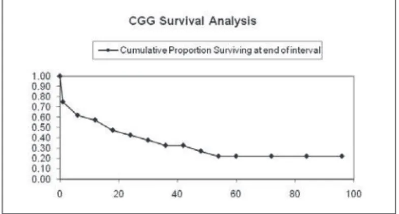
The success rate at 12 and 60 months were respectively, 92.5% and 79%. Kaplan-Meier survival analysis showed probability of success at 60 months of 71.5% (Figure 2). For patients with congenital glaucoma the cumulative proportion surviving at the end of 5 year was 22%, and for neovascular glaucoma was 94% (Figu-res 3 and 4).
Postoperative complications that occurred during the study are listed on Table 2. The two most commom complications, fibrin exudates (116 eyes, 21.5%) and hyphema (58 eyes, 10.7%) occurred in eyes with neovascular glaucoma. Among 13 eyes that went to phthisis bulbi 10 of them had neovascular glaucoma.
D
ISCUSSIONRefractory glaucoma is a subset of the disease where conventional surgical treatment has failed.(2,9) It
has been demonstrated that ECP is a safe alternative to achieve a satisfactory IOP control by decreasing the Figure 1- Mean + SD intraocular pressure (IOP) of 427 eyes with
a mean follow-up of 88.9 + 10.3 months (ranged from 60 to 108 months) plotted as a function of time after ECP
Figure 2 - Kaplan-Meier estimate of time to failure after ECP in all eyes
Figure 3 - Kaplan-Meier estimate of time to failure after ECP eyes with congenital glaucoma (CGC)
aqueous humor production without causing widespread damage to the ciliary body and adjacent structures as occurs with transscleral procedures.(10-12)
Previous reports suggest that ECP is a safe and effective procedure in refractory glaucoma patients after failure of conventional filtration surgery.(2,11,12,14) Lima
et al.(2) compared the use of Ahmed Valve and ECP in 68
patients with refractory glaucoma and found, in the ECP group, a success rate of 82.35% and 73.52% at 12 and 24 months, respectively. Ahmed Valve group had similar success rate. In our study, the overall success rate was 92.57% and 78.84% at 1 and 5 years of follow-up, respectively. Gayton et al.(13) compared cataract surgery
combined with trabeculectomy or ECP. The ECP cases did not achieve an immediate reduction in IOP, as the trabeculectomy cases did. However, by 1 month, both groups stabilized at about 16 mmHg and less complications were observed in ECP group.
Chen et al.(12) analyzed retrospectively 68 patients
with diverse forms of glaucoma who had undergone ECP. The follow-up period was 12.9 months and the mean IOP decreased 10.7 mmHg. The mean number of medication preoperatively was 3 and postoperatively was 2, and the visual acuity remained stable in 94% of the cases. The present study demonstrated a mean IOP reduction by 25.6 mmHg, the postoperative number of glaucoma medication and the visual acuity stability were similar to the above-mentioned study.
Uram(7) described the use of ECP in 10 intractable
neovascular glaucoma with the mean follow-up of 12 months. The IOP dropped 67.6% which was similar to the present study. It is important to notice that Uram(7)
just analyzed neovascular glaucoma which seems to be associated with the biggest index of success, as the survival curve shows. However, the great majority of phthisis bulbi were among eyes with neovascular glaucoma.
The most common complication in our study was fibrin exudates (21.5%), followed by hyphema (10.7%) and immediate postoperative IOP spikes (9.8%). Chen et al.(12) reported a 24% incidence of fibrin exudates, 12%
of hyphema and no phthisis bulbi. Probably the anterior chamber injection of dexamethasone performed as a routine in the present study has decreased the inflammation observed in the early postoperative period. In the present study, the sample that showed the lowest cumulative proportion surviving was congenital glaucoma.
Neely et al.(11) reported the use of the ECP in 36
eyes with pediatric glaucoma with a mean follow-up of
19.2 months. The survival curve for the first procedure in each eye demonstrated a high rate of early treatment failure in the first postoperative year, the survival curve flattened significantly after 14 months, similarly to the present study.
C
ONCLUSIONAlthough this is a retrospective study, the sample size and the long follow-up time provide reliable results. ECP not only resulted in satisfactory IOP, but also preserved visual acuity in the majority of the cases. These facts associated with low incidence of complications make ECP a safe and effective procedure in the treatment of refractory glaucomas.
R
ESUMOfo-ram observados intensa reação inflamatória em 116 olhos (21,5%), hifema em 58 eyes (10,7%), descolamento de coróide em 31 olhos (5,7%), phthisis bulbi em 13 olhos (2,4%), descolamento de retina em 8 olhos (1,4%) e hipotonia em 7 eyes (1,2%).Conclusão: Estes resulta-dos sugerem que CFE é uma técnica segura e eficaz para o tratamento de glaucomas refratários após longo tem-po de seguimento.
Descritores: Glaucoma/cirurgia; Coagulação por laser/métodos; Fotocoagulação/métodos; Acuidade visu-al; Pressão intra-ocular
R
EFERENCES1. Quigley HA, Broman AT. The number of people with glau-coma worldwide in 2010 and 2020. Br J Ophthalmol. 2006;90(3):262-7. Comment in: Br J Ophthalmol. 2006;90(3):253-4.
2. Lima FE, Magacho L, Carvalho DM, Susanna R Jr, Avila MP. A prospective, comparative study between endoscopic cyclophotocoagulation and the Ahmed drainage implant in refractory glaucoma. J Glaucoma. 2004;13(3):233-7. 3. Syed HM, Law SK, Nam SH, Li G, Caprioli J, Coleman A.
Baerveldt-350 implant versus Ahmed valve for refractory glaucoma: a case-controlled comparison. J Glaucoma. 2004;13(1):38-45.
4. Hampton C, Shields MB, Miller KN, Blasini M. Evaluation of a protocol for transscleral neodymium: YAG cyclophotocoagulation in one hundred patients. Ophthalmol-ogy. 1990;97(7):910-7.
5. Shields MB, Shields SE. Noncontact transscleral Nd: YAG cyclophotocoagulation: a long-term follow-up of 500 patients. Trans Am Ophthalmol Soc. 1994;92:271-83; discussion 283-7.
6. Shields MB, Chandler DB, Hickingbotham D, Klintworth GK. Intraocular cyclophotocoagulation. Histopathologic evalua-tion in primates. Arch Ophthalmol. 1985;103(11):1731-5. 7. Uram M. Ophthalmic laser microendoscope ciliary process
ablation in the management of neovascular glaucoma. Oph-thalmology. 1992;99(12):1823-8.
8. Uram M. Endoscopic cyclophotocoagulation in glaucoma management. Curr Opin Ophthalmol. 1995;6(2):19-29. 9. Allingham RR, Shields MB, Damji KF, Freedman S, Moroi
SE, Shafranov G. Shields’ textbook of glaucoma. 5th ed. Phila-delphia: Lippincott Williams & Wilkins; 2005.
10. Trevisani MG, Allingham RR, Shields MB. Histologic com-parison of contact transcleral diode cyclophotocoagulation and endoscopic cyclophotocoagulation. Invest Ophthalmol Vis Sci. 1995;36(4):144-8. [Annual meeting. Fort Lauderdale, Florida, May 14-19, 1995. Abstracts].
11. Neely DE, Plager DA. Endocyclophotocoagulation for man-agement of difficult pediatric glaucomas. J AAPOS. 2001;5(4):221-9.
12. Chen J, Cohn RA, Lin SC, Cortes AE, Alvarado JA. Endoscopic photocoagulation of the ciliary body for treatment of refrac-tory glaucomas. Am J Ophthalmol. 1997;124(6):787-96. 13. Gayton JL, Van Der Karr M, Sanders V. Combined cataract
and glaucoma surgery: trabeculectomy versus endoscopic la-ser cycloablation. J Cataract Refract Surg. 1999;25(9):1214-9. 14. Lima FE, Carvalho DM, Beniz J, Ávila M. Ciclofotocoagulação endoscópica em glaucomas refratários. Rev Bras Oftalmol. 1997;56(6):387-93.
Reprints requests to: Francisco E. Lima, MD