Rev Bras Anestesiol. 2013;63(2):209-212
Offi cial Publication of the Brazilian Society of Anesthesiology www.sba.com.br
REVISTA
BRASILEIRA DE
ANESTESIOLOGIA
Abstract
Background and objectives: The increased intraocular pressure (IOP) - which decreases perfusion
pressure on the optic nerve - increases by prone positioning (1). The aim of our study was to compare the effect of head rotation 45o laterally in prone position on the increase in IOP of upper
placed and lower placed eyes in patients undergoing percutaneous nephrolithotomy (PCNL). Methods: Forty-fi ve patients were randomly divided into 2 Groups. IOP of the patients were recorded bilaterally in supine position before the operation had started. Patients were turned to prone position. The head was placed on a prone headrest without external direct compression to both eyes. Patients in Group I were kept in strictly neutral prone position where as patients in Group II were placed prone with their heads rotated 45º laterally to the right side. At the end of the operation, patients were turned to supine position and their IOP was measured immediately. Results: There was no difference related to demographics, duration of surgery, blood loss and fl uid input data. IOP values after surgery in prone position increased signifi cantly compared to preoperative values in both groups (p < 0.05). After surgery in prone position IOP values of the upper positioned eyes in Group II were signifi cantly lower than Group I and lower positioned eyes in Group II (p < 0.05).
Conclusion: prone positioning increases IOP. In patients with prone position with a head rotation of 45o laterally, IOP in the upper positioned eye was signifi cantly lower.
© 2013 Sociedade Brasileira de Anestesiologia. Published by Elsevier Editora Ltda. All rights reserved.
The Effect of Head Rotation on Intraocular Pressure
in Prone Position: a Randomized Trial
M. Nuri Deniz*
1, Arzum Erakgün
1, Nezih Sertöz
1, Suzan Güven Yılmaz
2, Halil Ateş
2,
Elvan Erhan
11. MD; Ege University School of Medicine Department of Anesthesiology and Reanimation 2. MD; Ege University School of Medicine Department of Ophthalmology
Received fromEge University school of Medicine Department of Anesthesiology and Reanimation, Turkey.
Submitted on November 16, 2011. Approved on March 21, 2012.
Keywords:
Intraocular Pressure; Head Movements; Rotation; Prone position.
SCIENTIFIC ARTICLE
*Corresponding author: Ege University School of Medicine Department of Anesthesiology and Reanimation 35100 Izmir-Turkey. Phone: +902323902142, FAX: +902323757321
E-mail: [email protected]
ISSN/$ - see front metter © 2013 Sociedade Brasileira de Anestesiologia. Published by Elsevier Editora Ltda. All rights reserved.
Introduction
Studies have shown that hypotension, anemia, obesity, peripheral vascular disease, and middle-aged men may be risk factors for the development of postoperative vision loss after surgery 2,3. Prone positioning is also considered a risk
factor because it increases intraocular pressure (IOP), which decreases perfusion pressure to the optic nerve 1,4,5. The risk
of postoperative vision loss in patients undergoing spine sur-gery in prone position is 0.05%1. Some studies investigated
210 M. N. Deniz et al.
may cause a decrease in IOP 6. Although primary open angle
glaucoma is a bilateral disease, glaucomatous defects are usually asymmetrical between the two eyes and one eye is more affected than the fellow eye. There are also situations like diabetic retinopathy where the walls of the blood vessels are affected and the change in perfusion during perioperative
period may deteriorate the perfusion of the eye.
Percutaneous nephrolithotomy (PCNL) – performed in
prone position - has become the gold standard for managing large and complex renal calculi 7.The aim of our study was to
compare preoperative and immediately postoperative IOP in patients undergoing PCNL in prone position and the effect of head rotation 45º laterally in prone position on the increase in IOP of upper placed and lower placed eyes in patients undergoing PCNL. Our hypothesis is that head rotation 45º laterally in prone position may prevent the increase in IOP of the upper positioned eye and may protect the more severely affected eye in situations like glaucoma.
Materials and Methods
After approval by institutional ethics committee and ac-quiring written informed patient consent of 45 patients (American Society of Anesthesiologists physical status I–II),
aged between 18–80 years, they were scheduled for PCNL
in the prone position in the Department of Urology. Patients
with preexisting eye disease or previous eye surgery, Diabetes Mellitus, peripheral vascular disease, eye trauma or injury were excluded from the study. Patients were randomly al-located into 2 groups using a computer-generated list: Group I (n = 23) and Group II (n = 22). Before the study, intravenous catheters and standard anesthetic monitors (i.e., blood pressure cuff, electrocardiograph, and pulse oximeter) were placed in all patients. Anesthesia protocol was standardized for all study patients. After induction of anesthesia with propofol 2 mg.kg-1, remifentanil 1 µg.kg-1, and rocuronium 0.6 mg.kg-1
intravenously, and ventilation with O2-air-sevofl urane via a
facemask; the same anesthesiologist performed tracheal intu-bation. After intubation, the same ophthalmologist as before the operation had started (preoperative values) measured patient IOP bilaterally in supine position. IOP measurement was performed using Perkins Tonometer MK2 Clement clarke (Protected worldwide patents USA 5012812 ENGLAND). Urinary
catheters were placed and patients were turned to prone position. The head was placed on a silicone headrest without external direct compression to both eyes, and the neck was in the midline position. Patients in Group I were kept in this prone position whereas patients in Group II were placed in the prone position with their heads rotated 45º laterally to the right side. Anesthesia was maintained with O2-air-sevofl urane and remifentanil (0.25-1.0 μg.kg-1.min-1). Standard vital
pa-rameters, including blood pressure, ECG, SpO2, and EtCO2
(end-tidal carbon dioxide) were monitored throughout the perioperative period. Mean arterial pressure was measured from the arm every 5 minutes and was kept within 20% of awake value, and ventilation was adjusted to keep EtCO2 in the range of 30 to 35 mm Hg throughout the intraoperative period. All fl uids administered were recorded, and estimated blood loss and urine output were measured to prevent over hydration. At the end of the operation, patients were turned to supine position and their IOP was measured immediately (postoperative values).
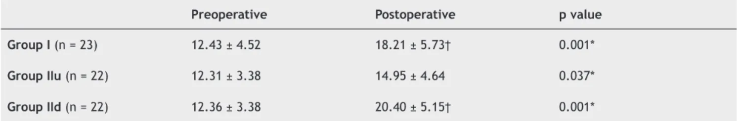
In Group I, bilateral measurements were recorded and IOP of the right eye was used for comparison. In Group II, IOP in upper (Group IIu) and lower (Group IId) positioned eyes were recorded separately. We noted the length of time in the prone position. We recorded hematocrit for each patient preo peratively and postoperatively. Prospective power analy-sis revealed that a group size of approximately 22 patients in each group would have been required to identify signifi cant difference between two groups with 95% power and p = 0.05. Paired Samples test was used for the comparison within each group whereas Independent Samples Test was used for the comparison between groups.
Results
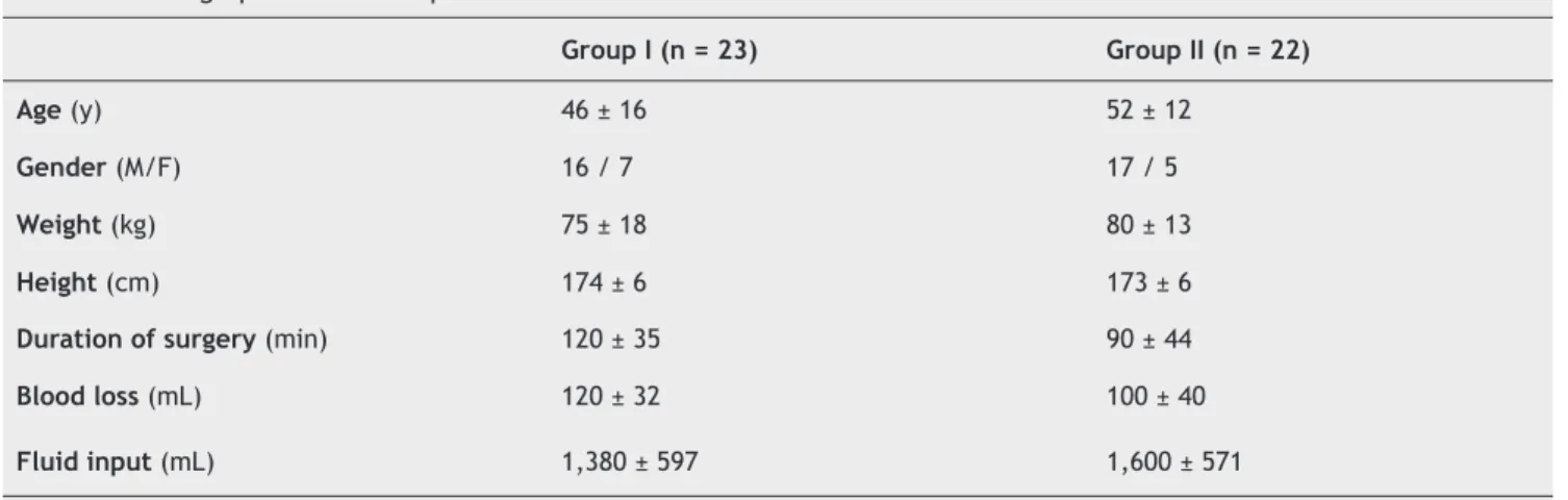
Every patient in each group completed the trial. Groups were comparable with respect to their demographic data, duration of surgery, fl uid input and blood loss (Table 1). None of the patients reported any eye complaints after surgery. None of the patients required blood transfusion. Vital pa-rameters were within normal limits through the surgery in both groups.
Table 1 Demographic and intraoperative data.
Group I (n = 23) Group II (n = 22)
Age (y) 46 ± 16 52 ± 12
Gender (M/F) 16 / 7 17 / 5
Weight (kg) 75 ± 18 80 ± 13
Height (cm) 174 ± 6 173 ± 6
Duration of surgery (min) 120 ± 35 90 ± 44
Blood loss (mL) 120 ± 32 100 ± 40
Fluid input (mL) 1,380 ± 597 1,600 ± 571
211 The Effect of Head Rotation on Intraocular Pressure in Prone Position: a Randomized Trial
Intraocular pressure values after the surgery in prone position increased signifi cantly compared to preoperative values in both groups (p < 0.05) (Table 2). After surgery in prone position IOP values in Group IIu (upper positioned eyes) were signifi cantly lower than Group I and Group IId (lower positioned eyes) (p < 0.05). After surgery in prone position there was no signifi cant difference in IOP values in Group I and Group IId (Table 2).
Discussion
The results of our study showed that upon prone positioning, the IOP signifi cantly increased in patients undergoing PCNL.
These fi ndings are in accordance with previous studies 8,9.
Our results have also suggested that when patients were positioned prone with their heads rotated 45o laterally, IOP
in the upper positioned eyes were signifi cantly lower than lower positioned eyes and IOP of the patients positioned prone without head rotation. The idea of head elevation being associated with less increase in IOP in prone position is not new, but our study is original in that it is the fi rst to compare IOP of patients in the prone fl at versus prone posi-tion with head rotaposi-tion laterally. Since upper posiposi-tioned eyes had signifi cantly lower IOP values compared with strictly neutral prone position and lower position eyes, head rotation may have advantages for patients with glaucoma to protect a more severely affected eye. However, excess rotation should be avoided to prevent twisting the internal jugular vein with subsequent increase in venous pressure and IOP. It is also important to use appropriate silicone headrests to avoid direct eyeball compression because there should be no contact with headrest and orbita.
The study by Cheng et al. 8 was the fi rst study to measure
IOP in prone anesthetized patients undergoing spinal surgery. They examined the combined effects of general anesthesia and the prone position on IOP in patients undergoing spine surgery. They found that prone positioning increased IOP dur-ing anesthesia. Since ocular perfusion pressure is commonly defi ned as the difference between mean arterial pressure (MAP) and IOP, the authors drew attention to the fact that ocular perfusion pressure could decrease despite mainte-nance of normotension during surgery in prone position 8. In
a similar study, Hunt et al. 9 also studied patients undergoing
spinal surgery and found that IOP increased when anesthe-tized patients are placed in prone position. Those in prone positions were signifi cantly higher than supine positions.
Some studies have shown that Trendelenburg position in prone resulted in further increase in IOP compared to prone position 1,6. Head elevation in prone position (reverse
Trendelenburg) ameliorated the increase in IOP in prone fl at 6. Ozcan et al. 6 studied the effect of body inclination
during prone positioning on intraocular pressure in awake
volunteers. They showed that a 10th reverse Trendelenburg
position ameliorated the increase in IOP caused by the prone position in awake volunteers. Although that study did not include all the factors that might have affected the IOP in an operative setting, it suggested a strategy to positively infl uence the IOP by changing the operating table inclination for patients in the prone position 6.
The current study is original in that it is the fi rst study to compare IOP of subjects in prone position with head rotation 45o laterally. This study’s main fi nding is that head rotation
has an advantage on the upper positioned eyes without caus-ing further disadvantage on the lower positioned eyes. This position may be particularly advantageous for patients with glaucoma to prevent the increase in IOP when head rotation 45o laterally in prone position is used and the more severely
affected eye is kept in the upper position. The asymmetry of visual fi eld loss between eyes in primary angle closure glaucoma (PACG) and primary open angle glaucoma (POAG)
was described by Wang et al. 10. A more marked thinning of
the neuroretinal nerve fi ber layer in the eye with the higher IOP has also been described in normal-tension glaucoma
patients 11. Interocular asymmetries in IOP must be kept
in mind especially in glaucoma patients in certain circum-stances, such as operations under prone position and the eye with more severe glaucoma damage can be protected during the operation.
In conclusion, prone positioning increases IOP. When pa-tients were positioned prone with heads rotated 45o laterally,
IOP in the upper positioned eyes were signifi cantly lower than lower positioned eyes and prone position without head rotation. This position may be advantageous for patients with bilateral glaucoma to protect the more severely affected eye as well as unilateral glaucoma.
References
1. Walick KS, Kragh JE Jr, Ward JA, Crawford JJ - Changes in Intraocular Pressure due to Surgical Positioning Studying Potential Risk for Postoperative Vision Loss, Spine, 2007;32:2591-2595.
Table 2 Intraocular pressure values (mmHg) in two groups before and after the surgery.
Preoperative Postoperative p value
Group I (n = 23) 12.43 ± 4.52 18.21 ± 5.73† 0.001*
Group IIu (n = 22) 12.31 ± 3.38 14.95 ± 4.64 0.037*
Group IId (n = 22) 12.36 ± 3.38 20.40 ± 5.15† 0.001*
212 M. N. Deniz et al.
2. Abraham M, Sakhuja N, Sinha S, Rastogi S - Unilateral visual loss after cervical spine surgery. J Neurosurg Anesthesiol, 2003;15:319-322.
3. Lee L, Lam A - Unilateral blindness after prone lumbar spine surgery. Anesthesiology, 2001;95:793-795.
4. Locastro A, Novak K, Biglan A - Central retinal artery occlusion in a child after general anesthesia. Am J Ophthalmol, 1991;112:91-92.
5. Roth S, Nunez R, Schreider B - Unexplained visual loss after lumbar spinal fusion. J Neurosurg Anesthesiol, 1997;9:346-348.
6. Ozcan MS, Praetel C, Bhatti MT, Gravenstein N, Mahla ME, and Seubert CN - The effect of body inclination during prone positioning on intraocular pressure in awake volunteers: a comparison of two operating tables. Anesth Analg, 2004;99:1152-1158.
7. Deane LA, Clayman RV - Advances in percutaneous nephrostolithotomy. Urol Clin North Am, 2007;34:383-395. 8. Cheng MA, Todorov A, Tempelhoff R, McHugh T, Crowder CM,
Lauryssen C - The effect of prone positioning on intraocular pressure in anesthetized patients. Anesthesiology, 2001;95:1351-1355.
9. Hunt K, Bajekal R, Calder I, Meacher R, Eliahoo J, Acheson JF - Changes in intraocular pressure in anesthetized prone patients. J Neurosurg Anesthesiol, 2004;16:287-290.
10. Wang JC, Gazzard G, Foster FJ, et al - Interocular asymmetry of visual fi eld defects in primary open angle glaucoma and primary angle-closure glaucoma. Eye, 2004;18:365-368.