Objective: To identify selection criteria for selective dorsal rhizotomy (SDR) in cerebral palsy, to analyze the instruments used for evaluation, and to describe the characteristics of physical therapy in postoperative protocols.
Data sources: Integrative review performed in the following databases: SciELO, PEDro, Cochrane Library, and PubMed. The terms in both Portuguese and English for “cerebral palsy”, “selective dorsal rhizotomy”, and “physical therapy” were used in the search. Studies whose samples enrolled individuals with cerebral palsy who had attended physical therapy sessions for selective dorsal rhizotomy according to protocols and describing such protocols’ characteristics were included. Literature reviews were excluded and there was no restriction as to period of publication.
Data synthesis: Eighteen papers were selected, most of them being prospective cohort studies with eight-month to ten-year follow-ups. In most studies, the instruments of assessment encompassed the domains of functions, body structure, and activity. The percentage of posterior root sections was close to 50%. Primary indications for SDR included ambulatory spastic diplegia, presence of spasticity that interfered with mobility, good strength of lower limbs and trunk muscles, no musculoskeletal deformities, dystonia, ataxia or athetosis, and good cognitive function. Postoperative physical therapy is part of SDR treatment protocols and should be intensive and speciic, being given special emphasis in the irst year.
Conclusions: The studies underline the importance of appropriate patient selection to obatin success in the SDR. Postoperative physical therapy should be intensive and long-term, and must necessarily include strategies to modify the patient’s former motor pattern. Keywords: Muscle spasticity; Rhizotomy; Physical therapy specialty; Postoperative care; Cerebral palsy.
Objetivo: Identiicar critérios de seleção para a rizotomia dorsal seletiva (RDS) na paralisia cerebral (PC), analisar os instrumentos de avaliação e descrever as características da isioterapia nos protocolos pós-operatórios.
Fontes de dados: Revisão do tipo integrativa nas bases de dados SciELO, PEDro, Cochrane Library e PubMed. Os termos em português e inglês “paralisia cerebral”, “rizotomia dorsal seletiva” e “isioterapia” foram utilizados na busca. Os critérios de inclusão foram: artigos que arrolaram indivíduos com PC, que realizaram isioterapia nos protocolos de RDS e que descreviam características desses protocolos. Foram excluídos artigos de revisão da literatura e não houve restrição de período de publicação.
Síntese dos dados: Incluíram-se 18 estudos, sendo a maioria coortes prospectivas, com acompanhamento dos pacientes de oito meses a dez anos. Os instrumentos das avaliações contemplam, na maior parte dos trabalhos, os domínios de funções e estruturas corporais e atividade. O percentual de secção das raízes posteriores foi próximo a 50%. A principal indicação para a RDS incluiu deambuladores com diplegia espástica, que preenchiam os seguintes critérios: espasticidade que interfere com a mobilidade, boa força muscular de membros inferiores e tronco, sem deformidades ortopédicas, distonia, ataxia ou atetose e boa função cognitiva. A isioterapia é parte integrante dos protocolos de tratamento com RDS, devendo ser intensiva, especíica e enfatizada principalmente no primeiro ano.
Conclusões: Os estudos salientam a importância da seleção adequada dos pacientes para o sucesso dos resultados. A isioterapia é intensiva e de longa duração, devendo necessariamente ter estratégias para modiicar o antigo padrão motor.
Palavras-chave: Espasticidade muscular; Rizotomia; Fisioterapia; Cuidados pós-operatórios; Paralisia cerebral.
ABSTRACT
RESUMO
*Corresponding author. E-mail: dagostinirenata@hotmail.com (R.D. Nicolini-Panisson).
aPontifícia Universidade Católica do Rio Grande do Sul, Porto Alegre, RS, Brazil. bInstituto de Neuro-Ortopedia, Caxias do Sul, RS, Brazil.
SELECTIVE DORSAL RHIZOTOMY IN CEREBRAL
PALSY: SELECTION CRITERIA AND POSTOPERATIVE
PHYSICAL THERAPY PROTOCOLS
Rizotomia dorsal seletiva na paralisia cerebral: critérios de indicação
e protocolos de reabilitação fisioterapêutica pós-operatória
Renata D’Agostini Nicolini-Panisson
a,*, Ana Paula Tedesco
b,
INTRODUCTION
Spasticity is the main clinical feature of patients with spastic cerebral palsy (CP) and is considered the most important cause of discomfort, gait abnormalities, and functional limitations.1 It also generates muscle shortenings that inluence bone growth and lead to deformities. Controlling it, therefore, is crucial to the treatment of CP.2
Selective dorsal rhizotomy (SDR) is a neurosurgical proce-dure performed in children with bilateral spastic CP to reduce lower limb spasticity.3 It is mostly performed at the lumbosacral level and consists of the interruption of the aferent stimulus by the monosynaptic stretch relex.3 In order to preserve the sensory and sphincter functions, the dorsal root is divided into radicles and only a portion of these is sectioned.3
SDR was irst described by Foerster in 1908, after he observed that the dorsal (sensory) radicles section could decrease spastic-ity; signiicant muscle weakness with sensory and proprioceptive losses was also observed after the procedure.2 hus, in 1978, Fasano presented the intraoperative electrophysiological stimu-lation and the section of a portion of dorsal radicles, and both techniques are currently used.2 he method was then adopted and popularized by Peacock and Arens in 1980.2
SDR results indicate spasticity reduction, muscle strength gain, gait speed and kinematics increase, and gross motor func-tion improvement.4-6 Patients submitted to SDR and physical therapy are compared to with those who only received physical therapy, signiicant reduction in spasticity and functional improve-ment are seen in the irst group.7,8 Speciic physical therapy plays a central role in the postoperative phase, as spinal bone proce-dures such as laminectomy or laminotomy require special care in the irst weeks of this period, in addition to formal conduct.9,10
he centers that ofer SDR follow special protocols for the postoperative period. In Brazil, this technique is starting to be disseminated and, due to peculiarities related to postoperative treatment, this review of protocols described in the literature aims to help professionals to better understand the role of phys-ical therapy in rehabilitation. he objectives of this study were, therefore, to identify SDR selection criteria and to describe the characteristics of physical therapy postoperative protocols.
DATA SOURCE
his is an integrative literature review. he electronic search for references was carried out in August 2016 in the databases SciELO, PEDro, Cochrane Library, and PubMed. he terms used in the search, both in Portuguese and in English, were: “ paral-isia cerebral”/“cerebral palsy”, “rizotomia dorsal seletiva” /“dor-sal selective rhizotomy”, and “isioterapia”/“physical therapy”. Headings, abstracts and, when necessary, the full study were
reviewed to determine whether they would match inclusion criteria: studies conducted with individuals with CP who had attended physical therapy sessions according to SDR protocols and depicting such protocols’ characteristics. No ilters were applied to search, as well as there were no restrictions as to age group of sample subjects or period of publication. Literature reviews were excluded. he lists of references of selected papers were also searched for other relevant manuscripts.
After selection, the authors made a critical reading to grasp the main information, which was then presented in the fol-lowing categories:
• characteristics of studies; • characteristics of study samples. • SDR selection criteria;
• characteristics of physical therapy protocols.
DATA SYNTHESIS
According to the pre-established criteria, 18 articles were selected for this review. Figure 1 is the lowchart of papers’ search and selection.
Characteristics of studies
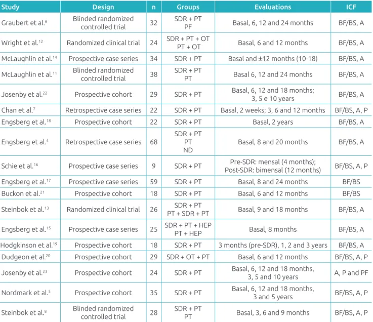
he studies included in our research are shown in Table 1. From 18 studies included, ive (27.8%) were randomized clinical tri-als,6,8,11-13 six (33.3%) were series of cases (four prospective14-17 and two retrospective4,7), and seven (38.9%) were prospective
Figure 1 Flowchart showing the search and selection of papers.
(37 in PubMed, 0 in SciELO, 6 in Cochrane Library, 1 in PEDro, 15 in reference lists)
59 papers found
Exclusion and inclusion criteria deined
52 papers selected, after duplicates were excluded
18 papers selected, 34 papers excluded:
1 spine lesion
13 literature reviews 15 no information about
2 opinions
2 systematic review 1 metanalysis
cohort studies.5,18-23 Sample sizes ranged from 9 to 68 individuals, most of them being distributed in groups of SDR intervention associated with physical therapy4-8,11,13,14,16-19,21-23 or only phys-ical therapy.4,6,8,11 Patient follow-up periods ranged from eight months15 to ten years.22,23 In most studies, the same physical therapist performed both pre- and postoperative evaluations.
Evaluation tools according to domains
of the International Classification of
Functioning, Disability and Health
he evaluation of SDR candidates should be as comprehensive as possible and encompass elements described by the International
Classiication of Functioning, Disability and Health (ICF). One of the studies7 used the quantitative classiication by ICF, and another one made evaluations considering ICF domains, as described below.
The literature brings a variety of information with respect to items to be evaluated, with domain, structure, and body function as per ICF considered in all studies but one.23 he instruments used in studies to evaluate domain, structure, and body function were: spasticity evaluation (Ashworth scale,5-8,11-14,16,22 clinical signs of spasticity,11,15 quantitative spasticity assessment (QSA)6,11,19 by isokinetic dynamometer4,17,18), motion range,5-8,11-15,19,22, relex range,12,14
Table 1 Characteristics of papers included in the review.
n: sample size; SDR: selective dorsal rhizotomy; PT: physical therapy; ND: no disability; OT: occupational therapy; HEP: home exercise program; ICF: International Classiication of Functioning, Disability and Health; BF/BS: body function, body structure; A: activity; P: participation; PF: personal factors.
Study Design n Groups Evaluations ICF
Graubert et al.6 Blinded randomized
controlled trial 32
SDR + PT
PF Basal, 6, 12 and 24 months BF/BS, A
Wright et al.12 Randomized clinical trial 24 SDR + PT + OT
PT + OT Basal, 6 and 12 months BF/BS, A
McLaughlin et al.14 Prospective case series 34 SDR + PT Basal and ±12 months (10-18) BF/BS, A
McLaughlin et al.11 Blinded randomized
controlled trial 38
SDR + PT
PT Basal 6, 12 and 24 months BF/BS, A
Josenby et al.22 Prospective cohort 29 SDR + PT Basal, 6, 12 and 18 months;
3, 5 e 10 years BF/BS, A
Chan et al.7 Retrospective case series 22 SDR + PT Basal, 2 weeks; 3, 6 and 12 months BF/BS, A, P
Engsberg et al.18 Prospective cohort 22 SDR + PT Basal, 2 years BF/BS, A
Engsberg et al.4 Retrospective case series 68
SDR + PT PT ND
Basal, 8 and 20 months BF/BS, A
Schie et al.16 Prospective case series 9 SDR + PT Pre-SDR: mensal (4 months);
Post-SDR: bimensal (12 months) BF/BS, A, P
Engsberg et al.17 Prospective case series 59 SDR + PT Basal, 8 and 24 months BF/BS
Buckon et al.21 Prospective cohort 18 SDR + PT Basal, 6 and 12 months BF/BS
Steinbok et al.13 Randomized clinical trial 26 SDR + PT
PT + SDR + PT Basal, 9 and 18 months BF/BS, A
Engsberg et al.15 Prospective case series 25 SDR + PT + HEP
PT + HEP Basal, 8 months BF/BS, A
Hodgkinson et al.19 Prospective cohort 18 SDR + PT 3 months (pre-SDR), 1, 2 and 3 years BF/BS, A
Dudgeon et al.20 Prospective cohort 29 SDR + OT + PT Basal, 6 and 12 months BF/BS, A, P
Josenby et al.23 Prospective cohort 24 SDR + PT Basal, 6, 12 and 18 months,
3, 5 and 10 years A, P and PF
Nordmark et al.5 Prospective cohort 35 SDR + PT Basal, 6, 12 and 18 months,
3 and 5 years BF/BS, A, P
Steinbok et al.8 Blinded randomized
controlled trial 28
SDR + PT
muscle strength,4,8,13,15,17-19, popliteal angle,22 musculoskel-etal deformities14 by hips and spine radiography,7 selectiv-ity assessment7, and isometric contraction assessment by electromyography.21
Only two studies17,21 did not measure the activity domain, and in those addressing it, the instruments used were: Gross Motor Function Measure (GMFM),4-8,11,12,14,16,18,22 walking sta-tus6,8,11,14, Gross Motor Classiication System (GMFCS),5,7,22,23 three-dimensional gait analysis,4,6,7,12,18 observational gait anal-ysis7,16 (Observational Gait Score7, Edinburgh Visual Gait Score16), urodynamics,7 Peabody Fine Motors Scale,8 self-care evaluation,8,20 walking distance in 60 seconds12, and Physiological Cost Index.8
Six studies5,7,8,16,20,23 addressed the domain participa-tion and its evaluaparticipa-tion instruments: Pediatric Evaluaparticipa-tion of Disability Inventory (PEDI),5,7,16,20,23 Canadian Occupational
Performance Measure (COMP)7, and self-care evaluation.8,20 SDR evaluation should follow more comprehensive proto-cols with postoperative analysis of the same instruments, thus allowing a more accurate evaluation of results and bet-ter conclusion-drawing.
Characteristics of study samples
Table 2 shows the characteristics of samples of the included studies. All of them enrolled individuals with spastic PC. he study by Chan et al mentioned a participant with hered-itary spastic paraparesis (HSP), in addition to 20 individuals with CP.7 Although CP diagnosis was one of the inclusion criteria of this review, SDR can also be indicated for patients with spasticity resulting from other diagnoses such as multi-ple sclerosis, Leigh syndrome,25 stroke,26 spinal cord injury24, and transverse myelitis.27
Study Age Topography GMFCS Level of section
Percentage
of section Surgical approach
Graubert et al.6 6.5 (3.3-14.5)* Diplegia – – – –
Wright et al.12 58.0
±12.7 (41-91) months Diplegia – L2-S2 50 Partial laminectomy L2-L5
McLaughlin et al.14 QE: 7.2±3.4; DE: 8.9±3.9** Diplegia,
quadriplegia – L2-S2 49 (29-60) Laminotomy T12-S2
McLaughlin et al.11 6.1
±3.0 (2.9-14.3)* Diplegia – 34 (20-56) Laminectomy or
laminotomy
Josenby et al.22 4.3 (2.6-6.7) Diplegia I-V – – –
Chan et al.7 8.6±2.6 (5.9-11.2) Diplegia,
quadriplegia I-IV L1-S2 49.7±2.2
Articulate laminotomy
L2-S1
Engsberg et al.18 8.8
±4.8 Diplegia I-III L1-S2 (60-65) Laminotomy L1
Engsberg et al.4 9.0
±5.3* Diplegia I-III L1-S2 65 Laminotomy L1
Schie et al.16 65 (43-82) months Diplegia II-III L2-S1 50 (31-68) Laminotomy L1-L5
Engsberg et al.17 8.5±4.4 (4-18)# Diplegia I-III L1-S2 – Laminotomy L1-L2
Buckon et al.21 63 (48-86) months# Diplegia – L2-S1 42 (36-48) Laminotomy L2-L5
Steinbok et al.13 (3-7) Diplegia – L2-S1 (33-62) Laminotomy L1-S1
Engsberg et al.15 9±4.2 (4-16)* Diplegia – L1-S2 (60-80) Laminectomy L2 and,
when needed, L1
Hodgkinson et al.19 9 (5.5-16.5) Quadriplegia – – 60 Laminotomy T12-L2
Dudgeon et al.20 8.1±4.1 (3.7-22) Diplegia,
quadriplegia – L2-S1 42 –
Josenby et al.23 4.1 (2.5-6.4) Diplegia I-V L2-S2 40 En-Bloc laminoplasty
L1-L5
Nordmark et al.5 4.5±1.1 (2.5-6.6) Diplegia I-V L2-S2 40 En-Bloc laminoplasty
L1-L5
Steinbok et al.8 50 (35-75) months# Diplegia I-IV L2-S2 45±5 Laminotomia L1-S1
Table 2 Characteristics of samples of papers included.
Age: mean±standard deviation (min. and max. interval) shown in years, unless indicated otherwise; *Group SDR + physical therapy; #Group CP; **Group
Only one study did not include individuals with CP due to spastic diplegia19 and four studies enrolled individuals with spastic quadriplegia.7,14,19,20 Regarding GMFCS levels, only half of the studies4,5,7,8,16-18,22.23 referred to this classifi-cation, and individuals presenting all levels are mentioned. Overall, SDR is the procedure of choice for spasticity of lower limbs in children with diplegia,9 since they have more involvement of the lower limbs and dystonia is not always present.9 Patients with spastic quadriplegia are more likely to present dystonia and involvement of both upper and lower limbs, and the treatment with continuous intrathe-cal baclofen infusion is more indicated,9 although some studies support SDR.7,14,19,20 Another aspect to be consid-ered when indicating SDR is ambulation potential,9 which includes GMFCS levels I, II, and III. However, investiga-tions have performed SDR for levels IV and V with spe-cific goals and suggested that this is an alternative to the
use of continuous intrathecal baclofen infusion, given the management and monitoring complexity of this method.28
Most studies had section of 50% of the selected posterior rootlets from L1 or L2 to S1 or S2. A meta-analysis showed direct relationship between the percentage of cut and function gain, that is, the decrease in spasticity helps in the acquisition of functional abilities.1
SDR selection criteria
As shown in Table 3, the studies usually have patients with spastic diplegia matching the selection criteria4,6,7,11-18,20 and the ive “s”:2,3,7 spastic – lower limb spasticity interfer-ing with functionality;4,6,7,11-20,22 strength – adequate lower limb muscle strength and control;7,12,22 straight – adequate trunk6,7,22 and head6 control without ixed orthopedic defor-mity;7,11,12,16,17,22 slim – being thin; and smart – not having sig-niicant cognitive deicits.4,6,7,11,18 Also, criteria including good
Table 3 SDR indication criteria in subjects with cerebral palsy.
Inclusion criteria Exclusion criteria
3-18 years6,11
3-21 years14
3-7 years8,13,16
4-17 years15
>2 years17
>4 years4,18
<7 years5
children, adolescents and young adult20
Bulbar involvement6
Dystonia, athetosis, rigidity, mild to severe hypotonia 4,14,17,18,22,23
Dystonia, athetosis, ataxia5,7,8,11,13
CNS malformation 4,18
Spastic diplegia4-8,11-18,20,21
Spastic quadriplegia with remarks7,14,17,20-23 Visual impairment limiting mobility11
Good head-trunk control6,7,22,23
LL reasonable muscle strength12,22,23 Depends on spasticity to stand up or walk22,23
Ability or potential to wander with and without supportive device,4,6,12,18 for three meters12
Able to walk barefoot for eight minutes, with or without support,4.18 to sit, kneel and crawl
independently for short periods,16 to crouch seven
times,16 sit on a bench with free arms and to stand
up with support8,13
Fixed LL contractures5,7
Severe ixed contractures:11,12,16,17,22,23 >30º on hips and knee;12 >15º
on hips and knee and >30º on ankle;11 >20º on hips, knee, and ankle
and >80º popliteal angle
GMFCS I – III,4,18 I-V,5 II-III16 Progressive subluxation of the hips 8,11
36-month or more intellectual function6,11
Minimum cognitive skills to actively participate4,5,18
Children with intellectual disabilities23
Spinal deformities, uncontrolled epilepsy, contraindication for prolonged anesthesia11
Spasticity of LL interfering with functional tasks such as sitting, standing, and walking4,5,7,8,12-14,16-20,22,23
Spasticity in at least six muscle groups of both LL16
Orthopedic surgery,4,12,18 in the previous year4,5,18 or near-term
planning8
Botulinum toxin or plaster in six months4,18
Availability for intensive physical therapy5,8,11
Good family and rehabilitation support11,16 Severe cognitive disability4,5,7,11,18
family support are cited,16,29 as well as good rehabilitation11,16 and the capacity to collaborate in rehabilitation (cognitively and emotionally).18 Even though this is not the population to whom SDR is ideally indicated, some studies indicate it for patients with spastic quadriplegia7,14,17,20 with the following criteria:3.7 signiicant spasticity interfering with positioning, care, and passive stretching; absence of other motor disorders; and absence of ixed contractures in multiple joints. In both topographies, abnormalities of movement (dystonia, ataxia, choreoathetosis, hypotonia, stifness),4,6,7,11,13,17,18,22 hips insta-bility,11 signiicant scoliosis,11 presence of signiicant ixed con-tractures,7,11,12,16,17,22 absence of antigravity muscle strength,11 and visual impairments limiting mobility11 are contraindica-tions for the procedure.
he correct indication of SDR is fundamental for the suc-cess of treatment.3,30 Criteria have been described and the lit-erature supports that it is important that this decision is made by a multidisciplinary team trained and specialized in the care of spasticity in CP patients and with access to all treatment options.1-3,10,31 his team should consist of a physical therapist, a pediatrician, an orthopedist, and a neurosurgeon, all of them trained and specialized.1,31 he whole staf, including patients’ family members, should agree with the SDR decision and with the individual treatment goals for each child.2,9 A recent sys-tematic review stated that these selection criteria vary across studies and are based more on clinical reasoning than on sci-entiic evidence, and it is important that specialists come to a consensus on the subject.3
Characteristics of physical
therapy protocols
Table 4 lists the characteristics of post-SDR physical ther-apy protocols, including start of sessions, length of hospital stay and frequency. Studies typically show that, after SDR, patients undergo intensive physical therapy rehabilitation lasting approximately one year, starting on the irst days after surgery and staying hospitalized from six days to six weeks. Two studies13,15 reported preoperative physical ther-apy and three12,20,21 mentioned postoperative occupational therapy as well.
Half of the studies report that after the in-hospital phys-ical therapy period, speciic treatment guidelines are sent to local therapists, with whom the responsible therapist had made prior contact, in order to maintain consistency of the treatment plan.
As for the physical therapy program itself, early mobi-lization of the lower limbs is made during the irst week after SDR to maintain a range of motion and positioning, including prone, supine and siting positions with extended
lower limbs.5,7,12,16 he irst ive days are speciic for muscle strength exercises with hip abductors and extensors, knee extensors, dorsilexors, and practice of normal orthostasis and gait patterns are initiated.16 he onset of orthostasis is described as initiated by the use of parapodium in the 8th day,12 or with the use of ixed or ground-reaction Ankle Foot Orthoses (AFO) to stimulate knee extension on the sixth day,16 and adaptation equipment.14 Muscle strength-ening is described as rehabilitation practice in most stud-ies,4,7,8,11-14,16,17,20,21 with emphasis on the lower limb extensor and hip abductors muscles, knee extensors and dorsilex-ors,8,12,13,16 in addition to upper limbs12 and trunk mus-cle.,4,12 he exercises are performed using isolated training,20 progressive resistance training,11 and selective or functional control.21 Gait training starts on the second7 or third week12 and focuses on normal motor pattern with the use of sup-portive devices17 if necessary. In addition, the use of normal movement pattern facilitation (neurodevelopmental theory) is also described,8,11-13,21 as well as ine motor skills training,12 functional activities,4,5,7,12,14,17,20-22 daily-living activities,5.7 posture control and alignment,8,13,14,22 and postural trans-fer training with emphasis to balance when siting, kneeling, crawling, standing from loor and chair, standing, and on gait.5,7,12,17,21,22 Hydrotherapy,5.16 equotherapy,5,16 and phys-ical activities5,22,23 are also mentioned.
According to the most recent recommendations by the National Institute for Health and Clinical Excellence (NICE), when it comes to treatment of spasticity in chil-dren and adolescents with non-progressive brain disorders, an intensive physical therapy program is essential after clin-ical approach to spasticity by SDR31 and also determinant for successful outcomes.30
FINAL REMARKS
Further prospective studies with long-term follow-up reha-bilitation protocols are suggested. he use of validated evaluation instruments for the analysis of both static/functional aspects and quality of life should be considered, aiming to clarify SDR
indication criteria and whether the current postoperative reha-bilitation conventions are appropriate.
hus, this literature review shows that physical therapy plays a key role in the rehabilitation of patients with CP who
Study PT start (day)
Length of
hospital stay Physical therapy frequency
Graubert et al.6 -- 4weeks de terapia:10 hours/week + 5months:
4-5 hours/ week + 6months: 1-3 hours/week
Wright et al.12 2nd or 3rd 6 weeks
6 weeks: 45 minutes/day of physical therapy e 2 sessions/week (45 minutes of occupational therapy); after discharge,
up to 1 year: 2 sessions/week (120 minutes)
McLaughlin et al.14 4th a6th 1 month 1st month: 2 hours/day for 5 days/week; following 5months:
3-5 hours/week; 6th month: normal therapy
McLaughlin et al.11 2nd 1 month 4 weeks: 2 hours/day for 5 days/week (40 hours) + 5 months:1 hour/
day for 4-5 days/week + 6months: 1 hour/day for 1-4 days/week
Josenby et al.22 1st -- 6months: twice/week (1 hour); 6th-18th month:
once/week and physical activities
Chan et al.7 2nd 4 weeks 4 weeks: 5 hours/day for 5 times/week;
2nd -12th month: 3-6 hours/week
Engsberg et al.18 5th 1 week 5th day-8th month: 4 times/week; 8th -16th month: 3 times/week
Engsberg et al.4 -- -- 8 months: 4 times/week + 12 months: 3 times/week
Schie et al.16 1st 1 week
5th day: sitting on WC and therapy 3 times/day (1 hour); 6th day:
orthostasis and, when possible, gait with GRO; 3 months: 5 times/ week (1 hour); 3rd-6th month: 4 times/week (1 hour);
6th -12th month: 3 times/week (30 minutes)
Engsberg et al.17 3rd 1 week 1st week: twice/day + 8 months: 4-5 times/week;
after 8th month: 3-4 times/week
Buckon et al.21 4th 1 month
1st month: twice/day + occupational therapy: 1time/day; 2th -6th month: 3-4 times/week, occupational therapy: 1-2 times/ week;
6th mês-1 year: 1-2 times/week
Steinbok et al.13 -- -- 3 months: 3 times/week + 6 months: twice/week
(9 months pre- and post-operative periods)
Engsberg et al.15 3rd
--Post-operative period, 6 months: twice/week; 3rd day post-operative period: 3 times/day;
up to 6 months: 4-5 times/week; 6th -8th month: 3-4 times/week
Hodgkinson et al.19 -- -- 6 months: once/day
Dudgeon et al.20 -- 4 weeks 4 weeks: 2 hours/day, 5 times/week;
occupational therapy: 3-5 hours/week + 5 months: 4-5 hours/week
Josenby et al.23 1st -- 6 months: 1 hour/2 times/week; up to 18 months: once/week and
physical activities.
Nordmark et al.5 5th 3-5 days ICU 1st week: 45 minutes/twice/day; 2nd -3rd week: 45 minutes/3 times/day;
2nd-6th month: 1 hour/twice/week; 6 months: 1 hour/once/week
Steinbok et al.8 2nd 6 days 6th day: weight support while standing up; 2nd week: gait;
3 months: 3 times/week + 6 months: 2 times/week
Table 4 Characteristics of physical therapy protocols following selective dorsal rhizotomy.
REFERENCES
1. McLaughlin J, Bjornson K, Temkin N, Steinbok P, Wright V, Reiner A, et al. Selective dorsal rhizotomy: meta-analysis of three randomized controlled trials. Dev Med Child Neurol. 2002;44:17-25.
2. Aquilina K, Graham D, Wimalasundera N. Selective dorsal rhizotomy: an old treatment re-emerging. Arch Dis Child. 2015;100:798-802.
3. Grunt S, Fieggen AG, Vermeulen RJ, Becher JG, Langerak NG. Selection criteria for selective dorsal rhizotomy in children with spastic cerebral palsy: a systematic review of the literature. Dev Med Child Neurol. 2014;56:302-12. 4. Engsberg JR, Ross SA, Collins DR, Park TS. Efect of selective
dorsal rhizotomy in the treatment of children with cerebral palsy. J Neurosurg. 2006;105:8-15.
5. Nordmark E, Josenby AL, Lagergren J, Andersson G, Stromblad LG, Westbom L. Long-term outcomes ive years after selective dorsal rhizotomy. BMC Pediatr. 2008;8:54. 6. Graubert C, Song KM, McLaughlin JF, Bjornson KF. Changes
in gait at 1 year post-selective dorsal rhizotomy: results of a prospective randomized study. J Pediatr Orthop. 2000;20:496-500.
7. Chan SH, Yam KY, Yiu-Lau BP, Poon CY, Chan NN, Cheung HM, et al. Selective dorsal rhizotomy in Hong Kong: multidimensional outcome measures. Pediatr Neurol. 2008;39:22-32.
8. Steinbok P, Reiner AM, Beauchamp R, Armstrong RW, Cochrane DD, Kestle J. A randomized clinical trial to compare selective posterior rhizotomy plus physiotherapy with physiotherapy alone in children with spastic diplegic cerebral palsy. Dev Med Child Neurol. 1997;39:178-84.
9. Steinbok P. Selective dorsal rhizotomy for spastic cerebral palsy: a review. Childs Nerv Syst. 2007;23:981-90.
10. Hendricks-Ferguson VL, Ortman MR. Selective dorsal rhizotomy to decrease spasticity in cerebral palsy. AORN J. 1995;61:514-8, 521-2, 525.
11. McLaughlin JF, Bjornson KF, Astley SJ, Graubert C, Hays RM, Roberts TS, et al. Selective dorsal rhizotomy: eicacy and safety in an investigator-masked randomized clinical trial. Dev Med Child Neurol. 1998;40:220-32.
12. Wright FV, Sheil EM, Drake JM, Wedge JH, Naumann S. Evaluation of selective dorsal rhizotomy for the reduction of spasticity in cerebral palsy: a randomized controlled tria. Dev Med Child Neurol. 1998;40:239-47.
13. Steinbok P, McLeod K. Comparison of motor outcomes after selective dorsal rhizotomy with and without preoperative intensified physiotherapy in children with spastic diplegic cerebral palsy. Pediatr neurosurg. 2002;36:142-7.
14. McLaughlin JF, Bjornson KF, Astley SJ, Hays RM, Hoinger SA, Armantrout EA, et al. The role of selective dorsal rhizotomy in cerebral palsy: critical evaluation of a prospective clinical series. Dev Med Child Neurol. 1994;36:755-69.
15. Engsberg JR, Olree KS, Ross SA, Park TS. Spasticity and strength changes as a function of selective dorsal rhizotomy. Neurosurg focus. 1998;4:e4.
16. Schie PE, Vermeulen RJ, Ouwerkerk WJ, Kwakkel G, Becher JG. Selective dorsal rhizotomy in cerebral palsy to improve functional abilities: evaluation of criteria for selection. Childs Nerv Syst. 2005;21:451-7.
17. Engsberg JR, Ross SA, Wagner JM, Park TS. Changes in hip spasticity and strength following selective dorsal rhizotomy and physical therapy for spastic cerebral palsy. Dev Med Child Neurol. 2002;44:220-6.
18. Engsberg JR, Ross SA, Collins DR, Park TS. Predicting functional change from preintervention measures in selective dorsal rhizotomy. J Neurosurg. 2007;106(4 Suppl):282-7. 19. Hodgkinson I, Berard C, Jindrich ML, Sindou M, Mertens P,
Berard J. Selective dorsal rhizotomy in children with cerebral palsy. Results in 18 cases at one year postoperatively. Stereotact Funct Neurosurg. 1997;69:259-67.
20. Dudgeon BJ, Libby AK, McLaughlin JF, Hays RM, Bjornson KF, Roberts TS. Prospective measurement of functional changes after selective dorsal rhizotomy. Arch Phys Med Rehabil. 1994;75:46-53.
21. Buckon CE, Thomas SS, Harris GE, Piatt JH Jr, Aiona MD, Sussman MD. Objective measurement of muscle strength in children with spastic diplegia after selective dorsal rhizotomy. Arch Phys Med Rehabil. 2002;83:454-60.
22. Josenby AL, Wagner P, Jarnlo GB, Westbom L, Nordmark E. Motor function after selective dorsal rhizotomy: a 10-year practice-based follow-up study. Dev Med Child Neurol. 2012;54:429-35.
23. Josenby AL, Wagner P, Jarnlo GB, Westbom L, Nordmark E. Functional performance in self-care and mobility after selective dorsal rhizotomy: a 10-year practice-based follow-up study. Dev Med Child Neurol. 2015;57:286-93.
was submitted to SDR. Such role takes place from the ini-tial selection of patients — along with the team —, pre- and postoperative evaluations, through rehabilitation. his review may assist health professionals in the post-SDR treatment of patients with bilateral spastic CP.
Funding
his study did not receive funding.
Conflict of interests
© 2017 Sociedade de Pediatria de São Paulo. Published by Zeppelini Publishers. This is an open access article under the CC BY license (http://creativecommons.org/licenses/by/4.0/). 24. Gump WC, Mutchnick IS, Moriarty TM. Selective dorsal
rhizotomy for spasticity not associated with cerebral palsy: reconsideration of surgical inclusion criteria. Neurosurg Focus. 2013;35:E6.
25. Mazarakis NK, Vloeberghs MH. Spasticity secondary to Leigh syndrome managed with selective dorsal rhizotomy: a case report. Childs Nerv Syst. 2016;32:1745-8.
26. Eppinger MA, Berman CM, Mazzola CA. Selective dorsal rhizotomy for spastic diplegia secondary to stroke in an adult patient. Surg Neurol Int. 2015;6:111.
27. Mazarakis NK, Ughratdar I, Vloeberghs MH. Excellent functional outcome following selective dorsal rhizotomy in a child with spasticity secondary to transverse myelitis. Childs Nerv Syst. 2015;31:2189-91.
28. Ingale H, Ughratdar I, Muquit S, Moussa AA, Vloeberghs MH. Selective dorsal rhizotomy as an alternative to intrathecal baclofen pump replacement in GMFCS grades 4 and 5 children. Childs Nerv Syst. 2016;32:321-5.
29. Reynolds MR, Ray WZ, Strom RG, Blackburn SL, Lee A, Park TS. Clinical outcomes after selective dorsal rhizotomy in an adult population. World Neurosurg. 2011;75:138-44. 30. Giuliani CA. Dorsal rhizotomy for children with cerebral
palsy: support for concepts of motor control. Phys Ther. 1991;71:248-59.