ECOLOGY, BEHAVIOR AND BIONOMICS
Filamentous Fungi Associated with Mosquito Larvae (Diptera: Culicidae)
in Municipalities of the Brazilian Amazon
E
LENY DAS P
EREIRA1, M
ARIAI
DEM S
ARQUIS2, R
UTHL
EILAF
ERREIRA-K
EPPLER1, N
EUSAH
AMADA1,
Y
AMILEB A
LENCAR11Coordenação de Pesquisas em Entomologia, Instituto Nacional de Pesquisas da Amazônia, C. postal 478, 69011-970, Manaus, AM; leny_spbr@yahoo.com.br; ruth@inpa.gov.br; nhamada@inpa.gov.br; yamile@inpa.gov.br 2Depto de Micologia, Instituto Oswaldo Cruz-Fiocruz, C. postal 926, 21045-900, Av. Brasil, 4365, Manguinhos, RJ;
sarquis@ioc.gov.br
Edited by Daniel Sósa-Gomez – EMBRAPA/Soybean Neotropical Entomology 38(3):352-359 (2009)
Fungos Filamentosos Associados a Larvas de Mosquitos (Diptera: Culicidae) em Municípios da Amazônia Brasileira
RESUMO - Algumas espécies da família Culicidae são importantes vetores de doenças em humanos e em outros animais. Estágios imaturos são fi ltradores não seletivos de partículas orgânicas e microrganismos. Estudos da diversidade microbiológica podem contribuir para a descoberta de novas substâncias que podem ser usadas em indústrias farmacêuticas, para alimentação ou para controle biológico. O objetivo deste estudo foi isolar e identifi car os fungos associados a larvas de Culicidae encontradas em diferentes tipos de criadouros (natural e artifi cial), como casca de frutos, buracos em pedras, lagoas, plantas aquáticas, bráctea de palmeira e potes de cerâmica, em vários municípios da Amazônia Brasileira, principalmente no Amazonas e em Rondônia. O total de 38 isolados foram obtidos a partir de larvas de Aedes aegypti (L.), Aedes fl uviatilis (Lutz),Trichoprosopon digitatun (Rondani), Anopheles argyritarsis argyritarsis Robineau-Desvoidy, Anopheles darlingi Root,Aedeomyia squamipennis (Lynch Arribálzaga),Mansonia titillans (Walker) e Uranotaenia sp. Os fungos que ocorreram nas larvas de Culicidae foram: Acremonium kiliense,Aspergillus sydowii,Fusarium sacchari var. sacchari,Fusarium merismoides var. merismoides,Gliocladium viride,Paecilomyces sp., Penicillium citrinum,Penicillium sclerotiorum,Penicillium melinii,Penicillium oxalicum. Características macro-microscópicas dos isolados foram apresentadas, assim como informações sobre a distribuição geográfi ca.
PALAVRAS-CHAVE: Fungo anamorfo, inseto aquático, microrganismo, Amazonas, Rondônia
ABSTRACT - Several species of the family Culicidae are important vectors of diseases in humans and other animals. Immature stages are fi lter-feeders of organic particulate matter and microorganisms. Studies on microbial diversity can contribute to the discovery of new substances that can be used in the pharmaceutical industry for food or for biological control. The aim of this study was to isolate and identify the fungi associated with Culicidae larvae found in different habitats (natural and artifi cial), such as fruit shells, rock holes, lakes, aquatic plants, palm bracts and ceramic pots, in several municipalities of Brazilian Amazonia, especially in the states of Amazonas and Rondônia. A total of 38 fungal lineages were isolated from larvae of Aedes aegypti (L.), Aedes fl uviatilis (Lutz), Trichoprosopon digitatum (Rondani), Anopheles argyritarsis argyritarsis Robineau-Desvoidy, Anopheles darlingi Root, Aedeomyia squamipennis (Lynch Arribálzaga), Mansonia titillans (Walker) and Uranotaenia sp. The following fungi occurred associated with the larvae of Culicidae: Acremonium kiliense,Aspergillus sydowii,Fusarium sacchari var. sacchari,Fusarium merismoides var. merismoides,Gliocladium viride, Paecilomyces sp., Penicillium citrinum,Penicillium sclerotiorum,Penicillium melinii and Penicillium oxalicum. Macro- and microscopic characteristics of the lineages are presented, as well as information on their geographical distribution.
Female Culicidae mosquitoes are haematophagous and some are vector of etiological agents of diseases such as yellow fever, malaria and fi laria to humans and other animals. The immature stages are non-selective fi lter-feeders of organic particles suspended in the water suspension and of microorganisms such as bacteria, viruses, protozoans and fungi (Forattini 2002).
The fungi are heterotrophic, fi lamentous and pluricellular organisms. They occur in all environments of the planet and are important parasites, decomposers or saprophytes. Some can be pathogenic due to toxin production (Mallozzi & Corrêa 1998), while others can be benefi cial and play an important ecological role in degrading organic matter (Putzke & Putzke 1998). In recent years, the interest in searching for persistent microorganisms that multiply easily and limit host resistance acquirement, as natural alternative ways to control pest populations without harming the environment has increased (Alves 1998).
Some fungi can occasionally attack insects or develop symbiotic relationships (Lichtwardt 1986). Studies on entomopathogenic fungi have shown their promise as biological control agents of mosquito vectors of tropical diseases (Messias 1989).There are many examples worldwide related to the interactions of fungi and Culicidae larvae (e.g. Agarwala et al 1999, Lucarotti & Shoulkamy 2000, Scholte et al 2004, Pereira et al 2005). However, the present paper is the fi rst study of Hyphomycetes fungi associated with Culicidae larvae in the Brazilian Amazon region.
In this investigation we isolated and identified Hyphomycetes associated with Culicidae larvae in different habitats in Amazonia, thereby contributing information on the distribution and taxonomy of these fungi. The taxonomic section includes descriptions of species with published names and others that are not named (Paecilomyces sp.) due to an insuffi cient number of collected specimens. These studies can be useful both in efforts to discover biological control agents of insect vector and in the indication of substances with larvicidal action for pest insects in agriculture.
Material and Methods
This study was conducted from April to December 2004 in different localities in the states of Amazonas and Rondônia. In Amazonas, the collection sites were located in Manaus municipality, in the vicinity of the AM 010 road at the Reserva Florestal Adolpho Ducke (02º57’S; 59º57’W) and Bairro Educandos (03º08’S; 60º00’W); in Iranduba municipality, at the lago Camaleão, ilha da Marchantaria (03º15’S; 59º58’W) and, in Rio Preto da Eva municipality at the Vale Piratininga - km78/AM-010 (02º42’S; 59º43’W). In Rondônia state, the collection was done in Porto Velho municipality (Rio Abunã, Cachoeira Fortaleza do Abunã) (09º46’S; 65º30’W).
Culicidae larvae were collected using forceps, pipettes, sieves and nets in different types of standing water (temporary, semi-permanent and permanent) on holes in stones, palm tree bracts, fruit shells, fi sh-culture tanks and
fl oating aquatic macrophytes, placed in containers and stored under refrigeration until being dissected in the laboratory.
The species were identifi ed according to Forattini (2002). Voucher specimens were deposited in the Invertebrate Collection of the Instituto Nacional de Pesquisas da Amazônia (INPA), Manaus, Amazonas, Brazil.
The collected insects were surface-sterilized by consecutive washing in sterile distilled water and 70% alcohol for 1 min, and then dissected, separating the head and the breathing siphon of the body under aseptic conditions on a vertical laminar fl ow hood following Alencar et al (2003). For each Culicidae species collected, the bodies of 10 last-instars were macerated in 0.2 ml saline solution (0.9%). The macerated samples were processed according to Alves (1998) and seeded onto Petri dishes containing Potato Dextrose Agar (PDA), Malt Extract Agar (MEA) or Czapeck Yeast Agar (CYA) (DIFCO), to which 0.05 g per l of chloranphenicol was added. Plates were incubated at 28ºC and examined every three days for 20 days. Selected colonies were then transferred to tubes with PDA. Cultures were identifi ed by microscopic characteristics (sexual and asexual) using slide culture techniques and specifi c literature (Raper & Fennell 1965, Samson 1974, Gerlach & Nirenberg 1982, Pitt 1985, Klich & Pitt 1998, Putzke & Putzke 1998, Humber 1998, Klich 2002). The material was mounted on semi-permanent slides using Amann lactophenol plus cotton blue and observed under oil immersion using an optical microscope.
The representative cultures studied were preserved in PDA under mineral oil (Putzke & Putzke 1998) and incorporated into the Fungus Culture Collection of The Coordenação de Pesquisas em Entomologia do Instituto Nacional de Pesquisas da Amazônia (INPA), Manaus (AM) and in the Coleção de culturas de Fungos do Departamento de Micologia do Instituto Oswaldo Cruz-Fiocruz (IOC), Rio de Janeiro (RJ).
Results and Discussion
A total of eight species of Culicidae larvae were collected: Aedes aegypti (L.), Ae. fl uviatilis (Lutz), Trichoprosopon digitatum (Rondani), Anopheles argyritarsis Robineau-Desvoidy, An. darlingi Root, Aedeomyia squamipennis (Lynchi Arribálzaga), Mansonia titillans (Walker) and Uranotaenia sp.
A total of 38 fungal lineages were isolated from the Culicid larvae. The semi-permanent slides with genera and species of fungi had a predominance of anamorphic fungi: Acremonium kiliense,Aspergillus sydowii,Fusarium sacchari var. sacchari,Fusarium merismoides var. merismoides, Gliocladium viride,Paecilomyces sp., Penicillium citrinum, Penicillium sclerotiorum,Penicillium melinii and Penicillium oxalicum.
necessary to confi rm the generic and specifi c status of these isolates (Borazjani et al1998, Tymon & Pell 2005).
Acremonium kiliense(Fig 1a)
Acremonium kiliense was isolated in MEA medium, and shows the following microscopic characteristics: septate hyphae; phialides solitary, erect; these are differentiated from hyphae by a septum, fi ne walls slightly tapering at the apex or intercalary, 13-30 m in length. At the apices of the phialides are the conidia, ellipsoidal to short-cylindrical, 4-7 × 2-3 m in size. This fungus is characterized microscopically by an agglomeration of conidia at the superior extremity of the phialides.
Species of this genus are fi lamentous and cosmopolitan, common in decomposing organic matter, fallen plants and soil. Some species are contaminants and parasitize live
fungi and algae (Bott & Rogenmuser 1980). Acremonium kiliense has been recognized as a human pathogen mainly in imunocompromised patients and in other animals such as dogs (Mendoza et al1985, Simon et al 1991, Fridkin 1996, Pastorinoet al 2005). It also has enzymatic properties on account of the production of Cephalosporium acremonium with proteolytic activity (Heyningen et al 1971). Rodrigues et al (2005) reported the association of A. kiliense with Atta sexdens rubropilosa Forel nests where this fungus can coexist in a dormant phase. The present study is the fi rst report of the association of this fungus with Culicidae larvae.
Mosquito host species.Mansonia titillans
Sampling site. Iranduba municipality, by Ferreira-Keppler RL and Pereira ES in 30/xi/2004.
Habitat. Mosquito larvae were collected from groups of Eichhornia crassipes in fl oodplain lakes. In this environment, the immature mosquitoes of this gender remain fi xed by
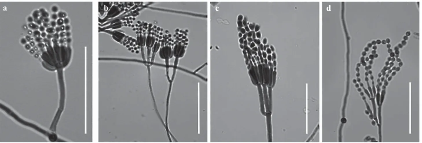
Fig 1 Microscopic characteristics of: a) Acremonium kiliense (general features); b) Aspergillus sydowii (general features); c) Fusarium sacchari var. sacchari (detail of conidiophore); d) Fusarium merismoides var. merismoides (detail of conidiophore); e) Gliocladium viride (detail of conidiophore: stipes, phialides and metulae with disposition detail to conidia; f) Penicillium citrinum (detail of conidiophore: phialides and metulae); scale = 30 m.
a
d
b
e
c
their respiratory siphon and attached to roots of aquatic macrophytes to obtain oxygen, food, and shelter (Ferreira 1999).
Aspergillus sydowii(Fig 1b)
Aspergillus sydowii was isolated in MEA and CYA media and had radiate conidial heads; stipes 127-387 m, thick-walled, smooth, colorless to pale brown, expanding to clavate vesicles 9-12 m in width; aspergilla biseriate; metulae and phialides present; conidia spherical, rough to spinose, 2-4 m in diameter; diminutive conidial structures similar to simple penicilliate heads.
The genus Aspergillius has more than 180 species. Some are rare and some are common. An important characteristic that distinguishes this fungus from others is that the phialides appear simultaneously from the vesicles (Klich 2002). Many species of Aspergillus have been used by industry for the production of enzymes and acids, amilase and citric acid, among others (Klich 2002). However, this group has problems in their use as some can degrade agricultural products (Mallozzi & Corrêa 1998). In addition, some species are pathogenic or allergenic to humans and other animals. Bioassays performed by Moraes et al (2001) showed high pathogenicity in the lineages Aspergillus ochraceus, A. kanagawaensis and A. sulphureus for larvae of Ae.fl uviatilis andCulex quinquefasciatus Say.
Aspergillus sydowii is widely distributed and has been reported from many substrates, but it is mainly found associated with soil (Moraes et al 1998, Klich 2002).
Isolated mosquito species.Aedesfl uviatilis
Sampling site. State of Rondônia, by Ferreira-Keppler RL and Silva JO, in 02/vi/2004.
Habitat. In this work, larvae were collected in stone holes, with water retention, at locations generally of low depth (3 to 20 cm), containing sand and little organic matter. These locations are placed in open areas along rivers or small streams. This species is known to colonize these environments, presenting adaptations for its development and being common in different conditions of temperature
according to the incident solar energy.
Fusarium spp.
Species of the genus Fusarium can be distinguished from other anomorphic genera by their two types of conidia: macro, moon shaped and micro, spherical or oval, both produced by phyalides (Putzke & Putzke 1998). These are
fi lamentous, cosmopolitan, saprophytic or opportunistic plant parasites, which can be found in fruit, seeds and soil (Putzke & Putzke 1998, Almeida et al 2005). Some species are considered human opportunistic pathogens causing superfi cial and systemic infections, mainly when the patient is immunodefi cient (Anaissie et al 1988).
Fusarium sacchari (Butler) var. sacchari (Fig 1c). The fungous was isolated in PDA medium displayed septate hyphae, conidiophores, phialides and macroconidia. Conidiophores arising laterally on hyphae, loosely ramose; monophialidic, being sometimes polyphialidic, with a strong tendency to proliferate, slender, almost cylindrical; macroconidia uniform, slightly, falcate, canoe form, with two or more cells measuring 10-15 × 2-3 m. We found two lineages of Fusarium sacchari.
Isolated mosquito species.Mansonia titillans
Sampling site. Iranduba municipality, by Ferreira-Keppler R L and Pereira E S in 30/xi/2004.
Habitat. See item 1.
Fusarium merismoides var. merismoides (Fig 1d). The fungous was isolated in PDA media, had septate hyphae, conidiophores, phialides and macroconidia. Conidiophores arising as single lateral phialides on hyphae, more or less irregularly branched; monophialidic almost cylindrical or obclavate (5-20 m long); macroconidia were observed with great variation in size (6-12 × 2-3m), straight to slightly curved in the extremities, canoe shaped in two or more cells. We found one lineage.
Isolated mosquito species.Mansonia titillans
Sampling site. Iranduba municipality, by Ferreira-Keppler
Fig 2 Microscopic characteristics of: a) Penicillium sclerotiorum (detail of conidiophore: stipes, metulae and disposition to conidia); b) Penicillium melinii (conidiophore with phialides); c) Penicillium oxalicum (detail of conidiophore: stipes, phialides and metulae); d) Paecilomycessp (detail of conidiophore: phialides, metulae with disposition detail to conidia); scale = 30 m.
RL and Pereira E S in 30/xi/2004.
Habitat. See item 1.
Gliocladium viride (Fig 1e). The fungous was isolated in MEA and CYA media and had septate hyphae, conidiophores, phialides and conidia. Conidiophores are erect, terminated by a dense brush-like branching system bearing tapered phialides; phialides (measuring 11-13 m long) of the terminal branches give rise to fl ask shaped; conidia measure 2-3m in diameter, they are one-celled, ovoid to cylindrical accumulating in the shape of a ball or in a loose column.
This is a widely distributed fi lamentous fungus that can be isolated from decomposing plants and soil (Itoh et al 1980, Pandeyet al 1990). It was not reported as causing disease in humans and animals; however, it is considered to be a “contaminant” fungus.
Isolated mosquito species. Anopheles darlingi
Sampling site. Rio Preto da Eva municipality, by Ferreira-Keppler R L, Pereira E S, Oliveira A F and Martins M S.
Habitat. Larvae were collected along a lake in locations with suffi cient suspended organic matter (e.g. small fruits, leaves, kindlings) between grasses. Females of An. darlingi, main malaria vector in the Amazon region, generally search for places with permanent clean water and relatively covered by vegetation for oviposition (Zeilhofer et al 2007).
Penicilliumspp.
Penicillium citrinum Thom (Fig 1f). The fungous was isolated in MEA and CYA media. Conidiophores borne from surface of hyphae; stipes 87-287 m long, smooth walls terminating in well-defi ned verticils of 3 divergent metulae and vesiculate; metulae usually of uniform length 15-23
m, less usually with metulae interleaving, spathulate or terminally in vesiculate; phialides ampulliform 10-14 m long; conidia spherical, 2 m diameter with smooth walls originating in long well-defi ned columns, one per metula.
Penicillium citrinum is found in decaying vegetation and in the air. It is also a biodeteriogen and causes losses principally in foods, textiles, paintings and plastics (Pitt 1985). Russel et al (2001) reported P. citrinum parasitizing eggs of Ae. aegypti in Australia by the production of mycotoxins that inhibit the complete development of the eggs during the dry season, thereby diminishing the incidence of mosquitoes during the rainy season. In Brazil, da Costa and Oliveira (1998) isolated P. citrinum from adults and larvae of Culicidae.
Isolated mosquito species. Aedeomyia squamipennis,An. darlingi and Ae.fl uviatilis.
Sampling sites. The Culicidae species were collected in Amazonas. In the municipality of Rio Preto da Eva from larvae ofAd. squamipennis and one An. darlingi were collected by Ferreira-Keppler R L, Pereira E S, Oliveira A F and Martins MS, in 30/ ix/ 2004. On the Abunã River, in Rondônia state, from larvae of Ae.fl uviatilis,Ad. squamipennis by Ferreira-Keppler, R L and Silva J O, in 02/ vi/ 2004.
Habitat. Aedeomyia squamipennis and An. darlingi were collected in a natural lake (item 4) and Ae.fl uviatilis in stone holes (item 2). In Ad. squamipennis, monotype species, immature forms were collected from a laminar water fl ow
at locations covered by aquatic macrophytes, frequently in artifi cial lakes, as fi sh tanks. They cohabit with species of Anopheles and Mansonia which does not have the same medical importance.
Penicillium sclerotiorum (Fig 2a). The fungous was isolated in MEA and CYA media presented conidiophores arising from surface or subsurface hyphae; stipes 20-42 m long, slender with thin and smooth walls, fi nishing in well defi ned verticillate, strictly monoverticillate; phialides numerous, ampulliform, 6-9 m long; conidia ellipsoidal 1-3 m in diameter, growing in well-defi ned columns and becoming irregular.
This fungus is commonly found in the soil but can occur in tissue biodeteriorative situations of tissue (Pitt 1985) and foods along with other species (Amoa-Awua et al 1997).Penicillium sclerotirum catalyzed the bioconversion of herbertenediol into dimers: mastigophorenes A and B, neurotrophically active compounds (Harinantenaina et al 2005). In this study we found one lineage.
Isolated mosquito species. Anopheles argyritarsis and An. darlingi.
Sampling sites. Both Culicidae species were collected in Rio Preto da Eva municipality by Ferreira-Keppler R L, Pereira, E S, Oliveira A F and Martins M S, in 30/ix/2004.
Habitat.The species op cit were collected in a lake, according to item 4. They can grow in a variety of locations, which can be related to reservoirs built for hydroelectric constructions (Tadei et al 1998, Forattini 2002). In the transmission of malaria,An. argyritarsis females, contrary to An. darlingi, may not be anthropophilous (Forattini 2002).
Penicillium melinii (Fig 2b). The fungous was isolated in MEA and CYA media had conidiophores arising from the surfaces of hyphae; stipes measuring 12-120 m with walls roughened, bearing terminal verticils of 2-4 metulae, integrated with short monoverticillate conidiophores; metulae rough walled, 9-16 m long; phialides ampulliform, 7-10
m long; conidia spherical, spinose, 1-2 m in diameter, arising in short to long chains, in disordered to well-defi ned columns.
Penicillium melinii appears to be exclusively a soil fungus. In general, the new colonies produce characteristic pigments (Pitt 1985).
Isolated mosquito species. Uranotaenia sp.
Smapling site. Rio Preto da Eva municipality, by Ferreira- Keppler R L and Pereira E S, in 30/xi/ 2004.
Habitat.Uranotaeniasp. was collected in a lake according to the description for the species Anopheles and Aedeomyia (items 4 and 5). Portions of this environment are mainly liquid collections as in waterlogged conditions where they were found in locations with abundant vegetation, at the superior water level. The medical and sanitary aspects of these species, at present, are unknown.
metulae; metulae 16-23 m long; phialides acerose, 12-17
m long, with short collula; conidia ellipsoidal, with walls smooth, 3-5 m × 1-3 m.
This fungus is widely distributed and although common in the soil its main habitat is rotting vegetation (Pitt 1985). In Brazil, da Costa & de Oliveira (1998) isolated P. oxalicum fromMansonia spp. (Culicidae) larvae and adults.
Santamarina et al (2002) extracted metabolites of P. oxalicum lineages demonstrating their antagonistic effect against bacteria, fungi and insects. Bioassays were undertaken to test the pathogenicity of P. oxalicum against Fusarium oxysporum lycopersici and Fusarium oxysporum gladioli.Penicillium oxalicum has shown good capability as a biocontrol agent against species of Fusarium (Santamarina et al 2003).
Species of Penicilliumcan be found in soil, rotting plants and fruits or in dry places such as seeds and wood; dry spores of this fungus are disseminated by wind and insects (Trabulsi et al 1999). Some species can play important roles in the natural processes of biological recycling (Pitt 1985) and in the control of infectious diseases. Others are contaminants or opportunistic pathogens due to their production of mycotoxins (Mallozzi & Corrêa 1998). According to Pitt (1985),Penicilliumis a large genus with at least 150 or maybe 300 species. Penicillium was found in larvae and adults of some species of Anopheles,Aedes,Culex and Mansonia in the southeast and northern regions of Brazil: P. canescens,P. chrysogenum,P. citrinum,P. corylophilum,P. decumbens, P. expansum,P. fellutanum,P. implicatum,P. janthinellum, P. oxalicum, P. purpurogenum, P. viridicatum and P. waksmanii. Bioassys were undertaken with P. corylophilum andP. janthinellum indicating that they were pathogenic to larvae of Ae.fl uviatilis,Ae. aegypti,An. aquasalis and Cx. quinquefasciatus, demonstrating that these lineages have a potential for use in biological control of Culicidae vectors (da Costa & de Oliveira 1998, da Costa et al 1998).
Isolated mosquito species. Mansonia titillans
Sampling site. Iranduba municipality by Ferreira- Keppler RL and Pereira ES, in 30/xi/ 2004.
Habitat.See items 1 and 3.
Paecilomyces sp. (Fig 2d)
Paecilomyces sp. was recorded and illustrated but not named. It was isolated in MEA and CYA media had the following microscopic characteristics: septate hyaline hyphae with fl at walls; conidiophores, phialides and conidia are observed; conidiophores 55-112 m wide, branched with phialides at the extremity; phialides measuring 8-12 m, swollen at their bases and tapering towards their apices; conidia are unicellular, hyaline, fl at, oval measuring 2-3
m in diameter. Paecilomyces sp. is distinguished from Penicillum by the irregular or verticillate forms of the phialides and the divergent conidial chains, and from Acremonium by verticillate conidiophores and fl ask-shaped phialides (Samson 1974). It is a cosmopolitan genus that can be found in decomposing plants and soil. Some species are considered to be insect parasites. Kalkar et al (2006) isolatedPaecilomyces reniformis from Orthoptera (Insecta) in Indonesia. Scholte et al (2004) reported the presence of
Paecilomyces farinosus andPaecilomyces lilacinus in larvae ofCx. pipiens andAe. aegypti.
Isolated mosquito species. Anopheles darlingi
Sampling site. Rio Preto da Eva municipality, by Ferreira-Keppler R L, Pereira E S, Oliveira A F and Martins M S in 30/ ix/ 2004.
Habitat.See items 4 and 5.
This paper is a contribution to a collection of insect-related microorganisms in the Brazilian Amazon. Five genera were identified: Acremonium, Aspergillus, Fusarium, Gliocladium and Penicilium. Species of these genera are known for their importance in the production of secondary metabolites, especially antibiotics and mycotoxins. Some of the lineages isolated, as cited above, have been reported in the literature as having biotechnological potential. Further studies in this area can be undertaken with the objectives of selecting industrially important biocontrol agents.
Acknowledgments
To Dr Barbara Robertson for the manuscript translation to English and to Dr Philip M Fearnside for reviewing it. This study received fi nancial support from the Fundação de Amparo à Pesquisa do Estado do Amazonas/FAPEAM (PIPT/827/03, DCR/954/03) and the Conselho Nacional de Desenvolvimento Científi co e Tecnológico-CNPq (CT-Amazônia, nº 553088/2005-0).
References
Agarwala S P, Sagar S K, Sehgal S S (1999) Use of mycelial suspension and metabolites of Paecilomyces lilacinus (Fungi: Hyphomycetes) in control of Aedes aegypti larvae. J Commun Dis 31: 193-196.
Alencar Y B, Rios-Velasquez C M, Lichtwardt R W, Hamada N (2003) Trichomycetes (Zygomycota) in the digestive tract of arthropods in Amazonas, Brazil. Mem Inst Oswaldo Cruz 98: 799-810.
Almeida C V, Yara R, Almeida M (2005) Fungos endofíticos isolados de ápices caulinares de pupunheira cultivada in vivo e in vitro. Pesq Agropec Bras 40: 467-470.
Alves S B (1998) Controle microbiano de insetos. Ed FEALQ, Piracicaba, 1163pp.
Amoa-Awua W K, Frisvad J C, Sefa-Dedeh S, Jakobsen M (1997) The contribution of moulds and yeasts to the fermentation of ‘agbelima’ cassava dough. J Appl Microbiol 83: 288-96.
Anaissie E, Kantarjian H, Ro J, Hopfer R, Rolston K, Fainstein V, Bodey G (1988) The emerging role of Fusarium infections in patients with cancer. Medicine 67: 77-83.
Bott T L, Rogenmuser K (1980) Fungal pathogen of Cladophora glomerata (Chlorophyta). Appl Environ Microbiol 40: 977-980.
Costa G L da, Moraes A M de, Oliveira P C de (1998) Pathogenic action of Penicillium species on mosquito vectors of human tropical diseases. J Basic Microbiol 38: 337-341.
Costa G L da, Oliveira P C de (1998) Penicillium species in mosquitoes from two Brazilian regions. J Basic Microbiol 38: 343-347.
Ferreira R L M (1999) Aspectos biológicos de três espécies de Mansonia(Mansonia) Blanchard, 1901 (Diptera, Culicidae) em Laborstório. Rev Bras Entomol 43: 29-34.
Forattini O P (2002) Culicidologia médica. v 2, Editora USP, São Paulo, 860p.
Fridkin S K, Kremer F B, Bland L A, Padhye A, McNeil M M, Jarvis J R (1996) Acremonium kiliense endophthalmitis that occurred after cataract extraction in an ambulatory surgical center and was traced to an environmental reservoir. Clin Infect Dis 22: 222-7.
Gerlach W, Nirenberg H (1982) Mitteilungenaus der Biologischen Bundesanstalt fur Land- und Forstwirtschaft. The genus Fusarium – a pictorial atlas. Berlin-Dahlem, 230p.
Harinantenaina L, Noma Y, Asakawa Y (2005) Penicillium sclerotiorum catalyzes the conversion of herbertenediol into its dimers: mastigophorenes A and B. Chem Pharm Bull 53 256-257.
Heyningen S V, Secher D S (1971) A new alkaline protease from Acremonium kiliense. J Biochem 125: 1159-1160.
Humber R A (1998) Entomopathogenic fungal identifi cation. APS/ ESA Joint Annual meeting, Ithaca, NY, 28p.
Itoh Y, Kodama K, Furuya K, Takahashi S, Haneishi T, Takiguchi Y, Arai M (1980) A new sesquiterpene antibiotic, heptelidic acid producing organisms, fermentation, isolation and characterization. J Antibiot 33: 468-73.
Kalkar O, Carner G R, Scharf D, Boucias D G (2006) Characterization of an Indonesian isolate of Paecilomyces reniformis. Mycopathologia 161: 109-118.
Klich M A (2002) Identifi cation of common Aspergillus species. Department of Agriculture, New Orleans, Louisiana, USA, 108p.
Klich M A, Pitt J I (1998) A laboratory guide to common Aspergillus species and their teleomorphs. Commonwealth Scientifi c and Industrial Research Organisation. 116pp.
Lichtwardt R W (1986) The trichomycetes: fungal associates of arthropods, Springer-Verlag, New York, 343p.
Lucarotti C J, Shoulkamy M A (2000) Coelomomyces stegomyiae infection in adult female Aedes aegypti following the fi rst, second, and third host blood meals. J Invertebr Pathol 75: 292-295.
Mallozzi M A B, Corrêa B (1998) Fungos toxigênicos e micotoxinas. Bol Tecn Inst Bio 5: 5-26.
Mendoza L, Donato A, Padhye A (1985) Canine mycotic
keratoconjuntivitis caused by Acremonium kiliense. Sabouraudia 23: 447-450.
Messias L C (1989) Fungos, sua utilização para controle de insetos de importância médica e agrícola. Mem Inst Oswaldo Cruz 84: 57-59.
Moraes A M, Costa G L da, Barcellos M Z, Oliveira R L de, Oliveira P C de (2001) The entomopathogenic potencial os Aspergillus spp. in mosquitoes vectors of tropical diseases. J Basic Microbiol 41: 45-49.
Pandey A, Agrawal G P, Singh S M (1990) Pathogenic fungi in soils of Jabalpur, India. Mycoses 33:116-125.
Pastorino AC, Menezes U P, Marques H H S, Vallada M G, Cappellozi V L, Carnide E M G, Jacob C M A (2005) Acremonium kiliense infection in a child with chronic granulomatous disease. Brazilian J Infect Dis 9: 529-534.
Pereira E S, Ferreira R L M, Hamada N, Lichtwardt R W (2005) Trichomycete fungi (Zygomycota) associated with mosquito larvae (Diptera: Culicidae) in natural and artifi cial habitats in Manaus, AM Brazil. Neotrop Entomol 34: 325-329.
Pitt J I (1985) A laboratory guide to common Penicillium species. Commonwealth Scientifi c and Industrial Research organization, Division of Food Research, 122p.
Putzke J, Putzke M T L (1998) Os reinos dos fungos. EDUNISC, Santa Cruz do Sul, (1): 606p.
Raper K B, Fennell D I (1965) The genus Aspergillus. The Williams & Wilkins Company, United States of América, 690p.
Rodrigues A, Pagnocca F C, Bacci M J, Hebling M J, Bueno O C, Pfenning L H (2005) Variability of non-mutualistic fi lamentous fungi associated with Atta sexdens rubropilosa nests. Folia Microbiol (Praha) 50: 421-5.
Samson R A (1974) Paecilomycesand some allied Hyphomycetes. Studies in mycology. Centraalbureau voor Schimmelcultures Baarn, the Netherlands., 6:120p.
Santamarina M P, Rosello J, Llacer R, Sanchis V (2002) Antagonistic activity of Penicillium oxalicum Corrie and Thom, Penicillium decumbens Thom and Trichoderma harzianum Rifai isolates against fungi, bacteria and insects in vitro. Rev Iberoam Micol 19: 99-103.
Santamarina S M P, Rosello C J, Barcelo C S, Marin S S (2003) Effect of water activity and temperature on competing abilities of Penicillium oxalicum against Fusarium oxysporum. Rev Iberoam Micol 20: 154-159.
Scholte E-J, Knol B G J, Samson R A, Takken W (2004) Entomopathogenic fungi for mosquito control: a review. J Insect Science 4: 1-24.
Simon G, Rakoczy G, Galgoczy J, Verebely T, Bokay J (1991) Acremonium kiliense in oesophagus stenosis. Mycoses 34: 257-60.
Trabulsi L R, Alterthum F, Gompertz O F, Candeias J A N (1999) Microbiologia. 3 ed, Atheneu, São Paulo, 576p.
Tymon A M, Pell J K (2005) ISSR, ERIC and RAPD techniques to detect genetic diversity in the aphid pathogen Pandora neoaphidis. Mycol Res 109: 285-293.
Zeilhofer P, Santos E S dos, Ribeiro A L M, Miyazaki R D, Santos M A dos (2007) Habitat suitability mapping of Anopheles darlingi in the surroundings of the Manso hydropower plant reservoir, Mato Grosso, Central Brazil. Int J Health Geogr 6: 7.