Morphological Description and Clinical Implications of Myocardial
Bridges: an Anatomical Study in Colombians
Luis Ernesto Ballesteros Acunã
1, Luis Miguel Ramírez Aristeguieta
2, Saldarriaga Bladimir Tellez
3Universidad Industrial de Santander1,2, Universidad Autónoma de Bucaramanga1,3, Universidad Santo Tomas de Aquino2, Bucaramanga - Colômbia
Summary
Background: Myocardial bridges represent a hotly debated research topic. Myocardial bridges are considered to be a vascular heart variation due to its intermittent or enduring reducing of the arterial lumen, with a possible ischemic effect.
Objective: This study aimed at determining the incidence of myocardial bridges in the Colombian half-caste population.
Methods: 154 hearts were studied, having been extracted as fresh autopsy material. Coronary arteries were injected with synthetic resin and cleaned by extracting the pericardial fat.
Results: 92 myocardial bridges were observed in 62 hearts (40.3%). Average myocardial bridge length was 19.9 mm. Myocardial bridges varied per artery from one myocardial bridge in 42 hearts (27.3%), two myocardial bridges in 11 hearts (7.2%), three myocardial bridges in 8 hearts (5.2%) and four myocardial bridges in 1 heart (0.7%). Most myocardial bridges coincided with the anterior interventricular artery in its proximal and intermediate segments in 61 cases (39.6%) and left diagonal artery in 11 cases (7.2%). Arteries emitted just before myocardial bridges (pre-bridge branch) in 50 cases (54.3%) of the sample, with an average caliber of 1.41 mm. A third coronary artery was present in 46 cases (29.8%) of total sample. A higher frequency of myocardial bridges was found in hearts with a third coronary artery.
Conclusion: It is necessary to consider the clinical importance of the variations considering the arterial distribution and associated clinical implications. (Arq Bras Cardiol 2009;92(4):242-248)
Key words: Coronary vessels; myocardial bridging; chest pain; myocardial ischemia.
Mailing address: Luis Ernesto Ballesteros •
Calle 45 Nº 33-17 - Edificio La Nacional - Apto 702B, Bucaramanga - Colômbia
E-mail: [email protected]
Manuscript received November 17, 2007; revised manuscript received February 13, 2008; accepted February 19, 2008.
lumen, with a possible ischemic effect. Precordial ischemic pain needs an appropriate diagnosis because of the several pathologies associated with obstructive cardiomyopathy: atherosclerosis, thrombosis, hypertrophic cardiomyopathy, paroxystic AV block, segmental hypokinesia, cardiac arrhythmias, ventricular tachycardia, coronary spasm, myocardial acute infarct and/or sudden cardiac death.
Cardiac muscle ischemia appears to occur during the first 1/3 of diastole due to prolonged coronary artery lumen recovery following its systolic compression (75%). This alters coronary flow speed and reserves, especially during enhanced heart rhythm during exercise and emotional stress8.
MB incidence variability depends on recording method, gender, ethnicity and the nature of the study; in this sense a minor 0.5-12% angiographies present MB but in cadavers they are present in 5.4%-85.7%9-12. MB generally originate in
the left coronary artery (LCA) trunk and branches, especially in the anterior interventricular artery (AIA) (12%-63%), circumflex artery (CA) (2.8%-6.7%) and its lateral branch (13%). They are also found in diagonal arteries (DA), marginal arteries (MA) and coronary veins13,14.
This research was aimed at determining MB incidence in the half-caste Colombian population and establishing
Introduction
Myocardial bridges (MB) represent a currently controversial research issue. MB have an embryological origin, and there are no gender differences regarding its incidence. A benign or a malignant interpretation of the presence of MB depends on contrasting physiopathological point of views; some arguments explain that MB generates atheromas but others as being atheromas prevention. Some authors consider MB as being normal intramural arteries and criticize their surgical management, implicitly considering them to be a risk factor1. MB were first reported in 1737 by Reyman as
transitorily submerged myocardial arteries2. MB have also
been shown in anatomical, physiological, biochemical and imagenological studies3-7.
MB frequency related to coronary dominance, vessels that emerge immediately before the MB of compromised arteries or pre-bridge branches (PBB), as well as MB correlation with the presence of a third coronary artery. Third coronary artery (originating in the right aortic sinus) irrigates the conus arteriosus and also supplies the upper and middle AIA’s right ventricular territory, particularly when the AIA’s middle third has MB15-17.
Methods
The Institute of Forensic Medicine in Bucaramanga, Colombia, provided 154 hearts for the study; these were extracted as fresh autopsy material. The sample inclusion criteria consisted of being a half-caste adult male or female heart without pathology or signs of trauma.
Coronary arteries were injected with synthetic resin (80% Palatal GP 41L and 20% Styrene)18 at 120 mm Hg of
pressure. The hearts were cleaned with 15% KOH solution to extract the pericardial fat. MB number and position in the coronary arteries were determined taking sample gender into account.
Samples were measured with an electronic caliper (Mitutoyo) regarding MB length, MB wall thickness (measured as the maximum thickness at mid-point of the MB course), LCA trunk length, artery caliber at several course points just before, at mid-point and just after the MB.
MB incidence was also correlated with coronary dominance, according to Schlesinger dominance criteria19. MB
and LCA trunk length correlation were also established. PBB were recorded just before MB artery involved. The correlation between the presence of MB and the third coronary artery was documented.
Means and standard deviations were calculated for continuous variables, whereas nominal variables were described in terms of percentages. Up to 5% alpha error was accepted for Chi(X2) square and Student-T statistical tests; Epiinfo 2002 was used to create the database and STATA 8.0 for performing the statistical analysis.
Results
The sample consisted of 154 hearts, being 125 of them from males (81.2%) and 29 from females (18.8%). Age ranged from 15 to 73 years (mean of 31.5 years).
MB were present in 62 hearts (40.3%), being 54 in males (43.2%) and 8 in females (27.6%). A higher frequency of MB in males was not statistically significant (p=0.122). Of a total of 92 MB, they varied per artery from one MB in 42 hearts (27.3%), two MB in 11 hearts (7.2%) (Figure 1 and Figure 2), three MB in 8 hearts (5.2%) and four MB in 1 heart (0.7%). Regarding MB location, most matched AIA proximal and intermediate segments in 61 cases (66.3% relative frequency, 39.6% absolute frequency) and left DA (Figure 3) in 11 cases (12% relative frequency, 7.2% absolute frequency) (Table 1). Right coronary dominance was observed in 117 hearts (76%), balanced circulation in 25 (16.2%) and left coronary dominance in 12 (7.8%) hearts. Balanced irrigation presented higher MB incidence (56%), followed by MB in 39.3% with
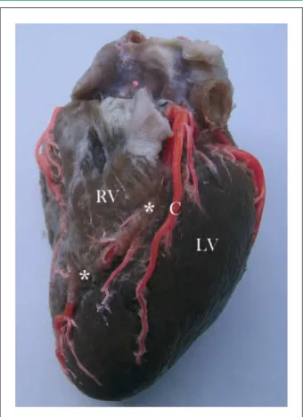
Figure 1 -Sternocostal heart surface. Anterior interventricular artery with two myocardial bridges (*). RV - right ventricle. LV - left ventricle. C - pre-bridge branch forming left anterior ventricular artery.
right irrigation dominance. There was no significant MB gender difference (female p=0.192, male p=0.295) (Table 2).
A third coronary was present in 46 cases (29.8%) of the total heart samples. A significant statistical association was found between MB and number of coronary arteries (p=0.024). The detection of three coronary arteries in hearts with MB was thus 2.34 times more common than in hearts with no MB.
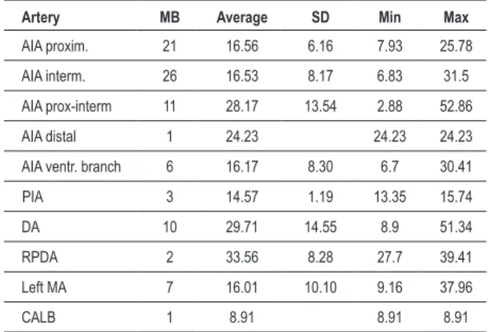
Average MB length were 19.9 mm (10.83 SD), 20.09 in males (10.81 SD) and 15.2 (8.3 SD) in females. The greater length in males was not statistically significant (p=0.21). The proximal AIA and 6 intermediate segments presented the longest (52.9 mm) (Figure 4) and shortest MB (2.88 mm). MB located in the right posterior DA presented (two cases) a higher average length (33.6 mm). The only MB present in the conus arteriosus measured 8.9 mm (Table 3).
Arterial caliber changed during MB arterial course. Arterial caliber just before the MB was 2.17 mm (0.61 SD), 1.92 mm (0.55 SD) at mid-point and 1.67 mm (0.57 SD) just after MB. These measurements showed 23.05% that has a reduced artery lumen during passage under the MB (from pre-bridge to post-bridge course). Supra-bridge myocardium thickness was 1.54 mm (0.73 SD), ranging from 0.27-3.8 mm.
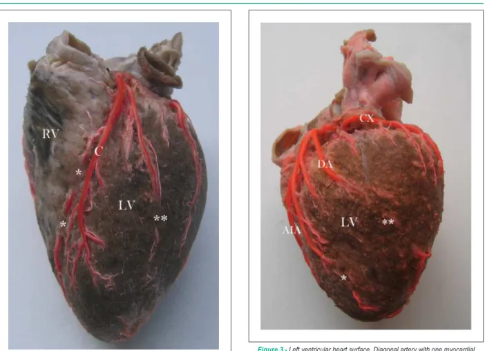
Figure 2 -Sternocostal heart surface. Anterior interventricular artery with two myocardial bridges (*). Anterior ventricular branch from the anterior interventricular artery with one myocardial bridge (**) RV - right ventricle. LV - left ventricle. C - pre-bridge branch forming left anterior ventricular artery.
Figure 3 -Left ventricular heart surface. Diagonal artery with one myocardial bridge (**) and anterior ventricular branch from anterior interventricular artery with one myocardial bridge (*) LV - left ventricle; DA - diagonal artery; AIA
- anterior interventricular artery; CX - circumlex artery.
Table 1 - Myocardial bridge artery location
Myocardial bridge artery location n %(₤) %(Ω)
Proximal AIA 61 66.30 39.6
Left DA 11 11.96 7.2
Left MA 8 8.70 5.2
AIA ventricular branch 6 6.52 3.9
PIA 3 3.26 1.9
RPDA 2 2.17 1.3
CALB 1 1.09 0.6
Total 92 100 59.7
AIA - anterior interventricular artery; PIA - posterior interventricular artery; DA - diagonal artery; MA - marginal artery; RPDA - right posterior diagonal artery; CALB - conus arteriosus left branch.
trunk and MB length in their middle AIA portion. When LCA trunk was less than 4.37 mm (25th percentile), MB length
was thus 21.49 mm and when LCA trunk was greater than or equal to 4.37 mm, MB length was 15.04 mm. Average MB length in mid-AIA portion was 14.92 mm when LCA trunk was greater than or equal to 6.81 mm (75th percentile). Such MB
length were 6.56 mm greater (p=0.059) in LCA trunk (at 25th
percentile) compared to those having 75th percentile. These
data thereby suggest that the shorter the LCA trunk, the longer the corresponding MB. The multiple correlation coefficient between the LCA trunk and MB length was 0.1158.
It was observed that compromised MB vessels emitted PBB just before MB in 50 cases (54.3%), with an average caliber of 1.41 mm (0.55 SD). This characteristic was more frequent in the AIA (60.7%) (Figure 5).
Discussion
A wide-range incidence of MB was observed, depending on the assessment method used; angiographic and anatomic results were therefore different. By contrast, the angiographical method returned very low prevalence. We think that this
Table 2 - MB side, gender and coronary arterial dominance
Coronary dominance
MB absent
n (%) MB presentn (%) n (%)Total
Right 71(60.7) 46(39.3) 117(76.)
Male 53(58.2) 38(41.8) 91(100)
Female 18(72) 7(28) 25(100)
Left 9(75) 3 (25) 12(7.8)
Male 7(70) 3(30) 10(100)
Female 2(100) 0(0) 2(100)
Balanced 11(44) 14(56) 25(16.2)
Male 10(43.5) 13(56.5) 23(100)
Female 1(50) 1(50) 2(100)
Total 92(59.7) 62 (40.3) 154(100)
MB - myocardial bridge.
Figure 4 -Sternocostal heart surface. Anterior interventricular artery with the longest myocardial bridge (*). RV - right ventricle; LV - left ventricle; C - pre-bridge branch forming left anterior ventricular artery; LA - sectioned left auricle.
Table 3 - MB length
Artery MB Average SD Min Max
AIA proxim. 21 16.56 6.16 7.93 25.78
AIA interm. 26 16.53 8.17 6.83 31.5
AIA prox-interm 11 28.17 13.54 2.88 52.86
AIA distal 1 24.23 24.23 24.23
AIA ventr. branch 6 16.17 8.30 6.7 30.41
PIA 3 14.57 1.19 13.35 15.74
DA 10 29.71 14.55 8.9 51.34
RPDA 2 33.56 8.28 27.7 39.41
Left MA 7 16.01 10.10 9.16 37.96
CALB 1 8.91 8.91 8.91
MB - myocardial bridge; AIA - anterior interventricular artery; PIA - posterior interventricular artery; DA - diagonal artery; MA - marginal artery; RPDA - right posterior diagonal artery; CALB - conus arteriosus left branch.
Figure 5 -Sternocostal heart surface. Anterior interventricular artery with one myocardial bridge (*). RV - right ventricle; LV - left ventricle; AIA - anterior interventricular artery; C - pre-bridge branch common trunk forming left anterior ventricular arteries.
compression (milking). Angiographic incidence ranged from 0.5% to 12%20-24.
Our anatomical MB incidence results (40.3%) partly coincided with other studies’ major and minor incidence findings. Polacek10, found a major incidence of 87.5%, Bezerra
etal25, 78%, Ferreira et al26, 58% and Von Ludinghausen 57%27.
Sahni and Jit9 found a minor incidence of 34.5%, Loukas et al28
34.5%, Reig et al29, 25% and Bohm et al30, 23%. We believe
that such differences are due to sample sizes, ethnicity and
other authors’ interpretation of complex differences amongst MB and intramural arterial segments (grooves) 9.
Regarding gender differences, our results revealed a male predominance with no significant statistical difference, in agreement with previous reports9,26,30-32. Polacek10 attained
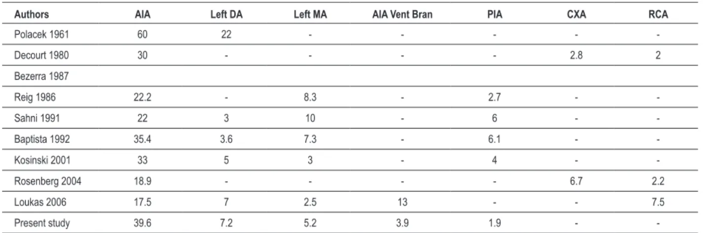
Table 4 - Percentage of MB location by different authors
Authors AIA Left DA Left MA AIA Vent Bran PIA CXA RCA
Polacek 1961 60 22 - - - -
-Decourt 1980 30 - - - - 2.8 2
Bezerra 1987
Reig 1986 22.2 - 8.3 - 2.7 -
-Sahni 1991 22 3 10 - 6 -
-Baptista 1992 35.4 3.6 7.3 - 6.1 -
-Kosinski 2001 33 5 3 - 4 -
-Rosenberg 2004 18.9 - - - - 6.7 2.2
Loukas 2006 17.5 7 2.5 13 - - 7.5
Present study 39.6 7.2 5.2 3.9 1.9 -
-AIA - anterior interventricular artery; DA - diagonal artery; MA - marginal artery; PIA - posterior interventricular artery; CXA - circumlex artery; RCA - right coronary artery.
sites33. Angiographic studies have shown AIA frequency
ranging from 0.4% to 3.5%24,34 and anatomical studies range
from 12%-60%10,29,30,35-38; our findings (39.6%) were in the
middle of such range. Different reports have pointed out that the MB presence in the diagonal and left marginal arteries are secondary MB locations. There is a consensus regarding rarer MB locations in the circumflex and right coronary branches, such as the posterior interventricular. MB were not found in circumflex artery and the frequency in the posterior interventricular artery was 1.9%. Table 4 shows our results and other authors’ findings.
A higher single MB frequency (27.3%) was found in this study, rather than two or more MB (13.1%). The single MB frequency (17-60%) is in agreement with Kosinski and Grzybiak38, Reig et al29, Bezerra et al, Sahni and Jit9 and
Loukas et al28. Our results are in agreement with the literature
regarding one MB; two or three MB have been reported at a lower frequency (5%-40%). However, Becerra et al25, have
obtained 38% for one MB and 40% for two or more26. Our
results are in agreement with Kosinzky and Grzybiak38, Sahni
and Jit9, and Loukas et al28, who have shown a 4% incidence
of two MB in AIA.
Great MB length variability has been reported, with dimensions ranging from a few millimeters to 50 millimeters11,29,39. Our results reported MB length as being
within a mid-range of previous reports. Higher averages (i.e. 31, 26, and 25 mm) have been reported by Loukas et al28,
Rozemberg et al39, and Lima et al11, respectively, whereas
lower averages (13, 17, and 16 mm) have been reported by Reig et al29, Kantarcy et al24, Kosinsky e Grzybiak38, respectively.
Kosinsky and Grzybiak38, did not find gender differences in
MB length, as observed in our results.
Supra-bridge myocardial thickness has been reported as being between 0.3 to 3.8 mm. Mean MB thickness in our study was 1.54 mm. This represents a mid-point amongst the results reported by other authors (3, 2.5, 1.8, 1.2, and 0.9 mm)24,28,29,38,39.
MB length, thickness and number are variables that must be considered in the cardiac pathophysiology of different diseases;
however, it is equally likely that MB does not produce any effects. It is understandable that a coronary artery with one or several MB might produce signs and symptoms such as angina or arrhythmias in situations requiring high cardiac demand (exercise and stress) and even sudden death, especially if such MB are large and deep. The anatomical characteristics of coronary arteries thus play a relevant protagonist role in the outcome of such situations.
Although it has not been reported in the literature, we found a high incidence of PBB (54%) in the AIA with considerable caliber (mean of 1.4 mm) compared to usual branches from the respective AIA. We consider that this PBB irrigation could act as compensation mechanism (angiogenesis) to irrigate neighboring territory, which might have suffered ischemia without such PBB and thus explain asymptomatic presentation in the presence of MB in some patients. Although this hypothesis is difficult to prove using conventional macroscopic morphological evaluation, perhaps the next step is to prove it through proteomics and genomics that characterize the morphogenesis protein involved in this vascular compensation model.
These PBB could behave as collaterals based on the general agreement that defines collateral vessels as emerging from another territory and irrigating an arterial territorial obstruction. These PBB actually could be generated currently by a MB. Rosenbergand Nepomnyashchikh39have stated that the
hemodynamics of coronary arteries with MB change due to obstructed flow and turbulence, producing collateral arterial compensation, which (according to the authors), prevent MB atheroma production40,41. It will be interesting to observe the presence of PBB and their biometrics in angiographic studies in the pediatric population.
reperfusion. This is consistent with LCA trunk stress during systolic turbulence against arterial walls. However, multiple correlation coefficient between LCA trunk and MB length (0.1158) showed a mild correspondence.
We also observed that a third coronary artery in AIA with MB is 2.34 times more frequent. This third coronary artery added to the AIA PBB compensation mechanism might work together to balance decreased coronary blood flow. The MB arterial stenosis was 23.5% in our study, almost concurring with that by Lima et al11, (28.2%). Although there is a controversy
regarding coronary dominance and MB, our results revealed balanced coronary irrigation in most observed MB. This is in agreement with Reig et al29; we found no statistically significant
difference between balanced coronary irrigation and right or left dominance29. MB in left coronary dominance has been
reported by Loukas et al28, Penthe et al37, Matonoha and
Zechmeister42, and Polacek10, although the last two groups
found statistically significant differences.Differently from our results, Loukas et al28, affirmed that the MB presence is directly
related to coronary artery dominance. The presence of MB in angiographic studies and the type of coronary irrigation may be taken into account for future studies.
MB remain a fascinating issue, leading to many hypotheses and some speculation. The presence of MB must certainly be carefully observed and studied from anatomical, pathophysiological, diagnostic and therapeutic viewpoints.
Potential Conflict of Interest
No potential conflict of interest relevant to this article was reported.
Sources of Funding
There were no external funding sources for this study.
Study Association
This study is associated with post-graduation program.
Referências
1. Lozano I, Baez JA, Lopez R, Pinar E, Pico F, Valdez M, et al. Pronostico a largo plazo de los pacientes con trayecto intramiocardico de la arteria descendente anterior con compresión sistólica. Rev Esp Cardiol. 2002; 55: 359-64.
2. Reyman H. Dissertatis de vasis cordis propriis. Bibl Anat. 1737; 2: 336.
3. Gow RM. Myocardial bridging: does it cause sudden death? Card Electrophysiol Rev. 2002; 6: 112–4.
4. Quagliano AP, Cury L, Pereira RAA, Prates NE. Puentes de miocárdio sobre las artérias coronárias y su importáncia practica. Archivos Anatómicos de Costa Rica. 1992; 1: 137-8.
5. Ying HL, Ying LC, Anraku AP, Prates NE. Myocardium bridges in fetuses, newborns and children: morphological aspects – the São Paulo study. In: 13º World Congress of Cardiology. Proceedings. Bologna: Monduzzi Editore; 1998. p. 767-75.
6. Shinjo SK, Prates NE, Oba SM, Sampaio LO, Nader HN. Distribution and composition of glycosaminogycans in the left human coronary arterial branches under myocardial bridge. Atherosclerosis. 1999; 143: 363-8.
7. Rychter K, Salanitri J, Edelman RR. Multifocal coronary artery myocardial bridging involving the right coronary and left anterior descending arteries detected by ECG-gated 64 slice multidetector CT coronary angiography. Int J Cardiovasc Imaging. 2006; 22: 713-7.
8. Downar J, Williams WG, McDonald C, Wigle ED, McCrindle BW. Outcomes after ‘‘unroofing’’ of a myocardial bridge of the left anterior descending coronary artery in children with hypertrophic cardiomyopathy. Pediatr Cardiol. 2004; 25: 390–3.
9. Sahni D, Jit I. Incidence of myocardial bridges in northwest Indians. Indian Heart J. 1991; 43: 431-6.
10. Polacek P. Relation of myocardial bridges and loops on the coronary arteries to coronary occlusions. AM Heart J. 1961; 61: 44-9.
11. Lima VJ, Cavalcanti JS, Tashiro T. Myocardial bridges and their relationship to the anterior interventricular branch of the left coronary artery. Arq Bras Cardiol. 2002; 79: 215-22.
12. Cay S, Ozturk S, Cihan G, Kisacik HL, Korkmaz S. Angiographic prevalence of myocardial bridging. Anadolu Kardiyol Derg. 2006 ;6: 9-12.
13. Sousa-Rodrigues CF, Alcantara F, Buarque LC, da Rocha AC, Alencar e Silva R, Olave E. Aspectos anatómicos y biométricos de los puentes de miocardio
y sus relaciones con la arteria interventricular anterior y venas adyacentes. Int J Morphol. 2006; 24: 270-84.
14. Pejkovich B, Bogdanovich D. The great cardiac vein. Surg Radiol Anat. 1992; 14: 23-8
15. Williams PL, Warwick R, Dyson M, Bannister LH. Gray’s anatomy. 37th ed. Edinburgh, Scotland: Churchill Livingston. 1989. p. 736-7.
16. Miyazaki M, Kato M. The developmental study on the third coronary artery of human being. Gegenbaurs Morphol Jahrb. 1986; 132: 195-204.
17. Miyazaki M, Kato M. Third coronary artery: its development and function. Acta Cardiol. 1988; 43: 449-57.
18. Nerantzis C, Antonakis E, Avgoustakis D. A new corrosion casting technique. Anat Rec. 1978; 191: 321-5.
19. Schlesinger MJ. Relation of anatomic pattern to pathologic conditions of the coronary arteries. Arch Pathol. 1940; 30: 403-15.
20. Noble J, Bourassa MG, Petitclerc R, Dyrda I. Myocardial bridging and milking effect of the left anterior descending coronary artery: normal variant or obstruction? Am J Cardiol. 1976; 37: 993-9.
21. Kramer JR, Kitazume H, Proudfit WL, Sones FM Jr. Clinical significance of isolated coronary bridges: benign and frequent condition involving the left anterior descending artery. Am Heart J. 1982; 103: 283-8.
22. Mohlenkamp S, Hort W, Ge J, Erbel R. Update on myocardial bridging. Circulation. 2002; 106: 2616-22.
23. Cay S, Ozturk S, Cihan G, Kisacik HL, Korkmaz S. Angiographic prevalence of myocardial bridging. Anadolu Kardiyol Derg. 2006; 6: 9-12.
24. Kantarci M, Duran C, Durur I, Alper F, Onbas O, Gulbaran M, et al. Detection of myocardial bridging with ECG-gated MDCT and multiplanar reconstruction. AJR Am J Roentgenol. 2006; 186: S391-4.
25. Bezerra AJ, Prates JC, DiDio LJ. Incidence and clinical significance of bridges of myocardium over the coronary arteries and their branches. Surg Radiol Anat. 1987; 9: 273-80.
26. Ferreira AG Jr, Trotter SE, Konig B Jr, Decourt LV, Fox K, Olsen EG. Myocardial bridges: morphological and functional aspects. Br Heart J. 1991; 66: 364-7.
Einbau in das Myokard. Dtsch Med Wschr. 1975; 100: 2448-51.
28. Loukas M, Curry B, Bowers M, Louis RG Jr, Bartczak A, Kiedrowski M, et al. The relationship of myocardial bridges to coronary artery dominance in the adult human heart. J Anat. 2006; 209: 43-50.
29. Reig J, Loncan MP, Martin S, Binia M, Petit M, Domenech JM. Myocardial bridges. Incidence and relation to some certain coronary variables. Arch Anat Histol Embryol. 1986; 69: 101-10.
30. Bohm J, Piot C, Warnke H, Lindenau KF, Portsman W. Zur Diagnostik und Operationstechnik bei intramuralem Koronararteienverlauf. Cor Vasa. 1980; 22: 319-26.
31. Ishi T, Hosoda Y, Osaka T. The significance of myocardial bridge upon atherosclerosis in the left anterior descending coronary artery. J Path. 1986; 148: 279-85.
32. Shiong SL, Ting LW. The role of the mural coronary artery in prevention of coronary aherosclerosis. Arch Pathol. 1972; 93: 32-5.
33. Baptista CA, DiDio LJ. The relationship between the directions of myocardial bridges and of the branches of the coronary arteries in the human heart. Surg Radiol Anat. 1992; 14: 137-40.
34. Greenspan M, Iskandrian AS, Catherwood E, Kimbiris D, Bemis CE, Segal BL. Myocardial bridging of the left anterior descending artery: evaluation using exercise thallium-201 myocardial scintigraphy. Cathet Cardiovasc Diagn.
1980; 6: 173-80.
35. Geiringer E. The mural coronary artery. Am Heart J. 1951; 41: 359-68.
36. Decourt LV, Carvalho VB, Martinez JRM. Pontes miocardica uma entidade controvertid. Rev Hosp Clin Fac Med S Paulo. 1980; 35: 157-60.
37. Penthe P, Bara JA, Blanc JJ. Etude anatomique descriptive des gros troncs coronariens et des principales collaterales epicardiques. Nouv Presse Med. 1976; 5: 71-5.
38. Kosinski A, Grzybiak M. Myocardial bridges in the human heart: morphological aspects. Folia Morphol. 2001; 60: 65-8.
39. Rozenberg VD, Nepomnyashchikh LM. Pathomorphology and pathogenic role of myocardial bridges in sudden cardiac death. Bulletin of Experimental Biology and Medicine. 2004; 138: 87-92.
40. Lovell MJ, Knight CJ. Invasive assesment of myocardial bridges. Heart. 2003; 89: 699-700.
41. Rozenberg VD, Nepomnyashchikh LM. Pathomorphology of myocardial bridges and their role in the pathogenesis of coronary disease. Bull Exp Biol Med. 2002; 6: 593-6.