e45
Mechanical Prosthetic Valve Thrombosis
Márcio Silva Miguel Lima e Marcelo Luiz Campos Vieira
Instituto do Coração (Incor) – HCFMUSP
A 51-year-old female patient with a double-disk mechanical mitral valve prosthesis for one year had a history of inadequate use of anticoagulation therapy and developed progressive dyspnea. On admission to the emergency room of InCor, Sao Paulo, she presented respiratory distress, tachypnea, BP of 80/40 mmHg, jugular venous distension and pulmonary crackles. Transesophageal echocardiography was performed one hour
Mailing Address: Márcio Silva Miguel Lima •
Av. Dr. Enéas de Carvalho Aguiar, 44 - 05403-900 - São Paulo, SP, Brasil E-mail: [email protected]
Manuscript received October 07, 2008; revised manuscript received February 06, 2009; accepted May 15, 2009
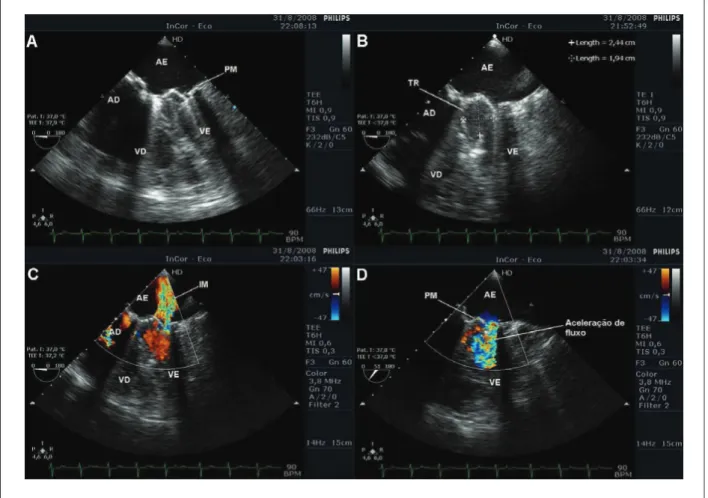
Fig. 1 – A and B - Transesophageal echocardiogram showing large thrombus adhered to the prosthetic mitral valve with reduced area; C - moderate paraprosthetic leak
on color-Doppler-low mapping; D - clear low acceleration consistent with reduced prosthetic valve area LA - left atrium, RA - right atrium, LV - left ventricle, RV - right ventricle, MP - mechanical prosthesis, TH - thrombus, MR - mitral regurgitation
after admission, and showed a large thrombus adhered to the mechanical prosthesis with restriction to disk motion; reduced prosthetic valve area (VA = 0.6 cm2); increased gradients
(peak gradient of 38 mmHg and mean gradient of 26 mmHg); and moderate paraprosthetic leak. Emergency surgery was indicated; however, the patient died immediately before the procedure due to cardiovascular collapse.
Key Words
Thrombosis; Heart Valve Prosthesis; Blood Coagulation.