Lower limb muscle strength is associated with functional
performance and quality of life in patients
with systemic sclerosis
Tatiana R. L. Lima1, Fernando S. Guimarães1,2, Mara N. Carvalho3, Thaís L. M. Sousa1, Sara L. S. Menezes1,2, Agnaldo J. Lopes1,4
ABSTRACT | Background: Complaints of peripheral muscle weakness are quite common in patients with systemic sclerosis (SSc). It is likely that the muscle impairments may reduce the patients’ exercise performance, which in turn may decrease their functional capacity and exert a direct impact on their quality of life. Objectives: To assess the peripheral and respiratory muscle strength in individuals with SSc and to investigate their correlation with the 6-min walk distance (6MWD) and quality of life measurements. Moreover, we aimed to characterize their nutritional status, pulmonary function, functional capacity, and quality of life compared to the controls. Method: The present cross-sectional study included 20 patients with SSc and 20 control subjects. All of the participants were subjected to isometric dynamometry, surface electromyography, bioelectrical impedance analysis, pulmonary function testing, and the 6-min walk test. Patients with SSc also responded to the Medical Outcomes Study 36-Item Short-Form Health Survey (SF-36) and the Health Assessment Questionnaire Disability Index (HAQ-DI). Results: The individuals with SSc exhibited a reduction in quadriceps strength (p=0.0001), increased quadriceps fatigability (p=0.034), impaired pulmonary function, and a reduced 6MWD (p=0.0001) compared to the controls. Quadriceps strength was signiicantly correlated with the 6MWD (Rho=0.719; p=0.0004) and the HAQ-DI (Rho=-0.622; p=0.003). We also found signiicant correlations between
quadriceps fatigability and maximal inspiratory (Rho=0.684; p=0.0009) and maximal expiratory (Rho=0.472; p=0.035) pressure. Conclusions: Patients with SSc exhibited reduced respiratory muscle and quadriceps strength and an increase in its fatigability. In these individuals, there was a relationship between quadriceps strength, functional capacity, and quality of life.
Keywords: systemic sclerosis; muscle strength; nutritional status; respiratory function tests; exercise; rehabilitation.
HOW TO CITE THIS ARTICLE
Lima TRL, Guimarães FS, Carvalho MN, Sousa TLM, Menezes SLS, Lopes AJ. Lower limb muscle strength is associated with functional performance and quality of life in patients with systemic sclerosis. Braz J Phys Ther. 2015 Mar-Apr; 19(2):129-136. http://dx.doi.org/10.1590/bjpt-rbf.2014.0084
1Programa de Pós-graduação em Ciências da Reabilitação, Centro Universitário Augusto Motta (UNISUAM), Rio de Janeiro, RJ, Brazil 2Departamento de Fisioterapia, Universidade Federal do Rio de Janeiro (UFRJ), Rio de Janeiro, RJ, Brazil
3Serviço de Pneumologia, Hospital Federal de Bonsucesso, Rio de Janeiro, RJ, Brazil
4Programa de Pós-graduação em Ciências Médicas, Universidade do Estado do Rio de Janeiro (UERJ), Rio de Janeiro, RJ, Brazil
Received: June 07, 2014 Revised: Sept. 30, 2014 Accepted: Nov. 18, 2014
Introduction
Systemic sclerosis (SSc) is a connective tissue
disease characterized by skin and organ ibrosis,
vascular obliteration, immune cell activation, and autoimmunity1,2. Its incidence is close to 20 cases per
million per year, and its prevalence is approximately 10 times higher1. SSc occurs two to eight times more
often among females compared to males2. Involvement
of the upper limbs is predominant, although SSc is a multisystemic condition associated with functional impairments3. In addition to the skin affection, which
can be quite disseminated and disabling, SSc can also affect multiple organs and systems, including the lungs, musculoskeletal system, kidneys, heart, and gastrointestinal tract, which worsens prognosis1-4.
Due to the multisystemic nature of SSc, several factors may potentially limit the functional capacity of individuals affected with SS, including cardiopulmonary compromise, osteoarticular abnormalities, and musculoskeletal disease5,6. It is likely that these
impairments may reduce the patients’ exercise performance, which in turn may decrease functional capacity and cause a negative impact on the quality of life4,5.
Muscle affection in SSc has been minimally investigated4. Its reported prevalence varies from 7% to 81%, thus relecting the wide heterogeneity of the criteria by which it is deined7-9. The muscle
are inlammatory and non-inlammatory myopathy,
which most often occur in areas close to the affected joints9. In some patients with SSc, the joint contractures
and the chronic nature of the disease result in disuse weakness and atrophy of the skeletal muscles. In addition, drug-induced muscle disease must be taken into consideration whenever muscle weakness appears concomitantly with the use of therapeutic agents known to cause myopathy, such as D-penicillamine, glucocorticoids, and antimalarials8,9.
Muscle weakness is a common occurrence and is reported by more than 80% of patients with SSc7,8.
Using a four-point scale, Clements et al.8 detected
muscle weakness in 30% of cases. In turn, Medsger Jr et al.7 found a reduction of >75% in muscle strength
relative to normal performance in 43% of patients. More objectively, Azevedo et al.10 found that in 100%
of patients, the mean peak torque of the elbow muscles was lower than expected as measured by isokinetic dynamometry. The 6-min walk test (6MWT) is a simple test to assess submaximal exercise capacity that is safe, inexpensive, noninvasive, highly reproducible,
and relects the effort required to perform daily living
activities11. Despite its feasibility, the 6MWT is non-speciic, making it dificult to identify which factors are
responsible for the functional impairment. In this way, the contributions of physical deconditioning, nutritional status, joint impairments, cardiopulmonary disease and peripheral muscle dysfunction for the functional disabilities in SSc have not been well documented10,12.
Thus, we hypothesize that lower limb muscle function
may signiicantly inluence performance on the 6MWT
in individuals with SSc. The objectives of the present study were to assess peripheral and respiratory muscle strength and endurance in individuals with SSc and to investigate their correlation with the 6MWT outcome and quality of life measurements. Moreover, we aimed to characterize their nutritional status, pulmonary function, functional capacity, and quality of life in comparison with a sample of healthy subjects. Given the scarcity of studies on the functional aspects of SSc, we believe that these results may subsidise future interventional studies on the rehabilitative strategies for these patients.
Method
Patients
This cross-sectional study was conducted between April 2013 and January 2014 and included 31 consecutive patients with SSc recruited at Hospital Federal de
Bonsucesso and Hospital Federal da Lagoa, Rio de Janeiro, RJ, Brazil. Adults (18 years old or older) were diagnosed with SSc based on the criteria formulated by the American College of Rheumatology/European League Against Rheumatism13. The patients were
categorized as having either limited or diffuse SSc13. The onset of disease was deined as the date when the irst symptom of SSc appeared, which is usually
Raynaud’s phenomenon13. The exclusion criteria were
peripheral oxygen saturation (SpO2) <90% at rest, a history of orthopaedic surgery of the chest or lower
limbs, and dificulty in walking.
A control group of 20 healthy volunteers (19 females) was recruited from Centro Universitário Augusto Motta (UNISUAM), Rio de Janeiro, RJ, Brazil. These individuals did not have a history of smoking and did not exhibit any evidence of cardiorespiratory or musculoskeletal disorders.
All of the participants signed an informed consent form, and the protocol was approved by the Research Ethics Committee of UNISUAM, under protocol number 300.826/2013.
Assessment of quality of life
The questionnaire known as the Medical Outcomes Study 36-Item Short-Form Health Survey (SF-36) was used to assess quality of life. This questionnaire comprises eight domains (physical functioning, physical role, bodily pain, general health perception, vitality, social functioning, emotional role, and mental health) that may be summarized into a physical and a mental component measure. The SF-36 total score may vary from 0 (poorest health status) to 100 (best health status). This questionnaire was translated into and validated in Portuguese14.
Assessment of body composition
Body composition was assessed using a bioelectrical impedance analysis device (BIA 310e, Biodynamics, Seattle, WA, USA). Individuals were instructed to rest for 5 min before the test. They then stood barefooted with their feet 15 to 30 cm apart, away from any metallic objects15. Two electrodes were placed on the
back of the right hand, and two others were placed on the top of the right foot. Resistance and reactance were calculated and then used to estimate the FFM using an equation previously validated for Brazilians: FFM = -4.104 + (0.518 × height(2)/resistance) +
(0.231 × weight) + (0.130 × reactance) + (4.229 × gender: male = 1, female = 0)15. Two estimates of
Assessment of pulmonary function
Pulmonary function testing consisted of spirometry, carbon monoxide diffusing capacity (DLco), and respiratory muscle strength. Measurements were conducted using an HDpft 3000 (nSpire Health, Inc., Longmont, CO, USA) following standard procedures and interpretation16. After three acceptable forced vital
capacity (FVC) maneuvers have been obtained, the maneuver with the largest sum of FVC and forced
expiratory volume during the irst second (FEV1) was
selected for interpretation. For DLco measurement, the average of two acceptable tests was reported. For maximal inspiratory pressure (MIP) and maximal expiratory pressure (MEP), the largest value from three acceptable efforts that vary less than 20% was recorded16. The pulmonary function testing results
were expressed as a percent of the predicted values for the Brazilian population16-19.
Assessment of peripheral muscle strength
Muscle strength was assessed using an isometric dynamometer (model DIN-TRO, EMG System do Brasil LTDA, Brazil) and an endurance test under surface electromyography (EMG model 810C, EMG System do Brasil LTDA, Brazil). The participants were instructed to cross their arms over their chests, and the seat of the dynamometer was adjusted to allow for
90-degree hip lexion. The knees were at 90 degrees,
and the dynamometer was positioned orthogonally
to the longitudinal axis of the tibia, ixed to the ankle
joint. The surface electromyography (EMG) electrodes were placed on the quadriceps (vastus medialis muscle) according to published recommendations20.
Each participant performed three maximal voluntary isometric contractions (MVIC) with two-minute rest intervals, and the highest value was selected for analysis. The endurance test consisted of a 60-second sustained contraction at 30% of the MIVC measured in the strength test. A median frequency and root mean square slopes (MDF and RMS, respectively) corresponding to the EMG signal during the isometric contraction over time were used to analyze quadriceps fatigability20.
Assessment of functional capacity
The degree of physical disability was assessed by means of the Health Assessment Questionnaire Disability Index (HAQ-DI) (short version). The HAQ-DI assesses an individual’s degree of disability through 20 items distributed across eight domains
of activities of daily living (dressing and grooming, arising, eating, walking, hygiene, reach, grip, and outside activity) that are scored from zero (without
any dificulty) to three (unable to do). The disability
index (DI) is calculated by adding the eight scores of the eight sections and then dividing the result by eight. The HAQ-DI has been validated for the Brazilian population21.
The 6MWT was performed in a 30-meter hallway. Heart rate, SpO2, and degree of dyspnoea on the
modiied Borg scale were measured before, at the
third minute, and at the end of the test. The tests were repeated twice, and the highest value was recorded12.
Each patient’s predicted value was calculated by means of the equations formulated by Gibbons et al.22, as
recommended by the American Thoracic Society12.
Data analysis
To assess the homogeneity of the sample, the Shapiro-Wilk test was used; if a meaningful number of variables did not have a normal distribution, nonparametric tests were used. The results were expressed as the median and interquartile range values or as frequencies (percentages). The Mann-Whitney test was used to compare the SSc and the control groups. Spearman’s rank correlations were calculated to investigate the associations between the peripheral and respiratory muscles measurements, the 6-min walk distance (6MWD), and quality of life parameters.
Correlation coeficients <0.30 (or -0.30) represent
little or no correlation; those in the range 0.30–0.49 (or -0.30–-0.49) represent a weak correlation; those in the range 0.50–0.69 (or -0.50–-0.69) represent a moderate correlation; those in the range 0.70–0.89 (or -0.70–-0.89) represent a strong correlation; and
those ≥0.90 (or -0.75) represent a strong correlation23.
Data analysis was performed using SAS 6.11 software (SAS Institute, Inc., Cary, NC, USA). The statistical
signiicance level was set at p<0.05.
Results
Of the 31 SSc patients initially recruited, 11 were excluded for the following reasons: refusal to participate in the study (n=6), oxygen desaturation at rest (n=3),
and dificulty in walking (n=2). Thus, the inal sample
diffuse form. Arthralgia and complaints compatible with gastrointestinal and heart disease were reported by 10, nine, and eight individuals with SSc, respectively. Six patients reported using glucocorticoids in the past,
ive reported use of methotrexate, four of chloroquine,
one of azathioprine, and one of cyclophosphamide. Three patients were ex-smokers.
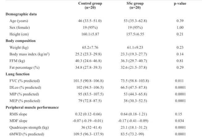
Table 1 shows the comparisons of demographic data, body composition, pulmonary function, peripheral muscle performance, and functional capacity between the SSc and control groups. The SSc group exhibited lower FFM and fat percentage values; however, no
signiicant differences were observed. Regarding
peripheral muscle performance, the SSc group exhibited lower MDF and quadriceps strength values (p=0.034 and p=0.0001, respectively). The pulmonary function
values and the 6MWD were signiicantly lower in the
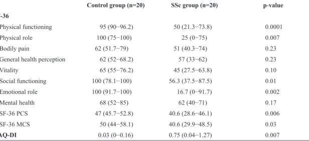
SSc group. Table 2 shows the results of the quality of life and physical capacity questionnaires between the SSc and control groups. The SSc group exhibited lower SF-36 physical component summary and SF-36 mental
component summary scores (p=0.006 and p=0.03, respectively) and higher HAQ-DI value (p=0.007).
The results of the univariate correlation analysis of the peripheral and respiratory muscle assessment, functional capacity, and quality of life in SSc patients are shown in Table 3. Quadriceps strength exhibited a strong correlation with the 6MWD. Quadriceps strength exhibited a moderate correlation with the HAQ-DI. The correlations of quadriceps strength with the 6MWD and the HAQ-DI are shown in Figures 1 and 2. The MDF exhibited a moderate correlation with MIP (Rho=0.684; p=0.0009) and a
weak correlation with MEP (Rho=0.472; p=0.035).
No signiicant correlations between these variables
were found in healthy subjects.
Discussion
The main indings in the present study are as
follows: (1) patients with SSc exhibited a reduction in the strength of the quadriceps and an increase in
Table 1. Comparison of demographic data, body composition, lung function, peripheral muscle performance, and functional capacity of healthy volunteers and SSc patients.
Control group (n=20)
SSc group (n=20)
p-value
Demographic data
Age (years) 46 (33.5–51.0) 53 (35.3–62.8) 0.39
Sex (female) 19 (95%) 19 (95%) 1.00
Height (cm) 160.1±5.87 157.5±6.55 0.21
Body composition
Weight (kg) 65.2±7.76 61.1±9.23 0.23
Body mass index (kg/m2) 25.2 (23.3–29.8) 23.3 (19.3–27.7) 0.14
FFM (kg) 40.3 (24.6–46.8) 36.3 (29.7–40.7) 0.81
Fat percentage (%) 34.8 (27.8–39.3) 32.6 (21.5–37.8) 0.29
Lung function
FVC (% predicted) 101.5 (90.8–106.8) 73.5 (58.8–103.8) 0.011
DLco (% predicted) 102 (94.5–106.5) 66.5 (47.5–87.8) 0.0001
MIP (% predicted) 95 (83.5–107.5) 53 (44.3–65.8) 0.0001
MEP (% predicted) 79 (72.8–87.5) 38 (30.3–52.5) 0.0001
Peripheral muscle performance
RMS slope 0.32 (0.12–0.66) 0.64 (0.18–1.21) 0.15
MDF slope -0.07 (-0.19–-0.01) -0.17 (-0.41–-0.09) 0.034
Quadriceps strength (kg) 36 (32–41.4) 23.1 (18.1–31.2) 0.0001
6MWD (% predicted) 109.5 (96.3–137.9) 83.5 (73.2–99) 0.0001
its fatigability, as well as reduced functional capacity;
(2) peripheral muscle strength exhibited a signiicant
correlation with the 6MWD and the HAQ-DI; and (3) fatigability exhibited a relationship with MIP and MEP. So far, no other study has focused on the
association of objective measures of lower limb muscle performance with the functional capacity and quality of life of individuals with SSc.
Compared to the control group, the SSc patients exhibited a reduction in the strength of the quadriceps
Table 2. Health-related quality of life and functional status (disability) measures in SSc patients.
Control group (n=20) SSc group (n=20) p-value
SF-36
Physical functioning 95 (90−96.2) 50 (21.3−73.8) 0.0001
Physical role 100 (75−100) 25 (0−75) 0.007
Bodily pain 62 (51.7−79) 51 (40.3−74) 0.23
General health perception 62 (52−68.2) 57 (33−62) 0.23
Vitality 65 (55−76.2) 45 (27.5−63.8) 0.10
Social functioning 100 (78.1−100) 56.3 (37.5−87.5) 0.01
Emotional role 100 (91.7−100) 16.7 (0−91.7) 0.002
Mental health 68 (52−85) 62 (40−71) 0.17
SF-36 PCS 47 (45.7−52.8) 40.6 (28.6−46.1) 0.006
SF-36 MCS 50 (44−58.1) 40.6 (29.9−48.5) 0.03
HAQ-DI 0.03 (0−0.16) 0.75 (0.04−1.27) 0.007
Values are medians (interquartile range) or number (%). SF-36 = Medical Outcomes Study 36-Item Short-Form Health Survey; PCS = Physical component summary score; MCS = physical component summary score; HAQ-DI = Health Assessment Questionnaire Disability Index.
Table 3. Spearman’s correlation coeficients between peripheral and respiratory muscles performance, functional capacity, and quality
of life in SSc patients.
RMS MDF Quadriceps strength MIP MEP
6MWD 0.054 0.116 0.719† 0.072 0.298
SF-36 PCS 0.143 0.111 0.045 0.163 0.135
SF-36 MCS -0.044 -0.056 -0.096 -0.010 0.046
HAQ-DI -0.084 0.017 -0.622* 0.282 0.235
RMS = angle of the linear regression line obtained with the values of root mean square electromyography signal over time during fatigability test of vastus medialis muscle; MDF = angle of the linear regression line obtained with the values of median frequency electromyography signal over time during the fatigability test of vastus medialis muscle; MIP = maximal inspiratory pressure; MEP = maximal expiratory pressure; 6MWD = 6-minute walk distance; SF-36 = Medical Outcomes Study 36-Item Short-Form Health Survey; PCS = physical component summary score; MCS = mental component summary score; HAQ-DI = Health Assessment Questionnaire Disability Index (HAQ-DI). *p<0.01; †p<0.001.
Figure 1. Relationship between quadriceps strength and the 6-min
walk distance (6MWD) (Rho=0.719; p=0.0004).
Figure 2. Relationship between quadriceps strength and the Health Assessment Questionnaire Disability Index (HAQ-DI)
and an increase in its fatigability. We were not able to locate any other study in the literature that assessed individuals with SSc by using isometric dynamometry and endurance testing of the quadriceps muscle. However, the characteristics of muscle affection in SSc are well established. According to Deuschle et al.5,
muscle biopsies of SSc patients show an increase in the connective tissue in the epimysium and perimysium, along with microangiopathy5. Interestingly, a higher
frequency of heart disease has been reported in association with SSc-related myopathy24, which suggests
that muscle affection is systemic and generalized in SSc patients. In accordance with other authors5,6,10, we
also found a reduced 6MWD in patients with SSc. The contribution of the muscular system impairment to the low aerobic capacity and poor exercise performance may be important, even in individuals with subclinical muscle affection10.
Similar to other studies14,25, we also observed lower
FFM and fat percentage values in patients with SSc
compared to controls, but no signiicant differences.
Reduced FFM in individuals with SSc may be attributed to several factors, including reduced physical activity, malnutrition, intestinal malabsorption, and use of glucocorticoids10. In addition to contributing to the pathogenesis of malnutrition, inlammation bears a
close relationship with muscle wasting14,25, which
at least partially explains the moderate correlation between FFM and quadriceps strength found in the present study. Therefore, we believe that individuals with SSc should be encouraged to improve their nutritional status and physical performance. Future prospective studies may contribute to the assessment of the effects of nutritional support and physical reconditioning on patients with SSc.
Exercise testing remains a robust and versatile tool that provides valuable diagnosis and prognostic information11,22. In patients with SS who frequently
experience musculoskeletal pain, the 6MWT may be more feasible and acceptable than tests that involve exercise equipment, such as the cycloergometer or treadmill. However, whether muscle affection in SSc patients exerts an impact on the 6MWT is still an open question. In the present study, we found a strong correlation between quadriceps strength and the 6MWD (Rho=0.719; p=0.0004). In addition to
myositis and myopathy, it is also believed that changes in the small blood vessels of skeletal muscles may
exert an inluence on the oxygen supply to cells, thus
contributing to poorer exercise performance8,24.
The HAQ-DI is a musculoskeletal-targeted questionnaire that assesses the impact of SSc on physical functioning and disability26. The DI values
we found are close to those reported by Singh et al.26
and de Achaval et al.27, but diverge from those
reported by Brower and Poole28 and Rannou et al.29, which possibly relects differences in the severity of
SSc among the patients tested. We also observed a moderate correlation between the HAD-QI values and quadriceps strength as reported by Steen and Medsger Jr.30, who applied clinical criteria to deine
the presence of proximal muscle weakness. According to Rannou et al.29, the HAQ-DI should be preferred
over the SSc HAQ (sHAQ) for assessing physical functioning, and the physical and mental component summary measures of the SF-36 are questionable for SSc patients.
In addition to reduced strength, we also found greater fatigability in the quadriceps muscle in the individuals with SSc. According to Hunzelmann and Brinckmann31, hypoxia is a decisive factor for modulating the inlammatory process in SSc, activating ibroblasts and changing their phenotype. The fact that type II ibers exhibit easy fatigability
in the presence of hypoxia may contribute to the reduced muscle endurance in individuals with SSc. We also observed a relationship between quadriceps fatigability and measures of respiratory muscle strength, which suggests that muscle weakness is generalized in individuals with SSc.
The strongest point of the present study is that it demonstrates that there are functional differences between SS patients and healthy subjects and that, although many factors have the potential to interfere with the functional capacity of SS patients, muscle strength has a strong association with performance in
6MWD. In addition, it is the irst study to investigate
understanding of the pathophysiological mechanisms of the disease.
To conclude, the present study showed that patients with SSc exhibited reduced respiratory muscles and quadriceps strength and an increase in its fatigability. In addition, quadriceps strength/endurance exhibited a relationship with the functional capacity and quality of life of these individuals. Although the nature of our results requires a more thorough investigation, our data suggest that quadriceps strength/endurance should be assessed in clinical practice.
Acknowledgements
This research was supported by Fundação de Amparo à Pesquisa do Estado do Rio de Janeiro (FAPERJ), Brazil.
References
1. Nikpour M, Stevens WM, Herrick AL, Proudman SM. Epidemiology of systemic sclerosis. Best Pract Res Clin Rheumatol. 2010;24(6):857-69. PMid:21665131.
2. Barnabe C, Joseph L, Belisle P, Labrecque J, Edworthy S, Barr SG, et al. Prevalence of systemic lupus erythematosus and systemic sclerosis in the First Nations population of Alberta, Canada. Arthritis Care Res (Hoboken). 2012;64(1):138-43. PMid:21972194.
3. Mouthon L. [Hand involvement in systemic sclerosis]. Presse Med. 2013;42(12):1616-26. PMid:24268960. 4. Krause L, Becker MO, Brueckner CS, Bellinghausen CJ,
Becker C, Schneider U, et al. Nutritional status as marker for disease activity and severity predicting mortality in patients with systemic sclerosis. Ann Rheum Dis. 2010;69(11):1951-7. PMid:20511612.
5. Deuschle K, Weinert K, Becker MO, Backhaus M, Huscher D, Riemekasten G. Six-minute walk distance as a marker for disability and complaints in patients with systemic sclerosis. Clin Exp Rheumatol. 2011;29(2 Suppl 65):S53-9. PMid:21586219.
6. Holland AE, Goh NS. The six-minute walk test in scleroderma: what should we measure and how should we measure it? Respirology. 2012;17(4):588-9. PMid:22394399.
7. Medsger TA Jr, Rodnan GP, Moossy J, Vester JW. Skeletal muscle involvement in progressive systemic sclerosis (scleroderma). Arthritis Rheum. 1968;11(4):554-68. PMid:5676926.
8. Clements PJ, Furst DE, Campion DS, Bohan A, Harris R, Levy J, et al. Muscle disease in progressive systemic sclerosis: diagnostic and therapeutic considerations. Arthritis Rheum. 1978;21(1):62-71. PMid:623695.
9. Ranque B, Bérezné A, Le-Guern V, Pagnoux C, Allanore Y, Launay D, et al. Myopathies related to systemic sclerosis: a case-control study of associated clinical and immunological features. Scand J Rheumatol. 2010;39(6):498-505. PMid:20726682.
10. Azevedo VF, Müller CS, Rinaldi L, Bredt MC, Giovanni K, Pereira MAC, et al. Avaliação nutricional e da capacidade funcional em doentes com esclerose sistémica progressiva. Acta Reumatol Port. 2009;34(2A):228-34. PMid:19569278. 11. ATS Committee on Proficiency Standards for Clinical
Pulmonary Function Laboratories. ATS statement: guidelines for the six-minute walk test. Am J Respir Crit Care Med. 2002;166(1):111-7. PMid:12091180.
12. Schoindre Y, Meune C, Dinh-Xuan AT, Avouac J, Kahan A, Allanore Y. Lack of specificity of the 6-minute walk test as an outcome measure for patients with systemic sclerosis. J Rheumatol. 2009;36(7):1481-5. PMid:19487260. 13. van den Hoogen F, Khanna D, Fransen J, Johnson SR, Baron
M, Tyndall A, et al. 2013 classification criteria for systemic sclerosis: an American College of Rheumatology/European League against Rheumatism collaborative initiative. Arthritis Rheum. 2013;65(11):2737-47. PMid:24122180.
14. Ciconelli RM, Ferraz MB, Santos W, Meinão I, Quaresma MR. Brazilian-Portuguese version of the SF-36. A reliable and valid quality of life outcome measure. Rev Bras Reumatol. 1999;39(3):143-50.
15. Kyle UG, Bosaeus I, De Lorenzo AD, Deurenberg P, Elia M, Gómez JM, et al, and the Composition of the ESPEN Working Group. Bioelectrical impedance analysis—part I: review of principles and methods. Clin Nutr. 2004;23(5):1226-43. PMid:15380917.
16. Miller MR, Hankinson J, Brusasco V, Burgos F, Casaburi R, Coates A, et al. Standardization of spirometry. Eur Respir J. 2005;26(2):319-38. PMid:16055882.
17. Pereira CAC, Sato T, Rodrigues SC. New reference values for forced spirometry in white adults in Brazil. J Bras Pneumol. 2007;33(4):397-406. PMid:17982531.
18. Neder JA, Andreoni S, Lerario MC, Nery LE. Reference values for lung function tests. II. Maximal respiratory pressures and voluntary ventilation. Braz J Med Biol Res. 1999;32(6):719-27. PMid:10412550.
19. Neder JA, Andreoni S, Peres C, Nery LE. Reference values for lung function tests. III. Carbon monoxide diffusing capacity (transfer factor). Braz J Med Biol Res. 1999;32(6):729-37. PMid:10412551.
20. Hermens HJ, Freriks B, Disselhorst-Klug C, Rau G. Development of recommendations for SEMG sensors and sensor placement procedures. J Electromyogr Kinesiol. 2000;10(5):361-74. PMid:11018445.
21. Shinjo SK, Gonçalves R, Kowalski S, Gonçalves CR. Brazilian-Portuguese version of the Health Assessment Questionnaire for Spondyloarthropathies (HAQ-S) in patients with ankylosing spondylitis: a translation, cross-cultural adaptation, and validation. Clin Rheumatol. 2007;26(8):1254-8. PMid:17205217.
22. Gibbons WJ, Fruchter N, Sloan S, Levy RD. Reference values for a multiple repetition 6-minute walk test in healthy adults older than 20 years. J Cardiopulm Rehabil. 2001;21(2):87-93. PMid:11314289.
24. Mimura Y, Ihn H, Jinnin M, Asano Y, Yamane K, Tamaki K. Clinical and laboratory features of scleroderma patients developing skeletal myopathy. Clin Rheumatol. 2005;24(2):99-102. PMid:15322944.
25. Souza RB, Borges CT, Takayama L, Aldrighi JM, Pereira RM. Systemic sclerosis and bone loss: the role of the disease and body composition. Scand J Rheumatol. 2006;35(5):384-7. PMid:17062439.
26. Singh MK, Clements PJ, Furst DE, Maranian P, Khanna D. Work productivity in scleroderma: analysis from the University of California, Los Angeles scleroderma quality of life study. Arthritis Care Res (Hoboken). 2012;64(2):176-83. PMid:22012885.
27. de Achaval S, Kallen MA, Mayes MD, Lopez-Olivo MA, Suarez-Almazor ME. Use of the Patient-generated Index in systemic sclerosis to assess patient-centered outcomes. J Rheumatol. 2013;40(8):1337-43. PMid:23772081. 28. Brower LM, Poole JL. Reliability and validity of the Duruoz
Hand Index in persons with systemic sclerosis (scleroderma). Arthritis Rheum. 2004;51(5):805-9. PMid:15478150. 29. Rannou F, Poiraudeau S, Berezné A, Baubet T, Le-Guern
V, Cabane J, et al. Assessing disability and quality of life
in systemic sclerosis: construct validities of the Cochin Hand Function Scale, Health Assessment Questionnaire (HAQ), Systemic Sclerosis HAQ, and Medical Outcomes Study 36-Item Short Form Health Survey. Arthritis Rheum. 2007;57(1):94-102. PMid:17266096.
30. Steen VD, Medsger TA Jr. The value of the Health Assessment Questionnaire and special patient-generated scales to demonstrate change in systemic sclerosis patients over time. Arthritis Rheum. 1997;40(11):1984-91. PMid:9365087.
31. Hunzelmann N, Brinckmann J. What are the new milestones in the pathogenesis of systemic sclerosis? Ann Rheum Dis. 2010;69(Suppl 1):i52-6. PMid:19995745. http://dx.doi. org/10.1136/ard.2009.117119.
Correspondence Agnaldo José Lopes
Rua Araguaia, 1266, bloco 1/405, Freguesia