VIEWS AND REVIEWS
Periodic EEG patterns: importance of their
recognition and clinical significance
Padrões eletrencefalográficos periódicos: importância do seu reconhecimento e
significado clínico
Maria Emilia Cosenza Andraus1, Cesar Fantezia Andraus2, Soniza Vieira Alves-Leon3
he term “periodic” was irst used by Cobb et al.1, in 1950,
to describe periodically occurring discharges in electroen-cephalograms (EEG) of 5 patients with subacute progres-sive encephalitis. Periodic EEG patterns consist of various forms discharges, usually epileptiform in appearance, and ply to waves or complexes occurring in sequence at an ap-proximately regular rate or intermittently regular intervals2,3.
hey are commonly classiied as periodic lateralized epilep-tiform discharges (PLEDs), bilateral independent PLEDs or BIPLEDs, generalized epileptiform discharges (GPEDs) and
triphasic waves2-7. Although not always epileptiform in
ap-pearance, triphasic waves are periodic and generalized, and the category of generalized periodic discharges also includes them2. PLEDs may be subclassiied into PLEDs-proper and
PLEDs-plus, and GPEDs into periodic short-interval difuse discharges (PSIDDs) and periodic long-interval difuse dis-charges (PLIDDs)3,5. Stimulus-induced rhythmic, periodic or
ictal discharges (SIRPIDs) consist on peculiar EEG patterns, which were described more recently, and may be present as periodic discharges2,3,8.
1 Electroencephalography Section, Service of Neurology of Prof. Sérgio Novis, 24th and 25th Infirmaries, Santa Casa da Misericórdia do Rio de Janeiro, Rio de
Janeiro RJ, Brazil. Permanent Professor of Strictu Sensu Post Graduation Program in Neurology, Universidade Federal do Estado do Rio de Janeiro (UNIRIO), Rio de Janeiro RJ, Brazil;
2 Neurosurgery Service, Hospital Universitário Clementino Fraga Filho, Universidade Federal do Rio de Janeiro (HUCFF-UFRJ), Rio de Janeiro RJ, Brazil; 3 Epilepsy Program, Service of Neurology, HUCFF-UFRJ, Rio de Janeiro RJ, Brazil. Associate Professor of Neurology and Permanent Professor of Strictu Sensu
Post Graduation Program in Neurology, UNIRIO, Rio de Janeiro RJ, Brazil.
Correspondence: Maria Emilia Cosenza Andraus; Santa Casa da Misericórdia do Rio de Janeiro – Setor de Eletroencefalografia, Serviço de Neurologia do Prof. Sérgio Novis, 25ª Enfermaria; Rua Santa Luzia 206; 20020-022 Rio de Janeiro RJ - Brasil; E-mail: [email protected]
Conflict of interest: There is no conflict of interest to declare.
Received 22 June 2011; Received in final form 19 October 2011; Accepted 26 October 2011 ABSTRACT
Periodic electroencephalographic (EEG) patterns consist of discharges usually epileptiform in appearance, which occur at regular intervals, in critical patients. They are commonly classified as periodic lateralized epileptiform discharges (PLEDs), bilateral independent PLEDs or BI-PLEDs, generalized epileptiform discharges (GPEDs) and triphasic waves. Stimulus-induced rhythmic, periodic or ictal discharges (SIRPIDs) are peculiar EEG patterns, which may be present as periodic discharges. The aim of this study is to make a review of the periodic EEG pat-terns, emphasizing the importance of their recognition and clinical significance. The clinical significance of the periodic EEG patterns is uncertain, it is related to a variety of etiologies, and many authors suggest that these patterns are unequivocally epileptogenic in some cases. Their recognition and classification are important to establish an accurate correlation between clinical, neurological, laboratorial and neu-roimaging data with the EEG results.
Key words: electroencephalography, PLEDs, BIPLEDs, GPEDs, triphasic waves, SIRPIDs.
RESUMO
Padrões eletrencefalográficos (EEG) periódicos consistem em descargas geralmente epileptiformes em aparência, que ocorrem a intervalos regulares, em pacientes críticos. Esses padrões são habitualmente classificados como descargas epileptiformes periódicas lateralizadas (PLEDs), PLEDs bilaterais e independentes ou BIPLEDs, descargas epileptiformes periódicas generalizadas (GPEDs) e ondas trifásicas. Des-cargas rítmicas, periódicas ou ictais induzidas por estímulos (SIRPIDs) são padrões eletrencefalográficos peculiares, que podem se apre-sentar como descargas periódicas. O objetivo deste estudo é fazer uma revisão dos padrões EEG periódicos, enfatizando a importância do seu reconhecimento e seu significado clínico. O significado clínico dos padrões EEG periódicos é incerto. Está relacionado a uma variedade de etiologias e muitos autores sugerem que tais padrões sejam inequivocamente de natureza epileptogênica em alguns casos. O seu reco-nhecimento e classificação são importantes para estabelecer uma correlação acurada entre dados clínicos, neurológicos, laboratoriais e de neuroimagem com os resultados de EEG.
Periodic EEG activity is commonly observed in critical patients, especially in the intensive care unit (ICU) setting, but, except for SIRPIDs, this terminology has been based pri-marily on routine 20-30 minute EEG recordings2,3,8. hey are
indicative of signiicant acute or subacute brain impairment and may be lateralized or generalized2,3. Periodic discharges
should direct the attention to the high potential for seizures and convulsive or nonconvulsive status epilepticus, and many authors recommend the use of conventional antiepileptic drugs (AEDs) to manage any occurrence of these discharges3,9.
Sometimes, however, the discharges are not actually epilepti-form in appearance nor epileptogenic in nature3,10. his is one
of the reasons for the “E” of “epileptiform” is no more recom-mended by the American Clinical Neurophysiology Society (ACNS) Critical Care Monitoring Committee nomencla-ture, that proposed the term lateralized periodic discharges (LPDs) to name PLEDs, bilateral independent periodic dis-charges (BIPDs) to name BIPLEDs, and generalized epilep-tiform discharges (GPDs) to name GPEDs10. his
nomencla-ture, however, is still undergoing testing and revision, and we preferred to keep the most known and consecrated nomen-clature. A ixed interval between the discharges is relatively rare, and some authors prefer to name them pseudoperiodic or quasi-periodicdischarges although this separation is not considered clinically relevant2,5,10-12.
A review was performed trying to show and understand the importance and clinical signiicance of the periodic EEG patterns, and to expose the strength of data on which man-agement of recommendations have been based. Because pe-riodic patterns are considered controversial EEG patterns, and are present in critical patients, who need therapeutic in-terventions based on decisions making, we considered it a relevant topic to review. We included SIRPIDs in this review for they are relatively new described EEG patterns which may present as periodic discharges.
Periodic Lateralized Epileptiform Discharges – PLEDs
PLEDs are the most common and the most periodic EEG pattern that have been studied2,5,13. his term was irst used by
Chatrian et al.14, in 1964, to describe a pattern consisting of
sharp waves and/or spikes, associated or not with slow waves, or other complex wave forms, occurring at periodic intervals. hey consist of lateralized complexes, usually recurring every 1.0 to 2.0 seconds, and often (but not always) appear as sharp waves or spikes which may be followed by a slow wave2-4. Cobb
et al.1, in 1950, attributed the periodicity of the discharges to
a disconnection of the cortex from the subcortical struc-tures, usually caused by a large white matter lesion. After that, Chatrian et al.14, through experimental studies with
patholog-ical specimens, showed that a lesion anywhere can be asso-ciated with PLEDs. Some authors hypothesized that PLEDs could be the EEG manifestation of an abnormal response of
neurons, which changes their excitatory neurotransmission in cases of acute brain lesions15. he cells would either
recov-er and respond normally or die aftrecov-er a precov-eriod of time, no lon-ger responding, and thereby leading to the disappearance of PLEDs recorded in the EEG15,16. hus, they could be considered
a nonspeciic result of acute partial and transient functional denervation in a localized area of the cortex15.
Reiher et al.17 proposed a subclassiication of PLEDs in two
major groups: PLEDs-proper, in which the periodicity of the discharges is relatively stable, the discharges are simply con-igured and uniform, and there are no associated rhythmic discharges; and PLEDs-plus, in which the periodicity of the discharges is variable and there is associated low amplitude rhythmic activity with the discharges. It seems that PLEDs-plus pattern is more associated with seizures, but few stud-ies suggested signiicant risk stratiication for it2,15,17. Chong et
al.2 reported their experience with 24-hour continuous EEG
recordings (coEEG) and observed that PLEDs-proper rarely occurs in isolation, with the EEG typically luctuating be-tween PLEDs-proper and PLEDs-plus, with high seizures risk in these patients.
PLEDs are indicative of an acute non-speciic brain dys-function or unilateral brain lesion, usually destructive, and they are most often present in cases of cerebral infarction2,5,15.
hey have also been described in the presence of fast-grow-ing brain tumors (as glioblastoma multiforme), brain abscess-es, viral encephalitis (especially related to the Herpes simplex
virus), Creutzfeldt-Jakob disease (CJD), hematomas and, less frequently, in demyelinating diseases, anoxia, primary epilep-sia, migraine and fat embolism syndrome, among others2,15,18-23.
Sekiguchi et al.24 described a case of PLEDs in an infant with
congenital protein C deiciency. In cerebral infarction, PLEDs are usually recorded on the area adjacent to infarction, which is partially afected by the disease process and is able to gen-erate electrical activity (unlike the area directly afected by in-farction), but whether these cells are apoptotic or regenerating is unknown2,3. Neufeld et al.20 and Chu et al.25 emphasized the
importance of the presence of an structural brain lesion associ-ated with metabolic disturbances in the production of PLEDs. PLEDs tend to be transient and resolve spontaneously within 2 to 3 weeks, the discharges tend to decrease in am-plitude, the repetition rate decreases and then the discharges cease2,3,9,11,26-28. Chronic PLEDs, however, have been reported
in patients with chronic epilepsy or old static lesions, espe-cially related to recent seizures, alcohol withdrawal or chron-ic toxchron-ic-metabolchron-ic disturbances4,7,9,11,14,15,26. he major
contro-versy about PLEDs is whether they are ictal, interictal or a postictal phenomenon2,3,9,15,23. Many authors consider that
speciic periodic EEG pattern9,15,28. Garzon et al.9 performed a
prospective and clinical study in 55 patients, with a total of
62 status epilepticus and 254 ictal/postictal EEG recordings,
and analyzed the relationship between PLEDs and status
epi-lepticus. hey demonstrated that, although PLEDs were not
always associated with seizures and status epilepticus, they can be unequivocally an ictal pattern. Increased focal glucose metabolism has been demonstrated associated with PLEDs, reinforcing their probable epileptogenic nature27. Although
they indicate an ictal pattern in some cases, PLEDs are usu-ally considered an interictal change or an unstable ictal-in-terictal continuum2,3.
PLEDs are usually associated with obtundation in 95% of patients, focal seizures and focal neurological signs may occur in 80%, and epilepsia partialis continua in 30% of the patients14,23. Clinical seizures or status epilepticus were seen
during the course of illness in 126 (90%) patients in a study per-formed by Snodgrass et al.29, with 50% presenting partial motor
status epilepticus, 22% partial motor seizures, 6% epilepsia
par-tialis continua, 6% isolated generalized seizures and 8%
gen-eralized status epilepticus. he prevalence of PLEDs in routine EEG laboratories ranges from 0.1% to 1%13-15,30.
Bilateral Periodic Lateralized Epileptiform Discharges – BIPLEDs
BIPLEDs occur when PLEDs are seen in both hemi-spheres, in an independent and asynchronous manner2,5,31.
his pattern is less common than PLEDs and is highly as-sociated with seizures in acutely ill patients2. In contrast to
PLEDs, BIPLEDs may present as asynchronous complexes, usually difering in morphology, amplitude, rate of repetition, and site of maximal involvement4. BIPLEDs are typically
as-sociated with acute structural lesions, with or without meta-bolic disturbances2,7,32. he most common causes of BIPLEDs
are anoxic encephalopathy and central nervous system in-fection, with a high incidence of coma state7,32. Few studies
demonstrated that clinical state and prognosis may be worse than with PLEDs, but a case of benign BIPLEDs has also been reported4,5,28,30. In a study with 21 patients with BIPLEDs,
Fitzpatrick et al.30demonstrated a mortality rate of 52%, and
De La Paz et al.32, studying 21 patients too, demonstrated a
mortality rate of 61%. he true prevalence and incidence of BIPLEDs are unknown, withstudies reporting an incidence of 4% to 22% in patients in the ICU, and a prevalence of 0.1% in routine EEG laboratories11,14,30.
Generalized Periodic Epileptiform Discharges – GPEDs
GPEDs are deined as periodic complexes occupying at least 50% of a standard 30-minute EEG, projected over the both hemispheres, in a symmetric, difuse and synchronous manner (although they may be more prominent in a given region, frequently the anterior regions)5,13. he discharges
vary in shape, but usually are characterized by spikes or sharp waves of high amplitude2,3. hey are usually classiied
as PSIDDs and PLIDDs2,5,7,32-34. According to the interval
be-tween the discharges, the burst suppression pattern may be present as GPEDs too7,33. PSIDDs have a periodicity less than
4.0 seconds and are more common and less speciic than PLIDDs5,33. Generally, PSIDDs are associated to
toxic-meta-bolic encephalopathies, anoxic brain injury, CJD and subclini-cal or nonconvulsive status epilepticus (Fig 1)5,33-39. Sometimes,
it is diicult to distinguish the etiology of GPEDs, even with history and clinical indings suggesting or not an epileptic or toxic-metabolic etiology34,38-40. Central nervous system
in-fection, as neurosyphilis, has been described in association with GPEDs in patients with cognitive and behavioral disor-ders41. In a study with 25 patients presenting GPEDs, Husain
et al.35 reported that 7 (28%) of them had toxic-metabolic
cephalopathy, 10 (40%) had anoxia and toxic-metabolic en-cephalopathy, 8 (32%) had a primarily neurological process, and 8 (32%) were in status epilepticus. In practice, a clinical trial with AEDs is almost always warranted to diferentiate nonconvulsive status epilepticus from severe encephalopa-thies although most patients will not respond clinically or electrographically39,40.
Gloor et al.42 suggested that the periodicity of the
discharg-es might be related to the recovery cycle of both cortical and subcortical structures, and that the subcortical discharges may be responsible for the surface discharges, but their repeti-tion rate depends upon cortical recovery time. hey proposed an abnormal functional state in the central nervous system, permitting rapid generalization of neuronal discharges.
In the CJD, the classic EEG patterns usually are character-ized by PSIDDs, with biphasic or triphasic sharp waves, with a duration of 100-300 milliseconds and recurring at 0.7-1.5 sec-ond interval between the discharges, with symmetric and syn-chronous widespread43. Traub et al.44 suggested, through
exper-imental evidence and computer modeling, that synchrony and periodicity in the CJD may be related to a virus-induced fusion of neuronal processes, particularly dendrites, leading to an ab-normal electrotonic coupling between cells, providing the ba-sis for powerful excitatory interaction whereby large neuronal aggregates burst in near synchrony. Cortical synchronous dis-charges would give rise to sharp waves in the EEG, whereas similar discharges in brainstem, spinal cord or elsewhere could lead to myoclonic jerks.
It is worth remembering, however, that the bioelectric cerebral activity is a dynamic process, with temporal varia-tions and transient patterns, which could diicult the cor-rect interpretation and show the necessity of serial EEGs in some cases, to establish the correct clinical and EEG correlation35,43. In the early course of the CJD, difuse
in the early stages of CJD may resemble nonconvulsive status
epilepticus too35. he clinical and EEG correlation, response
to AEDs and monitoring with serial EEG recordings may be helpful considerations in the diferential diagnosis35,45-47.
PLIDDs usually have a frequency of at least 4.0 seconds between the discharges48,49. hey are more speciic with
re-spect to the etiology and may be encountered in disorders like some toxic encephalopathies ( for baclofen or ketamine, for example), anoxic brain injury and SSPE3. In SSPE
(suggest-ed by the presence of PLIDDs in a child with dementia and myoclonic jerks), the stereotyped complexes, occurring at a regular interval and having a constant relationship to myo-clonus, make this as one of the most characteristic and dis-ease-speciic of all EEG patterns34,48-51. he early descriptions,
by Cobb48, in 1966, characterized the stage II of SSPE by
bi-laterally symmetrical and synchronous generalized, stereo-typed high amplitude delta waves, called Radermacker or “R” complexes, recurring at regular intervals of 5 to 15 seconds, although less speciic and atypical EEG changes have been described49. With advancing disease, the interval between
the discharges becomes progressively smaller2,49. Silva et al.51
described an atypical clinical and EEG pattern in a 14-year-old boy with SSPE, who presented an initial EEG character-ized by a left temporal focus which evolved to PLEDs. Typical GPEDs appeared during the 3rd and 4th weeks.
GPEDs are an uncommon EEG pattern with an overall in-cidence between 0.4% and 1% in EEG laboratory series3. It
seems that more than 50% of patients with GPEDs have dei-nite seizures during the acute illness3.
Triphasic waves
Generalized periodic discharges also include tripha-sic waves, a pattern initially described in 1950 by Foley et al.52, who named the waves as “blunted and spike waves”.
Triphasic waves are periodic and generalized, typically fron-tally predominant and not always epileptiform in appear-ance (a reason for they are often not included in the GPEDs categorization)2,5. his pattern can occur in any
toxic-meta-bolic or structural encephalopathy although the early descrip-tions associated its presence to hepatic encephalopathy3,6,35.
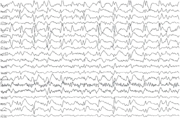
he three most common causes of triphasic waves are he-patic encephalopathy, renal failure (Fig 3) and anoxic injury6.
his term was coined by Bickford et al.53, in reference to the
typical morphology, characterized by three phases. hey con-sist of generalized periodic sharp waves or sharply contoured delta waves with a triphasic morphology (typically with a negative/positive/negativity polarity, with each phase lasting longer than the prior), recurring at 1.0 to 3.0Hz, with or with-out an anterior-posterior or posterior-anterior lag8. When
the term triphasic waves is used, it usually implies a pattern
seen with a variety of encephalopathies, particularly hepatic or renal4. It is worth to remember, however, that this term
can be used to describe the morphology of waveform in that sharp and slow wave three phases complexes4.
Sometimes, when patients present confused and obtund-ed and rhythmic sharp waves resembling triphasic waves ap-pear in the EEG, it is diicult to distinguish nonconvulsive
status epilepticus from toxic-metabolic encephalopathy4,54.
In these situations, some authors recommend the use of
benzodiazepines administration to verify the resolution of EEG changes in the cases of nonconvulsive status epilepticus
even though thriphasic waves may be abolished by benzodi-azepines administration, as demonstrated by Foutain et al.54.
Boulanger et al.55 compared the 87 EEGs of 71 patients with
triphasic waves and 27 EEGs of 13 patients with noncon-vulsive status epilepticus, and showed that, when compared to triphasic waves, epileptiform discharges associated with nonconvulsive status epilepticus had a higher frequency, a
Fig 3. Pseudoperiodic runs of triphasic sharp waves in a background generalized slowing EEG. This record corresponds to a metabolic (renal failure) encephalopathy in a 69-year-old man, presented with an altered mental status.
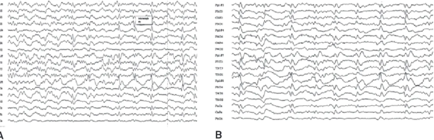
Fig 2. Periodic lateralized epileptiform discharges (PLEDs) in the right hemisphere, predominantly over the frontotemporal regions (A), with some widespread, evolving to generalized periodic epileptiform discharges (GPEDs) (B) in an EEG of a 50-year-old woman with rapidly progressive cognitive impairment, altered mental status and muscle spasms. The clinical picture, the neuroimaging studies, the absence of toxic-metabolic disorders and no improvement with antiepileptic drug allied to the EEG periodic patterns supported the presumptive diagnosis of Creutzfeldt-Jakob disease.
1. Cobb W, Hill D. Electroencephalogram in subacute progressive encephalitis. Brain 1950;73:392-404.
2. Chong DJ, Hirsch LJ. Which EEG patterns warrant treatment in the critically ill? Reviewing the evidence for treatment of periodic epileptiform discharges and related patterns. J Clin Neurophysiol 2005;22:79-91.
3. Hirsch LJ, Brenner RP. Periodic discharges and other controversial EEG patterns. In: Hirsch LJ, Brenner RP (Eds). Atlas of EEG in critical care. Chichester: Wiley-Blackwell, 2010: 129-160.
4. Brenner RP. EEG in convulsive and nonconvulsive status epilepticus. J Clin Neurophysiol 2004;21:319-331.
5. Brenner RP, Schaul N. Periodic EEG patterns: classification, clinical correlation and pathophysiology. J Clin Neurophysiol 1990;7:249-267.
6. Brigo F, Storti M. Triphasic waves. Am J Eletroneurodiagnostic Technol 2011;51:16-25.
7. Orta DSJ, Chiappa KH, Quiroz AZ, Costello DJ, Cole AJ. Prognostic implications of periodic epileptiform discharges. Arch Neurol 2009;66:985-991.
8. Hirsch LJ, Claassen J, Mayer SA, Emerson RG. Stimulus-induced rhythmic, periodic, or ictal discharges (SIRPIDs): a common EEG phenomenon in the critically ill. Epilepsia 2004;45:109-123.
9. Garzon E, Fernandes RM, Sakamoto AC. Serial EEG during human status epilepticus: evidence for PLED as an ictal pattern. Neurology 2001;57:1175-1183.
10. Hirsch LJ, Brenner RP, Drislane FW, et al. The ACNS subcommittee on research terminology for continuous EEG monitoring: proposed standardized terminology for rhythmic and periodic EEG patterns encountered in critically ill patients. J Clin Neurophysiol 2005;22:128-135.
11. Gross DW, Wiebe S, Blume WT. The periodicity of lateralized epileptiform discharges. Clin Neurophysiol 1999;110:1516-1520.
12. Markand ON, Daly DD. Pseudoperiodic lateralized paroxysmal discharges in electroencephalogram. Neurology 1971;21:975-981.
13. Kuroiwa Y, Celesia GG. Clinical significance of periodic EEG patterns. Arch Neurol 1980;37:15-20.
14. Chatrian GE, Shaw CM, Leffman H. The significance of periodic lateralized epileptiform dischages in EEG: an electrographic, clinical and pathological study. Electroencefalogr Clin Neurophysiol 1964;17:177-193.
15. García-Morales I, García MT, Galán-Dávila L, et al. Periodic lateralized epileptiform discharges: etiology, clinical aspects, seizures, and evolution in 130 patients. J Clin Neurophysiol 2002;19:172-177.
16. Gross DW, Quesney LF, Sadikot AF. Chronic periodic lateralized epileptiform discharges during sleep in a patient with caudate nucleus atrophy: insights into the anatomical circuitry of PLEDs. Electroencephalogr Clin Neurophysiol 1998;107:434-438.
17. Reiher J, Rivest J, Grand’Maison F, Leduc CP. Periodic lateralized epileptiform discharges with transitional rhythmic discharges: association with seizures. Electroencephalogr Clin Neurophysiol 1991;78:12-17.
18. Jacome DE. Periodic EEG patterns in cerebral fat embolism. Clin Electroencephalogr 1983;14:27-34.
19. Au WJ, Gabor AJ, Vijayan N, Markand ON. Periodic lateralized epileptiform complexes (PLEDs) in Creutzfeldt-Jakob disease. Neurology 1980;30:611-617.
20. Neufeld MY, Vishnevskaya S, Treves TA, et al. Periodic lateralized epileptiform discharges (PLEDs) following stroke are associated with metabolic abnormalities. Electroencephalogr Clin Neurophysiol 1997;102:295-298.
21. Pohlmann-Eden B, Hoch DB, Cochius JI, Chiappa KH. Periodic lateralized epileptiform discharges: a critical review. J Clin Neurophysiol 1996;13:519-530.
References
shorter duration of phase one, extra-spikes components and less generalized background slowing. Noxious or auditory stimulation frequently increased the triphasic waves and had no efect on the epileptiform pattern. he authors concluded that certain EEG criteria and the response to stimulation are very helpful in distinguishing triphasic waves from general-ized nonconvulsive status epilepticus.
Stimulus-induced Rhythmic, Periodic or Ictal Discharges – SIRPIDs
SIRPIDs were irst described in 2004 by Hirsch et al.8, who
recorded coEEG and digital video in critical patients in the ICU setting. hey have noted striking EEG patterns when stu-porous or comatose patients were stimulated and noted that many of these patterns appear ictal, but were consistently elicited by stimulation. hey named these EEG patterns as SIRPIDs and deined them as periodic, rhythmic or ictal ap-pearing discharges that were consistently induced by alerting stimuli, such as auditory, sternal rub, examination, suctioning, turning and other patient-care activity. hey consider SIRPIDs as periodic when the pattern consist of epileptiform discharg-es recurring at regular or nearly regular intervals, with an iden-tiiable interdischarge interval. he speciic periodic patterns were classiied as periodic epileptiform discharges (PEDs) and
subdivided in PLEDs, BIPLEDs, GPEDs and triphasic waves. Some patients present clinical seizures with SIRPIDs, especial-ly focal motor seizures, but this pattern is usualespecial-ly a pureespecial-ly elec-trographic change, with no obvious clinical manifestations2,8,10.
he pathophysiology, exact clinical, therapeutic and prognos-tic signiicance of SIRPIDs is still undeined2,8,10.
Final remarks
he clinical signiicance of the periodic EEG patterns remains uncertain. Many authors suggest that they are un-equivocally epileptogenic in some cases, and how aggressive to treat them with AEDs is still unclear. A clinical trial with AEDs to treat a possible nonconvulsive status epilepticus is indicated on the majority of the cases although most patients will not respond clinically or electroencephalographically.
22. de los Reyes EC, McJunkin JE, Glauser TA, Tomsho M, O’Neal J. Periodic lateralized epileptiform discharges in La Crosse encephalitis, a worrisome subgroup : clinical presentation, electroencephalogram (EEG) patterns, and long-term neurologic outcome. J Child Neurol 2008;23:167-172.
23. Gandelman-Marton R, Rabey JM, Flechter S. Periodic lateralized epileptiform discharges multiple sclerosis: a case report. J Clin Neurophysiol 2003;20(2):117-121.
24. Sekiguchi K, Akiyoshi K, Okazaki N, et al. PLEDs in an infant with congenital protein C deficiency: a case report. Clin Neurophysiol 2010;121:800-801.
25. Chu NS. Periodic lateralized epileptiform discharges with preexisting focal brain lesions. Role of alcohol withdrawal and anoxic encephalopathy. Arch Neurol 1980;37:551-554.
26. Westmoreland BF, Klass DW, Sharbrough FW. Chronic periodic lateralized epileptiform discharges: electrographic and clinical features. Arch Neurol 1986;43:494-496.
27. Handfort A, Cheng JT, Mandelkern MA, Treiman DM. Markedly increased mesiotemporal lobe metabolism in a case with PLEDs: further evidence that PLEDs are a manifestation of partial status epilepticus. Epilepsia 1994;35:876-881.
28. Silva AB, Bertolucci PHF. Descargas periódicas lateralizadas II. Aspectos evolutivos. Arq Neuropsiquiatr 1988;46:10-15.
29. Snodgrass SM, Tsuburaya K, Ajmone-Marsan C. Clinical significance of periodic lateralized epileptiform discharges: relationship with status epilepticus. J Clin Neurophysiol 1989;6:159-172.
30. Fitzpatrick W, Lowry N. PLEDs: clinical correlates. Can J Neurol Sci 2007;34:443-450.
31. Fushimi M, Matsubuchi N, Sekine A, Shimizu T. Benign bilateral independent periodic lateralized epileptiform discharges. Acta Neurol Scand 2003;108:55-59.
32. De La Paz D, Brenner RP. Bilateral independent periodic lateralized epileptiform discharges. Clinical significance. Arch Neurol 1981;38:713-715.
33. Yemisci M, Gurer G, Saygi S, Ciger A. Generalised periodic epileptiform discharges: clinical features, neuroradiological evaluation and prognosis in 37 adult patients. Seizure 2003;12:465-472.
34. Gaches J. Activités périodiques en EEG. Rev EEG Neurophysiol Clin 1971;1:9-33.
35. Husain AM, Mebust KA, Radtke RA. Generalized periodic epileptiform discharges: etiologies, relationship to status epilepticus, and prognosis. J Clin Neurophysiol 1999;16:51-58.
36. Fernández-Torre JL, Solar DM, Astudillo A, Cereceda R, Acebes A, Calatayud MT. Creutzfeld-Jakob disease and non-convulsive status epilepticus: a clinical and electroencephalographic follow-up study. Clin Neurophysiol 2004;115:316-319.
37. Wang PS, Wu YT, Hung CI, Kwan SY, Teng S, Soong BW. Early detection of periodic sharp wave complexes on EEG by independent analysis in patients with Creutzfeldt-Jakob disease. J Clin Neurophysiol 2008;25:25-31.
38. Drislane FW, Lopez MR, Blum AS, Schomer DL. Detection and
treatment of refractory status epilepticus in the intensive care unit. J Clin Neurophysiol 2008;25:181-186.
39. Korabathina K, Benbadis SR. EEG diagnosis of nonconvulsive status epilepticus: generalized periodic patterns – status or not? Expert Rev Neurother 2007;7:1643-1644.
40. Parkerson KA, Reinsberger C, Chou SH, Dworetzky BA, Lee JW. Lacosamide in the treatment of acute recurrent seizures and periodic epileptiform patterns in critically ill patients. Epilepsy Behav 2011;20:48-51.
41. Anghinah R, Camargo EC, Braga NI, Waksman S, Nitrini R. Generalized periodic EEG activity in two cases of neurosyphilis. Arq Neuropsiquiatr 2006;64:122-124.
42. Gloor P, Kalabay O, Giard N. The electroencephalogram in diffuse encephalopathies: electroencephalographic correlates of gray and white matter lesions. Brain 1968;91:779-802.
43. Ortega-Albás JJ, Serrano-García AL. Neurofisiología en la enfermedad de Creutzfeldt-Jakob. Rev Neurol 2003;36:376-380.
44. Traub RD, Pedley TA. Virus-induced electrotonic coupling: hypothesis on the mechanism of periodic EEG discharges in Creutzfeldt-Jakob disease. Ann Neurol 1981;10:405-410.
45. Lapergue B, Demeret S, Denys V, et al. Sporadic Creutzfeldt-Jakob disease mimicking nonconvulsive status epilepticus. Neurology 2010;74:1995-1999.
46. Bouwman NA, Verhagen WI, Meulstee J. EEG abnormalities in poikilothermia suggesting Creutzfeldt-Jakob disease. Clin EEG Neurosci 2009;40:196-199.
47. Hansen HC, Zschocke S, Stürenburg HJ, Kunze K. Clinical changes and EEG patterns preceding the onset of periodic sharp wave complexes in Creutzfeldt-Jakob disease. Acta Neurol Scand 1998;97:99-106.
48. Cobb W. The periodic events of subacute sclerosing leucoencephalitis. Electroencephalogr Clin Neurophysiol 1966;21:278-294.
49. Praaven-kumar S, Sinha S, Taly AB, et al. Electroencephalographic and imaging profile in a subacute sclerosing panencephalitis (SSPE) cohort: a correlative study. Clin Neurophysiol 2007;118:1947-1954.
50. Bertolucci PH, Silva AB. Descargas epileptiformes lateralizadas 1. Aspectos clínicos e eletrencefalográficos. Arq Neuropsiquiatr 1987;45:364-370.
51. Silva DF, Lima MM, Anghinah R, Zanoteli E, Lima JGC. Atypical clinical and electroencephalographic pattern in a patient with subacute sclerosing panencephalitis. Arq Neuropsiquiatr 1995;53:278-280.
52. Foley JM, Watson CW, Adams RD. Significance of the electroencephalographic changes in hepatic coma. Trans Am Neurol Assoc 1950;51:161-165.
53. Bickford RG, Butt HR. Hepatic coma: the electroencephalography pattern. J Clin Invest 1955;34:790-799.
54. Fountain NB, Waldman WA. Effects of benzodiazepines on triphasic waves: implications for nonconvulsive status epilepticus. J Clin Neurophysiol 2001;18:345-352.