ENZYMATIC ANALYSIS IN
Anopheles nuneztovari
GABALDÓN
(DIPTERA, CULICIDAE)
SCARPASSA, V. M.1 and TADEI, W. P.2
1Coordenação de Pesquisas em Entomologia, Instituto Nacional de Pesquisas da Amazônia, Avenida André Araujo,
2936, CEP 69011-970, Manaus, AM, Brazil
2Coordenação de Pesquisas em Ciências da Saúde, Instituto Nacional de Pesquisas da Amazônia, Avenida André
Araújo, 2936, CEP.: 69011-970, Manaus, AM, Brazil
Correspondence to: Vera Margarete Scarpassa, Coordenação de Pesquisas em Entomologia, Instituto Nacional de Pesquisas da Amazônia, Avenida André Araujo, 2936, CEP 69011-970, Manaus, AM, Brazil,
e-mail: vera@inpa.gov.br
Received January 4, 1999 – Accepted March 27, 2000 – Distributed November 30, 2000 (With 2 figures)
ABSTRACT
Enzymatic analysis in Anopheles nuneztovari was made using four populations from the Brazilian Amazon and two from Colombia. The enzymes ME and XDH presented a monomorphic locus in all of the studied populations. EST and LAP presented a higher number of loci. In EST, genetic varia-tion was observed in the five loci; LAP presented four loci, with allec variavaria-tion in two loci. In IDH, three activity regions were stained, with genetic variation for locus Idh-1 in the Brazilian Amazon populations. A locus for MDH was observed, with genetic variation in the six populations. A region was verified for ACON, with four alleles in Sitronela and three in the other popula-tions. PGM constituted one locus, with a high variability in the Brazilian Amazon populapopula-tions. A locus was observed for 6-PGD with allelic variation in all of the populations with the exception of Tibú. Enzyme PGI presented two loci, both with genetic variability in the Tucuruí population. The enzyme α-GPD showed an activity region with polymorphism in the Tucuruí, Tibú and Si-tronela populations. The phenotypic variations detected for these enzymes suggest that four (EST, LAP, ACON and PGM) possess monomeric structures and five (IDH, MDH, 6-PGD, PGI and α -GPD) dimeric structures in their proteins. These enzymes constitute in important markers to estimate variability and genetic divergence in natural populations of A. nuneztovari.
Key words: isozymes, electrophoretic profiles, genetic variation, neotropical anopheline, Amazonian.
RESUMO
Análise enzimática em Anopheles nuneztovari Gabaldón (Diptera, Culicidae)
ACON e PGM) possuem estrutura monomérica e cinco (IDH, MDH, 6-PGD, PGI e α-GPD), estrutura dimérica em suas proteínas. Essas enzimas constituem-se em importantes marcadores para estimar variabilidade e divergência genética em populações naturais de A. nuneztovari.
Palavras-chave: isoenzimas, perfis eletroforéticos, variação genética, anofelino neotropical, Amazônia.
INTRODUCTION
In Anopheles, Aedes and Culex geni, iso-zymes have been used in genetic variability analysis among populations within species, and also as genetic markers important in the sibling species separation, by diagnostic loci or by a combination of several loci that show differentiation (Narang & Seawright, 1990). In anopheline mosquitoes, diagnostic loci are frequently used to separate morphologically similar species in several species complexes. The identification of these spe-cies becomes relevant, as it allows separation of members that are involved in human malaria trans-mission (Coluzzi, 1988). Thus, interpretation of electrophoretic profiles of different enzymatic systems is a basic necessity for these studies.
In this study, we interpreted the electropho-retic profiles of eleven enzymes in six popula-tions of the human malaria vector Anopheles (Nyssorhynchus) nuneztovari, including the photos for each enzyme, the number of activity zones coded by different loci and their possible allelic variants. Population structure and genetic divergence among these populations were analyzed by Scarpassa et al. (1999).
MATERIAL AND METHODS
Samples of A. nuneztovari were collected at four sites in the Brazilian Amazon [km 206 of the BR-174 Highway (BR), Amazonas State (1º16’S, 60º23’W); Puraquequara (PUR), Ama-zonas State (3º6’7’’S, 60º1’30’’W); Tucuruí (TUC), Pará State (3º42’S, 49º27’W); and Nova Mazagão (NOMA), Amapá State (0º7’S, 51º17’W)], and two sites in Colombia [Tibú (TIBÚ), Norte de Santander Department (8º39’N, 72º42’W); and Sitronela (SIT), Valle Department (3º49’N, 77º4’W)]. The specimens were captured when feeding on pigs, cattle, resting on stable walls or human biting. After captures, blood-fed mosquitoes were individually isolated in plastic cups for egg laying, according to Scarpassa & Tadei (1990). Following oviposition and eclosion,
the offspring (F1) were reared until the 4th instar larvae, pupae and adults, when they were frozen at –70°C, until analyzed. Fourth instar larvae were used in the analysis of ten enzymes, except for α-glycerophosphate dehydrogenase (α-GPD), for which adults were used. An average of 2-4 individuals from each progeny were employed. Electrophoresis was carried out in starch (12%) and starch-agarose gels (1% and 0.8%, respectively). Buffer solutions and reactions mix-tures were according to Harris & Hopkinson (1976), Steiner & Joslyn (1979) and Scarpassa et al. (1999).
The electrophoretic conditions used for each enzyme are showed in the Table 1. Iden-tification of the specimens were done on eggs, adults and male genitalia (Cova-Garcia, 1961; Gorham et al., 1967; Savage, 1986). The denomination employed for loci and alleles was according to Manguin et al. (1995) and Scarpassa et al. (1999), frequently used in mosquitos, where the allele most common was considered as 100.
RESULTS AND DISCUSSION
The electrophoretic patterns of the eleven enzymatic systems were analyzed: malic enzyme (ME, E.C.1.1.1.40), xanthine dehydrogenase (XDH, E.C.1.2.1.37), esterase (EST, E.C.3.1.1.1), leucine aminopeptidase (LAP, E.C.3.4.11.1), isocitrate dehydrogenase (IDH, E.C.1.1.1.42), malate dehydrogenase (MDH, E.C.1.1.1.37), aconitase (ACON, E.C.4.2.1.3), 6 phosphoglu-conate dehydrogenase (6-PGD, E.C.1.1.1.44), phosphoglucomutase (PGM, E.C.5.4.2.2), phos-phoglucose isomerase (PGI, E.C.5.3.1.9) and α-glycerophosphate dehydrogenase (α-GPD, E.C.1.1.1.8). The number of involved loci and their allelic variants of each enzyme are described below.
Malic Enzyme and Xanthine Dehydrogenase
Buffer Enzymes
Bridge Gel
Gel type Migration time (hours)
V/cm
EST, LAP 0.3 M
Borate pH 8.0
0.17-0.0023 M Tris-citrate
pH 8.0
Starch 12 1.7
IDH, ME, MDH 0.245-0.15 M Phosphate-citrate pH
5.9
1:40 dilution (v:v) of the bridge
buffer
Starch 15 2.6
ACON, 6-PGD 0.135-0.040 M Tris-citrate
pH 6.90
0.009-0.003 M Tris-citrate
pH 7.10
Starch 16 2.3
PGM 0.22 M
TEMM pH 7.4
1:15 dilution (v:v) of the bridge
buffer
Starch-agarose 5 4.8
PGI 0.22 M
TEMM pH 7.4
1:15 dilution (v:v) of the bridge
buffer
Starch 16 2.3
α-GPD 0.1 M
Tris-phosphate pH 7.4
1:20 dilution (v:v) of the bridge
buffer
Starch 16 2.6
XDH 0.036-0.194 M Lithium-borate
pH 8.25
0.074-0.009 M Tris-citrate
pH 8.45
Starch 15 1.7
Similar results were obtained in several species of the Nyssorhynchus subgenus(Narang et al., 1979; Hii et al., 1991; Narang et al., 1991). For ME, however, one locus with two or more alleles was verified in the four species of A. qua-drimaculatus complex (Narang et al., 1989), in A. deaneorum and A. marajoara (Narang et al., 1993) and in A. pseudopunctipennis (Manguin et al., 1995). Two loci with genetic variation were found in the A. punctulatus complex (Foley et al., 1995). Genetic variation was detected for XDH in at least one locus in A. aquasalis (Steiner et al., 1981), in the A. quadrimaculatus complex (Narang et al., 1989), in A. albimanus (Narang et al., 1991), and in A. deaneorum and A. marajoara (Narang et al., 1993).
Esterase
Analysis of the 4th instar larvae presented an electrophoretic profile complex with five activity zones in the six populations. The allelic variants observed for all of these isozymes allow for the proposal that they are coded by independent loci: Est-1, Est-2, Est-3, Est-4 and Est-5. In the
Est-1 to Est-4 loci the number of alleles present cannot be quantified, due to overlapping of the alleles among these loci. Detailed analysis was possible only on the Est-5 locus (Fig. 1C). In the four Brazilian Amazon populations, five codominant alleles were observed, being the most frequents Est-5109,Est-5106 and Est-5100(Table 2). The population from NOMA presented the highest number of genotype combinations, with 12 phenotypes. The two Colombian populations presented only two alleles Est-5109 and Est-5106. The Est-5106 allele showed high frequency in both populations. The heterozygous individuals presented two bands, suggesting a monomeric structure for this isozyme.
Our data support previous studies, which indicate that esterase is the most variable enzyme in mosquitoes, as well as in other insects, small vertebrates and plants (Wagner & Selander, 1974). In anopheline mosquitoes such as in A. albimanus of the six stained loci, four were polymorphic (Vedbrat & Whitt, 1975), in A. aquasalis, five of the six stained loci presented genetic variation (Narang et al., 1979), in A. nuneztovari from Suriname and TABLE 1
TABLE 2
Frequencies of alleles at sixteen loci of Anopheles nuneztovari.
Population
Locus Allele BR PUR TUC NOMA TIBÚ SIT
Pgm n 141 158 52 136 85 85
113 0.039 0.013 0 0.026 0 0
108 0.429 0.516 0.019 0.371 0.265 0.412
100 0.362 0.263 0.875 0.445 0.735 0.588
94 0.149 0.196 0.096 0.011 0 0
91 0.021 0.013 0.010 0.121 0 0
89 0 0 0 0.026 0 0
6Pgd n 130 124 89 67 64 62
108 0 0.012 0 0.007 0 0
100 0.988 0.988 0.983 0.985 1 0.952
92 0.012 0 0.017 0.007 0 0.048
Acon n 112 124 76 51 64 60
106 0 0 0 0 0.102 0.275
103 0 0.004 0 0.108 0.156 0.042
100 0.991 0.960 1 0.892 0.742 0.667
98 0.009 0.036 0 0 0 0.017
Mdh n 104 152 99 71 72 73
113 0 0.026 0.005 0.014 0 0
100 1 0.947 0.949 0.986 0.319 0.322
94 0 0.026 0.035 0 0.681 0.678
78 0 0 0.010 0 0 0
Idh-1 n 138 169 99 83 66 54
106 0.029 0.012 0.030 0.102 0 0
Venezuela, three of the five loci presented variation (Steiner et al., 1980), as well as three of the six loci
TABLE 2 (Continued)
Population
Locus Allele BR PUR TUC NOMA TIBÚ SIT
100 0.964 0.988 0.934 0.898 1 1
93 0.007 0 0.035 0 0 0
αGpd n 55 66 56 75 44 97
107 0 0 0.036 0 0 0
100 1 1 0.964 1 0.989 0.052
90 0 0 0 0 0.011 0.948
Lap-1 n 156 145 130 80 31 69
100 1 0.976 1 1 1 1
98 0 0.024 0 0 0 0
Lap-5 n 160 147 21 120 87 72
100 1 1 0.857 0.967 0.937 0.938
98 0 0 0.143 0.033 0.063 0.063
Est-5 n 142 128 100 68 78 80
111 0.007 0.008 0.025 0.074 0 0
109 0.190 0.180 0.330 0.368 0.058 0.019
106 0.239 0.234 0.485 0.110 0.942 0.981
100 0.532 0.516 0.145 0.367 0 0
97 0.032 0.063 0.015 0.081 0 0
Pgi-1 n 73 123 28 75 69 69
100 1 1 0.911 1 1 1
96 0 0 0.089 0 0 0
Pgi-2 n 65 78 84 75 69 69
Also, ontogenetic analysis of the A. nunez-tovari population from Tucuruí, presented seven loci with genetic variation in six (Scarpassa, 1988). These results suggest that esterase isozymes are important markers for estimating genetic variability in natural populations of anopheline mosquitoes.
Leucine Aminopeptidase
Leucine aminopeptidase showed four elec-tronegative activity zones in the six populations. Lap-1 and Lap-5 presented a weak coloration, and Lap-2 and Lap-4 were intensely colored (Fig. 1D). In this study, Lap-3 was not visualized because it is exclusive for pupa and adult stages (Scarpassa et al., 1992). The Lap-1 locus was monomorphic in all populations, except in the
population from PUR, which showed two alleles, Lap-1100 and Lap-198 (Table 2). Lap-2 and Lap-4 loci were monomorphic in all the populations. The Lap-5 locus showed two codominant alleles, being the Lap-5100 allele fixed in the PUR, and it was more common in the populations from BR, TUC, NOMA, TIBÚ and SIT (Table 2). Two bands in the heterozygous individuals were verified,which allows to propose that the protein structure is mo-nomeric. We also observed in the Lap-5 locus two additional bands in heterozygous individuals and exclusive of the population from TUC. This results suggests that the presence of these bands may be a consequence of post-translational chan-ges. However, this hypothesis may be reinforced with additional studies.
TABLE 2 (Continued)
Population
Locus Allele BR PUR TUC NOMA TIBÚ SIT
100 1 1 0.952 1 1 1
93 0 0 0.024 0 0 0
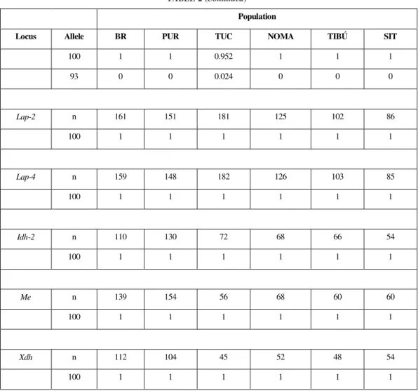
Lap-2 n 161 151 181 125 102 86
100 1 1 1 1 1 1
Lap-4 n 159 148 182 126 103 85
100 1 1 1 1 1 1
Idh-2 n 110 130 72 68 66 54
100 1 1 1 1 1 1
Me n 139 154 56 68 60 60
100 1 1 1 1 1 1
Xdh n 112 104 45 52 48 54
100 1 1 1 1 1 1
Low variability for this enzyme in A. nunez-tovari, is in accordance with results found for others species of Neotropical anopheline, such as A. aqua-salis (Narang et al., 1979), A. darlingi (Santos et al., 1996), A. oswaldoi (Scarpassa, V. N. 2, unpubl. data), among others. Agreeing with the above results, ontogenetic analysis in A. nuneztovari showed six loci with variation in only one (Scarpassa et al., 1992).
Isocitrate Dehydrogenase
Isocitrate dehydrogenase showed three elec-tronegative activity zones in the six populations (Fig. 1E). Idh-1 presented allelic variation in the four Brazilian Amazon populations. This suggests that it is genetically controlled by the Idh-1 locus, with three codominant alleles: Idh-1106, Idh-1100 and Idh-193. The Idh-1100 allele was the most com-A
D
B
E
C
F Me
Xdh
1
15 2
16 3
17 4
18
5
19 6
20 7
21 8
22
9 10
23 11
24 12
25 13
26 14
Est - 5111 Est - 51 0 9 Est - 51 0 6 Est - 51 0 0 Est - 59 7
Lap-1
Lap-2
Lap-4 Lap-51 0 0 Lap-59 8
Idh-11 0 6
Idh-11 0 0
Idh-19 3
Idh-3
Mdh1 0 0
Mdh9 4
Fig. 1 — Anopheles nuneztovari. A) Malic enzyme. Samples from PUR. B) Xanthine dehydrogenase. Samples from PUR. C) Esterase.
Samples from TUC (10, 11) and BR (12 to 14). Phenotypes of the Est-5 locus: Est-5111/106 (10), Est-5109/106 (11), Est-5109/100 (12),
Est-5100 (13), Est-5106/97 (14). D) Leucine aminopeptidase. Samples from TUC. Phenotypes of the Lap-5 locus: Lap-5100/98 (15,
17); Lap-5100 (16, 18). E) Isocitrate dehydrogenase. Samples from TUC. Phenotypes of the Idh-1 locus: Idh-1106/93 (19), Idh-1100/
93 (20), Idh-1106/100 (21), Idh-1100 (22). F) Malate dehydrogenase. Samples from TUC. Phenotypes of the Mdh locus: Mdh100 (23
mon in the Brazilian Amazon (Table 2). The com-bination of these alleles resulted in the following phenotypes: Idh-1106/100 and Idh-1100, detected in the populations from PUR and NOMA; Idh-1106/ 100, Idh-1100 and Idh-1100/93, in the population from BR; and Idh-1106/100, Idh-1106/93, Idh-1100 and Idh-1100/93, in the population from TUC.
In the Colombian populations the Idh-1100 allele was fixed. Idh-2 presented a weak intensity band, probably determined by one monomorphic locus in the six populations. Three bands in the heterozygous individuals suggest a dimeric protein structure. Idh-3, despite showing a strong intensity in coloration, included variable bands in its electrophoretic mobility. It was not possible to interpret the genetic mechanism involved for this zone.
Comparing these results with those obtained for other anophelines, it can be verified that only two loci have been described, with allelic variation occurring in at least one. Examples of this can be seen in A. stephensi (Van Driel et al., 1987), A. quadrimaculatus complex (Narang et al., 1989), A. albimanus (Narang et al., 1991), A. balabacensis (Hii et al., 1991), A. dirus complex (Green et al., 1992), A. darlingi (Santos et al., 1996). However, the Idh-3 observed in A. nuneztovari is probably a third locus detected for the first time in Neo-tropical anophelines.
Malate Dehydrogenase
Malate dehydrogenase showed an electro-negative zone in all studied populations (Fig. 1F). This zone presented four codominant alleles: Mdh113, Mdh100, Mdh94 and Mdh78. The Mdh100 allele had frequencies over 94% in the populations from the Brazilian Amazon, while the Mdh113, and Mdh78 alleles were observed low frequencies in the PUR, TUC and NOMA populations. On the other hand, the Mdh94 allele was the most frequent in the populations from TIBÚ and SIT (Table 2). The heterozygous individuals presented three bands, suggesting a dimeric enzyme structure. During analysis a less anodic region was observed. Its intensity varied according to the activity of the Mdh locus´s bands. We believe that the presence of this region may be due to an isozyme of secondary origin.
Similarly to A. nuneztovari, one or two loci have been revealed with genetic variation in at least
one, as in the A. quadrimaculatus complex (Narang et al., 1989), A. minimus (Green et al., 1990), A. albimanus (Narang et al., 1991), A. balabacensis (Hii et al., 1991), A. pseudopunc-tipennis (Estrada-Franco et al., 1993; Manguin et al., 1995), and A. punctulatus complex (Foley et al., 1995), among others.
Aconitase
A single activity zone of aconitase was detected in the six studied populations (Fig. 2A). In the Brazilian Amazon populations, the Acon100 allele was the most common, and Acon103 and Acon98 alleles were rare (Table 2). Elevated variation was verified in the SIT populatoin with four alleles: Acon106, Acon103, Acon100and Acon98. In SIT the Acon106 and Acon100 alleles were the most frequent; in TIBÚ were detected the first three. Acon103 and Acon100 were the most common. The heterozygous individuals showed two bands, indicating that the protein may be monomeric in structure.
However, Fritz et al. (1995) found two loci for Aconitase in A. nuneztovari from Venezuela, using adults. It is possible that the Acon-1 locus, detected by these authors, is the locus Acon in the present study. The differences between these two studies may be due to distinct ontogenetic patterns. In other anophelines, where adult individuals were analyzed, two loci were also revealed with genetic variation for the Acon-1 locus, such as in the A. punctulatus complex (Foley et al., 1995) and in A. rangeli and A. trinkae (Fritz et al., 1995).
6-Phosphogluconate Dehydrogenase
al., 1996; Maia, 1997; among others). The excep-tion is the A. quadrimaculatus complex, in which three loci were revealed with genetic variation in all of them (Narang et al., 1989).
Phosphoglucomutase
The phosphoglucomutase presented only one region (Fig. 2C). Genetic control for this zone
was interpreted as depending on one locus Pgm with six codominant alleles: Pgm113, Pgm108, Pgm100, Pgm94, Pgm91 and Pgm89 (Table 2). The population from NOMA presented six alleles, while in three other populations from the Brazilian Amazon only the first five were detected. In the populations from TIBÚ and SIT only the Pgm108 A
D
E
B C
1 2 3 4 5 6 7 8 9 10 11 12 13 14 15
Acon 98
16 17 18 19
20 21 22 23 24 25 26 27 28
and Pgm100 alleles were verified. Heterozygous individuals showed two bands that indicate monomeric protein structure. The PGM enzyme has been extensively investigated in different insect groups, including the genus Anopheles. The results of this study are similar to those studies performed in other anopheline species, which described elevated genetic variation, and usually more than three alleles (Narang et al., 1989; 1993; Manguin et al., 1995).
Phosphoglucose Isomerase
Phosphoglucose isomerase presented two activity regions (Fig. 2D). It is probable that these zones are coded by two loci Pgi-1 and Pgi-2. Gene-tic variation was exclusive to the TUC population, with two alleles in the first locus and three in the second (Table 2). Heterozygous individuals pre-sented a profile with three bands, allowing to pro-pose a dimeric structure for the protein.
Distinct results were obtained for A. nunez-tovari from Suriname and Venezuela. Steiner et al. (1980) described a monomorphic locus in adults possibly due to differences in the genic expression between larva and adult stage. One locus was also found in A. albitarsis (Maia, 1997), A. aquasalis (Steiner et al., 1981), A. quadrimaculatus complex (Narang & Seawright, 1988), A. punc-tulatus complex (Foley et al., 1995), A. deaneorum and A. marajoara (Narang et al., 1993), and A. pseudopunctipennis (Manguin et al., 1995).
α-Glycerophosphate Dehydrogenase
Analyzed in adults, the α-GPD presented four electrophoretic profiles, interpreted as resulting from the presence of three alleles in the α-Gpd locus: Gpd107, Gpd100 and Gpd90 (Fig. 2E; Table 2). The TUC population presented α-Gpd107/ 100 and α-Gpd100 phenotypes; the BR, PUR and NOMA populations, α-Gpd100 phenotype; the TIBÚ population, α-Gpd100 and α-Gpd100/90 phenotypes; and the population from SIT α-Gpd100, α-Gpd100/90 and α-Gpd90 phenotypes. Heterozygous individuals showed three bands, suggesting a dimeric protein structure. The α -Gpd90 phenotype, exclusive to the SIT population, presented one additional band, with slower migration in relation to the product of the α-Gpd90 allele. We suggest that its presence would have originated by post synthesis changes, implicating
structural modifications in the molecule that codes the allele α-Gpd90, as it was absent in the unique homozygote individual found for the allele α-Gpd100 (Scarpassa et al., 1996). Interesting, this additional band also was present in larvae, pupae and adults (Scarpassa et al., 1996). Interesting, this ontogenetic pattern differs from the Brazilian Amazon populations (Scarpassa & Tadei, 1993). Allelic variation in the α-Gpd locus was also noted in populations of A. nuneztovari from Suriname (Steiner et al., 1980) and from Tucuruí (Scarpassa & Tadei, 1993). However, in spite of the presence of genetic variation in populations of A. nuneztovari, one monomorphic locus has been observed in most Anopheles species(Narang et al., 1979; Lanzaro et al., 1995; Foley et al., 1995; among others).
The importance of the α-GPD in the flight of the insects led to the investigation of this en-zyme in various species, and the results showed that α-GPD varies little (Johnson, 1974). The low frequency of heterozygous individuals observed in the α-Gpd locus, indicates that most mutants are harmful (reviewed in Scarpassa & Tadei, 1993). These conclusions are based on the premise that in insects this enzyme exerts an important meta-bolic role, producing energy for the maintenance of flight (O’Brien & MacIntyre, 1972).
In summary, in this study a total of 21 loci was detected, 16 of them were analyzed due their better resolution and accuracy with the electro-phoretic conditions employed. Of the 16 loci, five (Lap-2, Lap-4, Idh-2, Me and Xdh) were mono-morphic in the six populations studied. The highest variability was found in the Pgm, Est-5 and Acon loci.
The data of this study are an important register of the electrophoretic profiles, including migration pattern in the gel, number of activity zones coded by different loci and their alleles of each enzyme studied in A. nuneztovari.
This makes it possible to compare them more accurately with patterns of other populations of A. nuneztovari, as well as of other anopheline species.
Acknowledgments — This research was supported by CNPq,
REFERENCES
COLUZZI, M., 1988, Anopheline mosquitoes: genetics meth-ods for species differentiation. In: W. H. Wernsdorfer &
I. McGregor (eds.), Malaria: Principles and Practise of Malariology. Edinburgh, Churchill Livingstone, pp.
411-430.
COVA-GARCIA, P., 1961, Notas sobre los anofelinos de Vene-zuela y suidentificación. Ed. Grafos, Caracas. ESTRADA-FRANCO, J. G., LANZARO, G. C.,
WALKER-AB-BEY, A., ROMANS, P., GALVAN-SANCHES, C., CES-PEDES, J. L., VARGAS-SAGARNAGA, R., LAU-GHINGHOUSE, A. & COLUMBUS, I., 1993, Charac-terization of Anopheles pseudopuntipennis sensu latu
from three countries of neotropical America from varia-tion in allozymes and ribosomal DNA. Am. J. Trop. Med. Hyg., 49(6): 735-745.
FOLEY, D. H., COOPER, R. D. & BRYAN, J. H., 1995, A new species within the Anopheles punctulatus complex in western province, Papua, New Guinea. J. Am. Mosq. Control Assoc., 11(1): 122-127.
FRITZ, G. N., BERMUDEZ, H. & SEAWRIGHT, J., 1995, Genetic differentiation and diagnostic loci of Anopheles nuneztovari, A. trinkae, and A. rangeli (Diptera: Culi-cidae). J. Med. Entomol., 32(5): 663-672.
GORHAM, J. R., STOJANOVICH, J. C. & SCOTT, H. G., 1967, Illustrated Keys to the Anopheline Mosquitoes of Eastern South America. Centers for Disease Control, U. S. Department of Health, Education and Welfare, Pub-lic Health Service, Atlanta, GA.
GREEN, C. A., GASS, R. F. & MUNSTERMANN, L. E., 1990, Population-genetic evidence for two species in
Anopheles minimus in Thailand. Med. Veter. Entomol., 4: 25-34.
GREEN, C. A., MUNSTERMANN, L. E., TAN, S. G., PANYIM, S. & BAIMAI, V., 1992, Population genetic evidence for species A, B, C and D of the Anopheles di-rus complex in Thailand and enzyme electromorphs for
their identification. Med. Veter. Entomol.,6: 29-36. HARRIS, H. & HOPKINSON, D. A., 1976, Handbook of
En-zymeElectrophoresis in Human Genetics. North-Holland,
Amsterdam.
HII, J. L. K., CHEW, M., SANG, V. Y., MUNSTERMANN, L. E., TA, S. G., PANYIM, S. & YASOTHORNSRIKUL, S., 1991, Population genetic analysis of host seeking and resting behaviors in the malaria vector, Anopheles bala-bacensis (Diptera: Culicidae). J. Med. Entomol., 28(5):
675-684.
JOHNSON, G. B., 1974, Enzyme polymorphism and metabo-lism. Science,184: 28-37.
LANZARO, G. C., ZHENG, L., TOURÉ, Y. T., TRAORE, S. F., KAFATOS, F. C. & VERNICK, K. D., 1995, Microsatellite DNA and isozyme variability in a West African population of Anopheles gambiae. Insect Mol. Biol., 4(2): 105-112.
MAIA, J. F., 1997, Variabilidade genética em populações naturais de Anopheles (Nyssorhynchus) albitarsis Lynch-Arribálzaga, 1878 (Diptera: Culicidae). Master’s thesis.
INPA/UFAM. Manaus, Amazonas. 118p.
MANGUIN, S., ROBERTS, D. R., PEYTON, E. L., FERNANDEZ-SALAS, I., BARRETO, M., LOAYZA, R. F., SPINOLA, R. E., GRANAOU, R. M. & RODRIGUES, M. H., 1995, Biochemical systematics and population ge-netic structure of Anopheles pseudopunctipennis, vec-tor of malaria in Central and South America. Am. J. Trop. Med. Hyg., 53(4): 362-377.
NARANG, S. K. & SEAWRIGHT, J. A., 1988, Electropho-retic method for recognition of sibling species of ano-pheline mosquitoes a practical approach. Florida Ento-mol., 71(3): 303-311.
NARANG, S. K. & SEAWRIGHT, J. A., 1990, Genetic dif-ferentiation among members of species complexes in anopheline mosquitoes (Diptera: Culicidae). In: R. C.
Sobti & G. Obe (eds.), Eukaryotic Chromosomes. Struc-tural and Functional Aspects. Norosa Publishing House,
pp. 59-96.
NARANG, S. K., KLEIN, T. A., PERERA, O. P., LIMA, J. B. & TANG, A. T., 1993, Genetic evidence for the exis-tence of cryptic species in the Anopheles albitarsis com-plex in Brasil: allozymes and mitochondrial DNA restric-tion fragment lenght polymorphisms. Biochem. Genet.,
31(1-2): 97-112.
NARANG, S. K., KITZMILLER, J. B., GALLER, R., RIOS, R. I. & NARANG, N., 1979, Genética de populações de anofelinos. III. Análise eletroforética de Anopheles aqua-salis (Diptera: Culicidae). Rev. Brasil. Pesq. Med. Biol., 12(4-5): 303-309.
NARANG, S. K., SEAWRIGHT, J. A. & SUAREZ, M. F., 1991, Genetic structure of natural populations of Anophe-les albimanus in Colombia. J. Am.Mosq. Control Assoc., 7(3): 437-445.
NARANG, S. K., TONIOLO, S. K., SEAWRIGHT, J. A. & KAISER, P. E., 1989, Genetic differentiation among sibling species A, B and C of the Anopheles quadrima-culatus complex (Diptera: Culicidae). Ann. Entomol. Soc. Am., 82(4): 508-515.
O’BRIEN, S. J. & MACINTYRE, R. J., 1972, The ∝ -Glyce-rophosphate cycle in Drosophila melanogaster. I.
Bio-chemical and developmental aspects. Biochem. Genet., 7: 141-161.
SCARPASSA, V. M., 1988, Estudo do ciclo biológico e de isoenzimas na ontogênese de Anopheles (Nyssorhynchus)
nuneztovari Gabaldón, 1940 (Diptera: Culicidae). Mas-ter’s thesis. INPA/UFAM, Manaus, Amazonas, 172p. SCARPASSA, V. M. & TADEI, W. P., 1990, Biologia de
Anofelinos Amazônicos. XIII. Estudo do ciclo biológico de Anopheles nuneztovari (Diptera: Culicidae). Acta Amaz., 20(único): 95-117.
SCARPASSA, V. M. & TADEI, W. P., 1993, Biology of Ama-zonian Anophelines. XIX. α-Glycerophosphate dehydro-genase in Anopheles nuneztovari (Diptera: Culicidae): ontogeny and genetic variation. Brazil. J. Genet., 16(2):
297-306.
SCARPASSA, V. M., TADEI, W. P. & CONTEL, E. P. B., 1992, Biologia de Anofelinos Amazônicos. XV. Leucina aminopeptidase em Anopheles (Nyssorhynchus) nunez-tovari: ontogenia e variação genética. ActaAmaz., 22(2): 229-238.
SCARPASSA, V. M., TADEI, W. P. & SUAREZ, M. F., 1996, Allozyme differentiation among allopatric populations of Anopheles nuneztovari (Diptera: Culicidae). Brazil. J. Genet., 19(2): 265-269.
SCARPASSA, V. M., TADEI, W. P. & SUAREZ, M. F., 1999, Population structure and genetic divergence in Anopheles nuneztovari (Diptera: Culicidae) from Brazil and
Colom-bia. Am. J. Trop. Med. Hyg., 60(6): 1010-1018.
STEINER, W. W. M. & JOSLYN, D. J., 1979, Electrophoretic techniques for the genetic study of mosquitoes. Mosq. News, 39(1): 35-54.
STEINER, W. W. M., KITZMILLER, J. B. & OSTERBUR, D. L., 1980, Gene differentiation in chromosome races of Anopheles nuneztovari Gabaldón. Mosq. Syst., 12(3):
306-319.
STEINER, W. W. M., KITZMILLER, J. B. & OSTERBUR, D.L., 1981, On the genetic identity and evolution of the malaria vectors Anopheles aquasalis Curry and Anophe-les emilianus Komp. In: R. Pal, J. B. Kitzmiller & T. Kanda (eds.), Cytogenetics and Genetics of Vectors.
Elsevier Science, Publishing Company INC., New York, pp. 75-90.
VAN DRIEL, J. W., SLUITERS, J. F. & VAN DER KAAY, H. J., 1987, Allozyme variation in Anopheles stephensi
Liston from Pakistan (Diptera: Culicidae). Biochem. Genet., 25(11-12): 789-802.
VEDBRAT, S. S. & WHITT, G. S., 1975, Isozyme of the mosquitoes Anopheles albimanus. In: Isozymes III – De-velopmental Biology. C. L. Markert Academic Press, New
York, San Francisco, London, pp. 131-143.