Introduction
Multidetector Computed Tomography (MDCT) has revolutionized the diagnostic approach of Coronary Artery Disease (CAD), allowing the noninvasive assessment of the coronary anatomy, ventricular function, myocardial perfusion and viability1-4.
Few studies have used MDTC to assess myocardial perfusion (MP)4-6, and in Brazil, there have been no reports describing the dynamic and quantitative MP by MDTC.
We describe the use of a second-generation equipment of dual-source computed tomography to carry out a study of dynamic and quantitative myocardial perfusion (SOMATOM Definition Flash, Siemens Healthcare, Forchheim, Germany)in a patient with a previous history of CAD.
Case Report
The patient was a 44-year-old male individual, with a previous infarction that had occurred three weeks before and had been submitted to angioplasty and stent implantation in the descending (DA) and circumflex (Cx) arteries. The DA angioplasty was considered a failure and Cx stent migration occurred after its expansion. The MDCT was indicated to evaluate Cx artery late stent positioning and residual myocardial ischemia.
The patient was requested to maintain a four-hour fast prior to the angiotomography, as well as a xanthine-free diet for 24 hours before the pharmacologic stimulus was carried out. The patient signed the Free and Informed Consent Form after the procedure was explained in detail and any questions the patient had were answered. The procedure was approved by the Ethics Committee of the institution.
The study protocol consisted in the prospective and quantitative analysis of MP, using a dynamic approach, followed by the coronary lumen evaluation and the presence or absence of myocardial Late Enhancement (LE).
The time for image acquisition was planned after the bolus test was performed to calculate the aortic peak time, with a 15 mL volume, followed by 50 mL of saline at 6 mL/s.
Pharmacologic stress was performed with an injection of dipyridamole at a dose of 0.56 mg/kg, for four minutes and the perfusion was carried out two minutes after the end of the drug injection.
The dynamic sequence started six seconds before contrast arrival in the ascending aorta, with nine phases and a total duration of 30 seconds. The acquisition was carried out with a continuous movement of the scanning table, coupled to the ECG. Data were acquired for 30 seconds through both 100 kV tubes, rotation time of 0.28 seconds, 350 mAs, slice thickness of 0.6 mm and 73-mm anatomic coverage. The volume of Optiray 350 iodinated contrast (Ioversol 350 mg/ mL-Mallinckrodt-USA) injected was 50 mL, followed by 50 mL of saline solution at 6 mL/s. The MP protocol was carried out without complications. After stress image acquisition, 480 mg of aminophylline diluted in 20 mL of saline solution was administered to attain the pharmacologic stimulus reversion. After the perfusion, 15 mg of metoprolol IV was administered to reduce heart rate to 60 bpm.
Maintaining the electrocardiographic coupling and using 3.4 pitch acquisition (flash mode), coronary anatomy images were acquired with 120 kV, rotation time of 0.28 seconds, 320 mAs and slice thickness of 0.6 mm. LE assessment was carried out seven minutes after the contrast injection, with 100 kV, rotation time of 0.28 seconds, 370 mAs, slice thickness of 0.6 mm and pitch of 3.4 (flash mode).
Perfusion image reconstruction was carried out with a 3-mm thickness, 2-mm increment, B23 filter and temporal resolution of 75 ms. Data were collected at the end of the systole (250 ms of the RR). The MP evaluation and quantification were carried out using the “Body Volume Perfusion CT” software (Syngo VE36A, Siemens Healthcare, Forchheim, Germany). The coronary anatomy and LE analyses were performed in a
Keywords
Myocardial reperfusion; computed tomography; coronary disease; evaluation.
We report a dual-source computed tomography study of dynamic and quantitative myocardial perfusion in a 44-year-old patient with previous documented coronary artery disease. Quantitatively, the tomography showed myocardial perfusion deficit in the territories with significant coronary stenosis, confirmed by computed tomography angiography and conventional angiography. Dual-source computed tomography allowed dynamic perfusion and anatomic evaluation in a single study during the follow-up of this patient.
Dynamic Myocardial Perfusion Imaging by Dual-Source Computed
Tomography
José Rodrigues Parga Filho, Cintia Souza Lima Moraes Lima, Felipe Gallego Lima, Tiago da Silveira Jaques, Luiz
Francisco Rodrigues de Ávila, Roberto Kalil Filho
Sociedade Beneficente de Senhoras - Hospital Sírio Libanês, São Paulo-SP - Brazil
Mailing Address: José Rodrigues Parga Filho •
Alameda Ministro Rocha Azevedo, 647 / 41 – Cerqueira César - 01410-001 – São Paulo, SP - Brazil
E-mail: [email protected], [email protected]
Seven minutes after the contrast injection in the coronaries, the LE assessment was carried out, which did not show area of myocardial fibrosis (figure 3).
Discussion
The present study reports on a new method of dynamic and quantitative assessment of MP using dual-source computed tomography MDCT in a 44-year-old patient with a previous history of CAD, which disclosed areas of myocardial ischemia, coronary stenosis and preserved myocardial viability.
The angiotomography associated with the MP assessment allows us to attain the diagnosis of coronary stenoses, ischemia detection and assess myocardial viability rapidly, noninvasively and with a low dose of radiation. Cury et al4, using 64-detector row computed tomography, demonstrated a sensitivity and specificity (88.0% and 79.3%), comparable to those of scintigraphy (68.8% and 76.1%) at the assessment of MP with dipyridamole4.
The literature reports a case on the use of MP with adenosine using dual-source computed tomography, demonstrating a very good association between the coronary anatomy and myocardial flow7. The advantage of using a dual-source is the dynamic acquisition during the first passage of contrast, associated to the anatomy and LE, with an acceptable dose of radiation. The MP studies through single-source MDCT, either with 64 detector row or more, workstation (Aquarius iNtuition ver.4.4 - Terarecon Inc – USA
and Leonardo Circulation, Siemens Healthcare, Forchheim, Germany) using multiplanar and curved reformatting.
The patient did not report any symptoms during the dipyridamole injection. The radiation doses of the MP, angiotomography and late enhancement were 8.2 mSv, 1.0 mSv and 0.8 mSv, respectively, resulting in a total dose of 10 mSv.
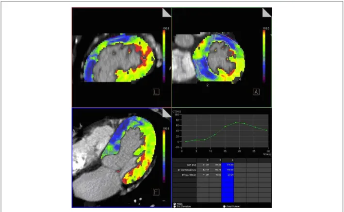
At the MP assessment using dipyridamole, an infero-septal and antero-lateral MP deficit was demonstrated. Quantitatively, the area with no perfusion deficit (normal area) had a myocardial flow of 119 mL/100 mL/min and total volume of 23 mL/100 mL (lateral wall), whereas the areas with ischemia had a flow of 52 mL/100 mL/min and volume of 11 mL/100 mL (infero-septal wall) and 50 mL/100 mL/min and volume of 10 mL/100 mL (antero-lateral wall) (Figure 1) (Chart 1).
The angiotomography showed arterial ectasia throughout their extension, suggesting a possible arteritis. The DA artery showed ectasia, diffuse calcifications and severe lesion in its mid-third after the stent (Figure 2A). The Cx artery showed calcified plaques, several ectatic areas and stent in its mid-third. The third marginal branch showed a moderate/significant lesion in its mid-third. The stent was not adhered to the arterial wall and was located in the dilated area (Figure 2B). The right coronary artery (RCA) showed multiple calcified plaques and severe decrease of luminal area in its mid-third (figure 2C). The angiotomography findings were similar to those found in the coronary angiography, which was used as reference.
Figure 2 - (A) Circumlex artery with stent in its mid-third. (B) Anterior descending artery with stent in its mid-third. (C) Right coronary artery (severe lesion in its
mid-third). LCT - left coronary trunk; ADA - anterior descending artery; Dg - diagonal branch; Cx - circumlex artery; Mg - marginal branch; RCA - right coronary artery.
Chart 1 - HU values according to time (in seconds) in the normal myocardial segments and with perfusion deicit (infero-septal and antero-lateral segments).
Time
Infero-septal
Antero-lateral
acquire a single phase for perfusion assessment with anatomic information, either during isolated stress or associated with rest4. The radiation dose used in this report was 10 mSv, comparable to the studies obtained with other equipment for anatomic assessment. We emphasize the scant literature on the use of second-generation dual-source computed tomography equipment1,8. The evaluation of myocardial viability by tomography did not require additional contrast infusion, as well as a small increase in total radiation dose (0.8 mSv of LE). The clinical and electrocardiographic picture was acute myocardial infarction, with early rescue angioplasty, which eventually preserved a considerable myocardial area. It was not possible to demonstrate the eventual presence of subendocardial infarction by another method such as magnetic resonance, which would have given more precise information on myocardial fibrosis, not detected by tomography.
The dual-source computed tomography MDCT allowed us to assess MP with dipyridamole and we detected a clear decrease in myocardial flow in sites with severe stenosis,
when compared with normal areas. Additionally, the angiotomography demonstrated significant stenoses and the final stent position.
This report confirms the perspective of use of the MDCT as a diagnostic tool in patients undergoing investigation for CAD, allowing the assessment of the coronary anatomy, perfusion and myocardial viability.
Potential Conflict of Interest
No potential conflict of interest relevant to this article was reported.
Sources of Funding
There were no external funding sources for this study.
Study Association
This study is not associated with any post-graduation program.
Figure 3 - Image showing absence of myocardial late enhancement. LV - left ventricle; RV - right ventricle; S - interventricular septum.
References
1. Feuchtner G, Cury RC, Plass A, Marincek B, Alkadhi H, Leschka S. Case of the issue Dec 14 th 2009. [Cited in 2010 Dec 10]. Available from: http:// www.diagnosticimaging.com/display/article/113619/1498426
3. Nagao M, Matsuoka H, Kawakami H, Higashino H, Mochizuki T, Ohshita A, et al. Detection of myocardial ischemia using 64-Slice MDCT. Circ J. 2009; 73(5): 905-11.
4. Cury RC, Magalhães TA, Borges AC, Shiozaki AA, Lemos PA, Júnior JS, et al. Dipyridamol stress and rest myocardial perfusion by 64-detector row computed tomography in patients with suspected coronary artery disease. Am J Cardiol. 2010;106(3):310-5.
5. Nieman K, Shapiro MD, Ferencik M, Nomura CH, Abbara S, Hoffmann U, et al. Reperfused myocardial infarction: contrast-enhanced 64-Section CT in comparison to MR imaging. Radiology. 2008;247(1):49-56.
6. Schuleri KH, Centola M, George RT, Amado LC, Evers KS, Kitagawa K, et al. Characterization of peri-infarct zone heterogeneity by contrast-enhanced multidetector computed tomography: a comparison with magnetic resonance imaging. J Am Coll Cardiol. 2009;53(18):1699-707.
7. Bamberg F, Klotz E, Flohr T, Becker A, Becker CR, Schmidt B, et al. Dynamic myocardial stress perfusion imaging using fast dual-source CT with alternating table positions: initial experience. Eur Radiol. 2010;20(5):1168-73. 8. Weininger M, Schoepf UJ, Ramachandra A, Fink C, Rowe GW, Costello P, et al.