173
Bazzi Jr JL et al. Poland’s syndrome: radiologic findings
Radiol Bras. 2012 Mai/Jun;45(3):173–174
Poland’s syndrome: radiologic findings
*
Achados radiológicos na síndrome de Poland
João Lourenço Bazzi Junior1, Eduardo Simões da Matta2, Luciano De Bortoli3, Felipe Raasch De Bortoli4
Poland’s syndrome is a rare non-inherited congenital anomaly. The authors describe the classic radiologic findings of Poland’s syndrome by reporting the case of a male four-year old patient with asymmetry of hands and chest, illustrating the fundamental imaging criteria for a conclusive diagnosis.
Keywords: Poland’s syndrome; Chest asymmetry; Congenital malformation.
A síndrome de Poland é uma anomalia congênita rara não hereditária. Os autores descrevem os achados radiológicos clássicos da síndrome de Poland através de um relato de caso de um paciente masculino de quatro anos de idade com assimetria torácica e das mãos, ilustrando os critérios imaginológicos fundamentais para a conclusão diagnóstica.
Unitermos: Síndrome de Poland; Assimetria torácica; Malformação congênita.
Abstract
Resumo
* Study developed at Clínica Via Imagem, Xanxerê, SC, Brazil. 1. MD, Radiologist at Clínica Via Imagem, Xanxerê, SC, Brazil. 2. MD, Vascular Surgeon and Vascular Echographist at Pró Circulação – Clínica de Angiologia, Cirurgia Vascular e Ecografia Vascular, Xanxerê, SC, Brazil.
3. MD, Pediatrician at Materclínica Materno Infantil, Xanxerê, SC, Brazil.
4. Graduate Student of Medicine, Universidade Católica de Pelotas (UCPel), Pelotas, RS, Brazil.
Mailing Address: Dr. João Lourenço Bazzi Jr. Rua Celestino do Nascimento, 573. Xanxerê, SC, Brazil, 89820-000. E-mail: [email protected]
Received October 1st, 2011. Accepted after revision Febru-ary 9, 2012.
Bazzi Jr JL, da Matta ES, De Bortoli L, De Bortoli FR. Poland’s syndrome: radiologic findings. Radiol Bras. 2012 Mai/Jun;45(3):173– 174.
0100-3984 © Colégio Brasileiro de Radiologia e Diagnóstico por Imagem
CASE REPORT
with normal caliper as well as normal ar-terial and venous blood flow. Radiography of the hands for comparative analysis con-firmed subtle hypoplasia of the left hand bones, with no sign of syndactyly or other local skeletal deformity (Figure 4).
DISCUSSION
Poland’s syndrome is a rare congenital disorder fundamentally characterized by unilateral agenesis of the pectoral muscles associated with ipsilateral brachydactyly(1– 4). Individuals affected by this syndrome
may also present several other alterations, such as hypoplasia or agenesis of the nipple and breast parenchyma, hypoplasia or apla-sia of the costal arches, shortening of up-Clinical examination detected axillary
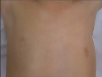
folds asymmetry, left nipple depression, volumetric reduction of the pectoral region and of the left hand as compared with the right side (Figure 1).
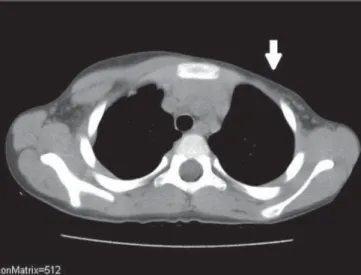
Non-contrast-enhanced helical com-puted tomography was performed with the objective of evaluating the soft tissues and bone structures, demonstrating pectoralis major and minor muscles at the left (Fig-ure 2). Vertebral bodies, costal arches, mediastinum and pulmonary parenchyma were found within normality parameters. A supplementary ultrasonography study (Fig-ure 3) was performed, demonstrating the same findings. Doppler ultrasonography of the subclavian and axillary vessels demon-strated symmetrical vascular structures, INTRODUCTION
Poland’s syndrome is characterized by pectoralis muscle agenesis associated with ipsilateral brachydactyly(1–4). Although
more rarely, other alterations may be asso-ciated with the syndrome whose early di-agnosis may contribute for the therapeutic management of the condition.
CASE REPORT
A male, four-year-old patient with a his-tory of no particular finding at prenatal follow-up and term birth with Apgar 9/10 was referred for study with chest computed tomography for deformity of the left ante-rior chest wall associated with asymmetry between his hands (smaller at left) detected at puericulture consultation. The patient did not present any motor, sensorial or neuro-logical deficits.
174
Bazzi Jr JL et al. Poland’s syndrome: radiologic findings
Radiol Bras. 2012 Mai/Jun;45(3):173–174 per limb due to hypoplasia of the radius and
ulna, syndactyly, among other less fre-quently described abnormalities, such as dextrocardia, single transverse palmar crease, scapular anomalies, vertebral anomalies, and anomalies of the urinary and digestive systems(3,5–9).
With a still unknown etiology, Poland’s syndrome affects a higher percentage of boys than girls, with the right side of the body being most frequently affected. In spite of the eminently clinical diagnosis, the radiological evaluation is justifiable for staging of the alterations found in addition to the classical findings, thus directing the
treatment. The early diagnosis may contrib-ute to pediatric, orthopedic, psychomotor and aesthetic management (the latter, with greater impact on women with amastia), as well as to the awareness of parents and fam-ily members, and to the social and school life of the child(1,2,4,6).
REFERENCES
1. Araújo ALN, França JCQ, Vieira SC, et al. Sín-drome de Poland. Brasília Med. 2009;46:395–8. 2. Caldas FAA, Isa HLVR, Trippia AC, et al. Sín-drome de Poland: relato de caso e revisão da lite-ratura. Radiol Bras. 2004;37:381–8.
3. Dilmann JR, Sanchez R, Ladino-Torres MF, et al. Expanding upon the unilateral hyperlucent hemi-thorax in children. Radiographics. 2011;31:723–41.
4. Pearl M, Chow TF, Friedman E. Poland’s syn-drome. Radiology. 1971;101:619–23.
5. Chung EM, Cube R, Hall GJ, et al. From the ar-chives of the AFIP: breast masses in children and adolescents: radiologic-pathologic correlation. Radiographics. 2009;29:907–31.
6. Freitas RS, Tollazi ARD, Martins VDM, et al. Poland’s syndrome: different clinical presentations and surgical reconstructions in 18 cases. Aesthetic Plast Surg. 2007;31:140–6.
7. Friedman T, Reed M, Elliott AM. The carpal bones in Poland syndrome. Skeletal Radiol. 2009;38: 585–91.
8. Jeung MY, Gangi A, Gasser B, et al. Imaging of chest wall disorders. Radiographics. 1999;19:617–37. 9. Mutlu H, Sildiroglu O, Basekim CC, et al. A
vari-ant of Poland syndrome associated with dextropo-sition. J Thorac Imaging. 2007;22:341–2.
Figure 2. Computed tomography. Axial section and soft-tissue window show-ing agenesis of the left pectoral muscle (arrow). Compare with the right chest wall, where the major and minor pectoral muscles present a normal tomo-graphic appearance.
Figure 3. Ultrasonography showing agenesis of the left pectoral muscle as compared with the normal right side.