http://doi.org/10.1590/0004-282X20170153
ARTICLE
Phrenic nerve conduction studies: normative
data and technical aspects
Aspectos técnicos e normatização da neurocondução do nervo frênico
Analucia Abreu Maranhão1, Sonia Regina da Silva Carvalho1, Marcelo Ribeiro Caetano2,
Alexandre Hofke Alamy3, Eduardo Mesquita Peixoto4, Pedro Del Esporte Peçanha Filgueiras 5
1Universidade Federal do Estado do Rio de Janeiro, Faculdade de Medicina, Departamento de Pneumologia, Rio de Janeiro RJ, Brasil; 2Universidade Federal do Estado do Rio de Janeiro, Faculdade de Medicina, Departamento de Neurocirurgia, Rio de Janeiro RJ, Brasil; 3Private Practice, Rio de Janeiro, Rio de Janeiro, Brazil;
4Universidade Federal do Estado do Rio de Janeiro, Faculdade de Enfermagem, Rio de Janeiro RJ, Brasil; 5Universidade Federal do Estado de Rio de Janeiro, Faculdade de Medicina, Rio de Janeiro RJ, Brasil.
Correspondence: Analucia A Maranhão; Rua Maris e Barros, 775; 20270-001 Rio de Janeiro RJ, Brasil; E-mail: [email protected]
Conflict of interest: There is no conflict of interest to declare.
Received 05 June 2017; Received in final form 26 July 2017; Accepted 21 August 2017. ABSTRACT
Objective: The aim of the present study was to define normative data of phrenic nerve conduction parameters of a healthy population. Methods: Phrenic nerve conduction studies were performed in 27 healthy volunteers. Results: The normative limits for expiratory phrenic nerve compound muscle action potential were: amplitude (0.47 mv - 0.83 mv), latency (5.74 ms - 7.10 ms), area (6.20 ms/mv - 7.20 ms/mv) and duration (18.30 ms - 20.96 ms). Inspiratory normative limits were: amplitude (0.67 mv - 1.11 mv), latency (5.90 ms - 6.34 ms), area (5.62 ms/mv - 6.72 ms/mv) and duration (13.77 ms - 15.37 ms). Conclusion: The best point of phrenic nerve stimulus in the neck varies among individuals between the medial and lateral border of the clavicular head of the sternocleidomastoid muscle and stimulation of both sites, then choosing the best phrenic nerve response, seems to be the appropriate procedure.
Keywords: electrodiagnosis; reference values; phrenic nerve; spirometry; neural conduction. RESUMO
Objetivo: O objetivo do presente estudo foi definir os dados normativos de condução do nervo frênico de uma população saudável. Métodos: Foram realizados estudos de condução do nervo frênico em 27 voluntários saudáveis. Resultados: Os limites normais do potencial de ação muscular composto do nervo frênico durante a expiração foram: amplitude (0.47 mv - 0.83 mv), latência (5.74 ms - 7.10 ms), área (6.20 ms/mv - 7.20 ms/mv) e duração (18.30 ms - 20.96 ms). E durante a inspiração os limites normais foram: amplitude (0.67 mv - 1.11 mv), latência (5.90 ms - 6.34 ms), área (5.62 ms/mv - 6.72 ms/mv) e duração (13.77 ms - 15.37 ms). Conclusão: O melhor ponto de estímulo do nervo frênico no pescoço varia entre a borda medial e lateral da cabeça clavicular do músculo esternocleidomastóideo. Estimular ambos os locais e escolher a melhor resposta do nervo frênico parece ser o procedimento mais adequado.
Palavras-chave: eletrodiagnóstico; valores de referência; nervo frênico; espirometria; condução nervosa.
Phrenic nerve conduction has found increasing applica-tion in the diagnosis of respiratory dysfuncapplica-tion associated with surgical, neuromuscular, and pulmonary disorders1,2,3,4,5,6,
which are important causes of respiratory failure and
fre-quently contribute to diiculties in weaning patients of the
ventilator in the critical care unit7. To determine a
neuromus-cular cause of hypercapnic respiratory failure, respiratory electrodiagnostic studies are often used8.
Recently, many authors have correlated phrenic nerve con-duction abnormalities with chronic obstructive pulmonary
disease. hese studies demonstrated abnormal phrenic com -pound motor action potential (CMAP) amplitudes and laten-cies in chronic obstructive pulmonary disease patients9,10,11,12.
Innervated by the phrenic nerve, the diaphragm is the
principal respiratory muscle in humans. he diaphragmatic
CMAPs are recorded with chest surface electrodes following phrenic nerve stimulation in the neck. Amplitude, latency, and area are measures used to evaluate phrenic nerve integrity1,2,3,4,5,6,7,13.
he amplitude measure of the CMAP is commonly used to deine neuropathy of the phrenic nerve and is an impor -tant parameter in patient selection for pacing the diaphragm. However, the range of the amplitude among healthy
here are several approaches to stimulate the phrenic nerve
in the neck, but a stimulation in the supraclavicular fossa, just above the clavicles, is considered to obtain the best results14. herefore, it remained to be determined if the best point to
stimulate is between the two heads of the sternocleidomastoid muscle or lateral to the clavicular head of this muscle.
he aims of the present study were to deine norma -tive data of phrenic nerve neuroconduction parameters of a Brazilian healthy population, and discuss some of the techni-cal aspects of the procedure.
METHODS
he study group consisted of 27 volunteers (15 men),
21–62 years old (median, 30 years), with no respiratory or neu-romuscular disorders, all of whom had normal spirometry tests
and chest X-rays. he participants’ data were as follows: height,
155–186 cm (mean, 171 cm); and weight, 52–100/ kg (mean,
73 kg). he study recruited students and employees (with diferent degrees of physical activity) from the university hospital. he study was approved by the Gafrée Guinle University Hospital Ethics
Committee, and all participants provided informed consent.
he spirometry test procedure used the forced vital capacity
(FVC) technique in which the participant performs a full inspi-ration and then a forceful expiinspi-ration, as rapidly and completely
as possible. Each participant performed, in the sitting position,
at least three trials and the best performance was used for anal-ysis. An adequate test required a minimum of three acceptable
FVC maneuvers. he test was considered acceptable when the diference between the largest and the next largest FVC and the irst second of forced expiratory volume (FEV1)was 0.150 L or
less15. he prediction equations of Knudson16 were used for the
Time-Volume and Flow-Volume curves. Parameters analyzed
were: FVC, FEV1 and the FEV1/FVC ratio. A Spiron (Physis, Rio
de Janeiro, Brazil) spirometer was used.
Postero-anterior and lateral chest X-ray ilms were obtained at maximal inspiration. he radiographs were
acquired by a trained radiographer and were read by the chest physician.
Phrenic neuroconduction was performed with partici-pants lying in a supine position, with a bipolar stimulating electrode (Neurosoft, Ivanovo, Russia) between the sternal and clavicular heads of the sternocleidomastoid muscle, just above the clavicle as described by Resman-Gaspersc and Podnar14; however, in almost one third of participants, we had
to stimulate lateral to the clavicular head, as described by Chen et al.7, to get a better CMAP. We used two disposable
self-adhesive disk recording electrodes (Viasys Healthcare,
Madison, Wisconsin). he active electrode (G1) was ixed
5 cm above the xiphoid process, and the reference electrode (G2) 16 cm from G1, on the chest margin ipsilateral to the stimulated phrenic nerve. An electromyography system
(NEURO-Mep-Micro, Neurosoft, Ivanovo, Russia) with stan
-dard settings ( ilters: 2 Hz to 10 kHz) was used. he gain was
set to 0.5 mv and the sweep speed to 2 ms/division. Bilateral
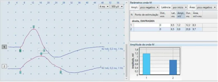
studies were performed on all participants. Electrical stimu -lation was carried out with rectangular pulses of 0.1 ms or 0.2 ms duration. Measurements were made separately during normal inspiration and expiration (Figure 1). Phrenic nerve CMAP onset latency (ms), amplitude (mV), duration (ms), and area of the negative phase were obtained at supramaxi-mal stimulation (10%–20% above maxisupramaxi-mal stimulation).
he measurements from 27 healthy nonsmoking volun
-teers were organized and analyzed in a Microsoft Excel spread
-sheet. he analysis was done by R, freeware statistical analytics
software. All participants had body mass index values under
normality limits. he distribution of the numeric variables was tested using the Shapiro-Wilk normality test at 5% signiicance levels. he variables: age, FEV1% and FVC% did not have
nor-mal distribution, so they were analyzed using a nonparametric approach. All other variables, considered normally distributed
Inspiratory CMAP is sharper and higher–reduced duration and increased amplitude comparing to expiratory CMAP (compound muscle action potential).
by the test, were analyzed with a parametric approach at 95%
conidence intervals. No signiicance was found between the
left and right side regarding the measurements made
relat-ing to the phrenic nerve, usrelat-ing the paired t-test. herefore, the
average values obtained from both sides were calculated for each participant, and common normative data were obtained.
Categorization for inferential analysis: Gender: male and female; Height: 1.55-1.72m, 1.73-1.86m; Weight: 52-75.99kg, 76-100kg; Age: under 30 years, 30 years or more.
RESULTS
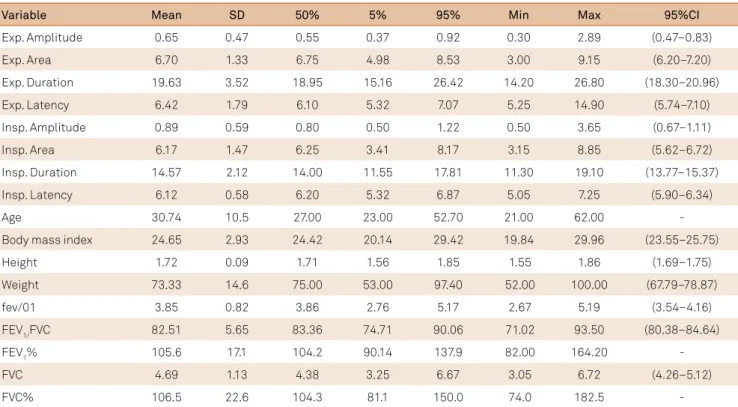
he summary of neuroconduction parameters, the pro
-ile of the 27 participants studied and the spirometric param
-eters are shown in Table 1. he CMAP amplitudes, latencies, duration, and areas, as FEV1, FVC and VEF1/CVF had normal limits deined according to the conidence interval at 95%. Expiratory CMAP normative limits were: amplitude (0.47
mv–0.83 mv), latency (5.74 ms–7.10 ms), area (6.20 ms/mv–7.20 ms/mv) and duration (18.30 ms–20.96 ms). Inspiratory CMAP
normative limits were: amplitude (0.67 mv–1.11 mv), latency
(5.90 ms–6.34 ms), area (5.62 ms/mv–6.72 ms/mv) and dura-tion (13.77 ms–15.37 ms). Lower and upper limits of
spiromet-ric parameters: FEV1 (3.54 L–4.16 L), FVC (4.26 L–5.12 L) and FEV1/FVC (80.38–84.64). Age did not have a normal
distribu-tion and its median was 27 years, ranging from 21–62 years.
he FEV1% and FVC% did not have normal distributions either
and showed medians equal to 104.20, ranging between 82.00 and 164.20, and equal to 104.3, ranging between 74.00 and 182.50, respectively.
he Pearson correlation is shown in Table 2 and strong correlations among the variables: FEV1, FVC, height,
inspira-tory latency and weight are illustrated in Figure 2.
Table 1. Numerical summaries of phrenic nerve neuroconduction, spirometric and general parameters.
Variable Mean SD 50% 5% 95% Min Max 95%CI
Exp. Amplitude 0.65 0.47 0.55 0.37 0.92 0.30 2.89 (0.47–0.83)
Exp. Area 6.70 1.33 6.75 4.98 8.53 3.00 9.15 (6.20–7.20)
Exp. Duration 19.63 3.52 18.95 15.16 26.42 14.20 26.80 (18.30–20.96)
Exp. Latency 6.42 1.79 6.10 5.32 7.07 5.25 14.90 (5.74–7.10)
Insp. Amplitude 0.89 0.59 0.80 0.50 1.22 0.50 3.65 (0.67–1.11)
Insp. Area 6.17 1.47 6.25 3.41 8.17 3.15 8.85 (5.62–6.72)
Insp. Duration 14.57 2.12 14.00 11.55 17.81 11.30 19.10 (13.77–15.37)
Insp. Latency 6.12 0.58 6.20 5.32 6.87 5.05 7.25 (5.90–6.34)
Age 30.74 10.5 27.00 23.00 52.70 21.00 62.00
-Body mass index 24.65 2.93 24.42 20.14 29.42 19.84 29.96 (23.55–25.75)
Height 1.72 0.09 1.71 1.56 1.85 1.55 1.86 (1.69–1.75)
Weight 73.33 14.6 75.00 53.00 97.40 52.00 100.00 (67.79–78.87)
fev/01 3.85 0.82 3.86 2.76 5.17 2.67 5.19 (3.54–4.16)
FEV1/FVC 82.51 5.65 83.36 74.71 90.06 71.02 93.50 (80.38–84.64)
FEV1% 105.6 17.1 104.2 90.14 137.9 82.00 164.20
-FVC 4.69 1.13 4.38 3.25 6.67 3.05 6.72 (4.26–5.12)
FVC% 106.5 22.6 104.3 81.1 150.0 74.0 182.5
-The summary of neuroconduction parameters, the profile of the 27 participants studied and the spirometric parameters are presented here. We also present general information such as age, height, weight and body mass index. The confidence interval was supressed for the variables considered as not normal distribution. Those using 5% and 95% measures are more representative. Exp: expiration; Insp: inspiration; FEV1: Forced expiratory volume in one second, FVC:
forced vital capacity; Min: minimum; Max: maximum.
Table 2. Pearson correlation matrix represented as percentage for normal distribution variables: Height, spirometric
parameters and neuroconduction parameters.
Variable BMI fev/01 FVC Height Weight
Expiratory amplitude 30.07% 35.79% 39.42% 22.42% 29.04%
Expiratory area -21.52% -1.49% -2.00% 0.02% -15.36%
Expiratory duration -37.50% -14.94% -19.02% -25.72% -34.27%
Expiratory latency 43.40% 56.47% 58.98% 42.56% 47.83%
fev/01 55.05% 100% 95.80% 85.20% 76.00%
FVC 54.08% 95.80% 100% 81.94% 73.68%
Height 65.26% 85.20% 81.94% 100% 88.24%
Inspiratory amplitude 28.47% 34.65% 38.42% 23.40% 27.97%
Inspiratory area -10.73% 6.62% 8.15% 13.37% -3.11%
Inspiratory duration -19.69% -4.94% -2.86% -13.28% -16.13% Inspiratory latency 50.88% 77.66% 76.00% 79.88% 69.49% The values in bold are considered strong positive correlations. No strong correlations were found using the Spearman’s method relating to age, FVC% or FEV1%. Correlation between spirometric parameters and inspiratory
latency is shown. There was also strong correlation of both spirometric and neuroelectrical parameters with height. BMI: body mass index; FEV1: forced
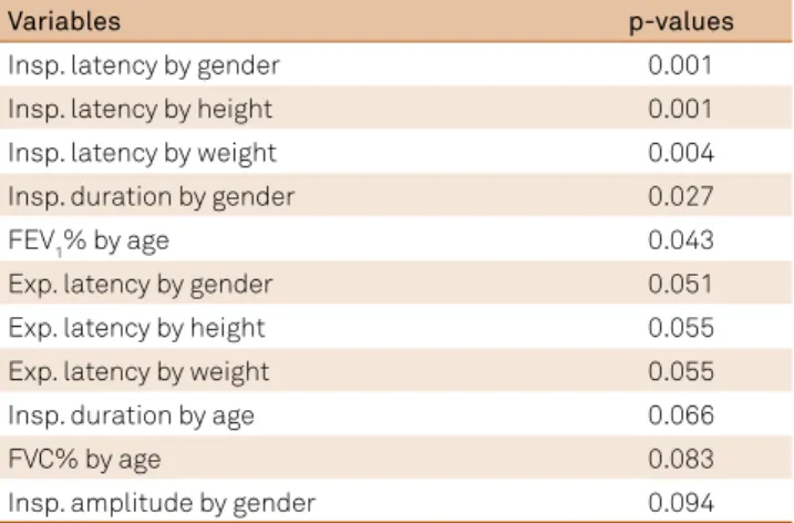
Inferential analysis by categorization of gender, height,
weight and age, disclosed statically signiicant diferences between eight variables and borderline diferences in six,
by using the t-test for parametric variables and Wilcoxon test
for nonparametric ones. he signiicant results can be seen in
Table 3 and the categories in the study of gender and height are further analyzed below.
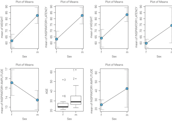
Gender Analysis (Figure 3):
1) Weight–Women were 20.85% (mean) lighter than men; 2) Inspiratory latency–Men had results 11.67% (mean) higher than women;
3) Height–Men were 6.6% (mean) taller than women; 4) Expiratory latency–Men were 18.02% (mean) higher
than women;
5) Inspiratory duration–Women were 13.23% (mean) lon-ger than men;
6) Age– Men were 17.24% (median) older than women; 7) Inspiratory amplitude– Men were 34.07% (mean) higher than women.
Height Analysis (Figure 4):
a) Inspiratory latency: was 10.49% (mean) higher in the taller group;
b) Expiratory latency: was 19.67% (mean) higher in the taller group.
DISCUSSION
Although the technique described by Resman-Gaspersc and Podnar14 was the best approach in the majority of the
participants, in one third of them, stimulating the lateral
5.0 3
6
5
4
3
5.
0
3.0 3.5 4.0 4.5 5.0 1.85 60 70 80 90 100
100
1.85
5.
0
4.
0
3.
0
1.85
1.65
1.
75
80
60
1.55 1.65 1.85
6.
07
.0
4 5 6 5.0 5.5 6.0 6.5 7.0
3
6
5
4
3
5
.0
3.0 3.5 4.0 4.5 5.0 1.85 60 70 80 90 100
1
00
1.
85
5
.0
4.
0
3
.0
1.
85
1.
6
5
1.
7
5
80
60
1.55 1.65 1.85
6
.0
7.
0
4 5 6 5.5 6.0 6.5 7.0
F.E.V1
FVC
HEIGHT
PIRATORY.LATEM
WEIGHT
The strong correlations found in Table 2 among the variables: FEV, FVC, height, inspiratory latency and weight are illustrated here. As the correlation increases, the dispersion graph tends to look more like a linear function. FEV1: forced expiratory volume in one second, FVC: forced vital capacity.
Figure 2. Dispersion diagram for strongly correlated variables.
Table 3. Inferential analysis: Hypothesis tests by categorization of gender, height, weight and age.
Variables p-values
Insp. latency by gender 0.001
Insp. latency by height 0.001
Insp. latency by weight 0.004
Insp. duration by gender 0.027
FEV1% by age 0.043
Exp. latency by gender 0.051
Exp. latency by height 0.055
Exp. latency by weight 0.055
Insp. duration by age 0.066
FVC% by age 0.083
Insp. amplitude by gender 0.094
The values in bold were obtained by the Wilcoxon test, while the other values were obtained by the t-test. The most significant relationships were related to inspiratory latency. The significance level adopted was 5% and the interval 5%-10% was considered borderline. The table only shows p-values < 0.10, the other variables were tested but they were not significant enough. Exp: expiration; Insp: inspiration; FEV1: forced expiratory volume in one second,
side of the clavicular head of the sternocleidomastoid mus-cle, as reported by Chen et al.7, showed reproducible CMAPs
with higher amplitudes and reliable morphology. We could avoid inadvertent brachial plexus stimulation (detected by
arm movement) by positioning the stimulator irmly in a
more medial direction, which produces a “hiccup” sensation.
We also had greater diiculty in stimulating the left side in
some participants, as described by Resman-Gaspersc and Podnar14, probably due to anatomic diferences.
he mean latency (inspiration 6.42 ms, expiration
6.12 ms) obtained was very close to that reported by Chen et al.7 (6.54 ms), Resman-Gaspersc and Podnar14
(inspira-tion 6.55 ms, expira(inspira-tion 6.59 ms) and that of Swenson and Rubenstein1 (right 6.28 ms, left 6.30 ms). he other reports by The mean of the variables with significant differences by gender are shown with a confidence interval at 95% for variables with normal distribution and boxplots for variables without normal distribution.
Figure 3. Graphic representation of the significant differences found by gender: general and neurophysiologic parameters. Plot of Means
Sex Sex Sex
Sex Sex
AG
E
mean of INSPIRA
TO
RY
AMPLITUDE
Sex
f m
Sex
Plot of Means Plot of Means
Plot of Means Plot of Means
Plot of Means
3
6 2 1
60
50
40
30
20
60
50
40
30
20
mean of INSPIRA
TO
RY
AMPLITUDE
17
16
15
14
13
mean of WEIGHT
90
85
80
75
70
65
60
mean of INSPIRA
TO
RY
LA
TENC
Y 90
85
80
75
70
65
60
mean of WEIGHT
90
85
80
75
70
65
60
mean of INSPIRA
TO
RY
LA
TENC
Y 90
85
80
75
70
65
60
f m
f m
f m f m f m f m
The mean of the variables with significant differences by height are showed with a confidence interval at 95% for variables with normal distribution and boxplots for variables without normal distribution. Lower height tends to result in lower latencies measurement both inspiratory and expiratory.
Figure 4. Graphic representation of the significant differences found by height categories: neurophysiologic parameters. Plot of Means
Plot of Means
heightcat heightcat
mean of INSPIRA
TO
RY
LA
TENC
Y
mean of INSPIRA
TO
RY
LA
TENC
Y
6.8
6.2
5.6
[1.55,1.71] (1.71,1.86]
8.5
7.
0
5.5
Newsom Davis2, MacLean and Mattioni3, Markland et al.4 and
Mckenzie and Gandevia5 had a diferent stimulation site, at the
level of the thyroid cartilage, which may explain the higher latencies (7.70 ms, 7.44 ms, 7.77 ms, and 7.68 ms, respectively).
he average amplitude we obtained (inspiration 0.78 mv,
expiration 0.57 mv) was close to that obtained by Chen et al.7
(0.66 mv) and Markland et al.4 (right 0.79 mv, left 0.77 mv); lower
than that obtained by Resman-Gaspersc and Podnar14
(inspira-tion 1.0 mv, expira(inspira-tion 0.71 mv); and higher than Swenson and Rubenstein1 (0.35 mv). he wide range of the phrenic nerve
amplitude creates a great problem in determining a lower nor-mal limit. Swenson and Rubenstein1 found 0.10 mv, Chen et al.7
0.30 mv, Johnson et al.13 0.12 mv and our data analyses showed 0.50 mv (inspiration) and 0.30 mv (expiration). he mean right
and the mean left CMAP amplitudes were nearly identical, but there was a lack of consistent right-to-left correlation, very simi-lar to that found by Swenson and Rubenstein1.
We found a substantial diference between genders and phrenic nerve parameters. We had signiicant diferences in
amplitude (p = 0.001), duration (p = 0.002), expiratory latency
(p = 0.005) and inspiratory amplitude (p= 0.094). his has
only previously been mentioned by Resman-Gaspersc and Podnar14, who found a signiicant diference only in relation
to amplitude (p = 0.03). It is not clear if the anthropometric
variance between genders found in our study was the
deter-minant for these substantial diferences.
he correlation between phrenic nerve parameters and spiro
-metric measures has not been reported previously. he FEV1 and
FVC showed strong correlation with phrenic nerve inspiratory
latencies. El-Tantawi et al.10 did not ind signiicant correlation
between spirometric parameters and phrenic nerve conduction in chronic obstructive pulmonary disease patients but they did not compare them with the data from normal individuals.
Height was strongly correlated with inspiratory latencies, as described by Resman-Gaspersc and Podnar14, and with FEV1 and FVC, as described by Knudson et al.
16.
Further studies with a larger number of individuals will be needed to better understand the relationship between these spirometric parameters and inspiratory phrenic nerve CMAP latencies.
In conclusion, the normative data obtained in our partici-pants were very similar to those available in recent articles using the same technique. In relation to the precise point of phrenic nerve stimulation in the neck, we propose that the best approach is to try both techniques, stimulating at the lateral and the medial border of the clavicular head of the sternocleidomastoid muscle in all patients and choosing the best CMAP response.
References
1. Swenson MR, Rubenstein RS. Phrenic nerve conduction studies. Muscle Nerve. 1992;15(5):597-603. https://doi.org/10.1002/mus.880150511
2. Newsom Davis J. Phrenic nerve conduction in man. J Neurol Neurosurg Psychiatry. 1967;30(5):420-6. https://doi.org/10.1136/jnnp.30.5.420
3. MacLean IC, Mattioni TA. Phrenic nerve conduction studies: a new technique and its application in quadriplegic patients. Arch Phys Med Rehabil. 1981;62(2):70-3.
4. Markand ON, Kincaid JC, Pourmand RA, Moorthy SS, King RD, Mahomed Y et al. Electrophysiologic evaluation of diaphragm by transcutaneous phrenic nerve stimulation. Neurology. 1984;34(5):604-14. https://doi.org/10.1212/WNL.34.5.604
5. McKenzie DK, Gandevia SC. Phrenic nerve conduction times and twitch pressures of the human diaphragm. J Appl Physiol. 1985;58(5):1496-504.
6. Mier A, Brophy C, Moxham J, Green M. Phrenic nerve stimulation in normal subjects and in patients with diaphragmatic weakness. Thorax. 1987;42(11):885-8. https://doi.org/10.1136/thx.42.11.885
7. Chen R, Collins S, Remtulla H, Parkes A, Bolton CF. Phrenic nerve conduction study in normal subjects. Muscle Nerve.
1995;18(3):330-5. https://doi.org/10.1002/mus.880180311
8. Bolton CF, Chen R, Wijdicks EFM, Zifko UA. Neurology of breathing. Amsterdam: Elsevier; 2004.
9. Podnar S, Harlander M. Phrenic nerve conduction studies in patients with chronic obstructive pulmonary disease. Muscle Nerve. 2013;47(4):504-9. https://doi.org/10.1002/mus.23617
10. El-Tantawi GAY, Imam MH, Morsi TS. Phrenic nerve conduction abnormalities correlate with diaphragmatic descent in chronic obstructive pulmonary disease, COPD:.Journal of Chronic Obstructive Pulmonary Disease. 2015;12(5):516-24. https://doi.org/10.3109/15412555.2014.993465
11. Lu Z, Tang X, Huang X. Phrenic nerve conduction and diaphragmatic motor evoked potentials: evaluation of respiratory dysfunction. Chin Med J. 1998;111(6):496-9.
12. Hopkinson NS, Sharshar T, Ross ET, Nickol AH, Dayer MJ, Porcher R et al. Corticospinal control of respiratory muscles in chronic obstructive pulmonary disease. Respir Physiol Neurobiol. 2004;141(1):1-12. https://doi.org/10.1016/j.resp.2004.04.003
13. Johnson NE, Utz M, Patrick E, Rheinwald N, Downs M, Dilek N et al. Visualization oh the diaphragm muscle with ultrasound improves diagnostic accuracy of phrenic nerve conduction studies. Muscle Nerve. 2014;49(5):669-75. https://doi.org/10.1002/mus.24059
14. Resman-Gaspersc A, Podnar S. Phrenic nerve conductions Studies: technical aspects and normative data. Muscle Nerve.
2008;37(1):36-41. https://doi.org/10.1002/mus.20887
15. Miller MR, Hankinson J, Brusasco V, Burgos F, Casaburi R, Cates A et al. Standardization of spirometry. Eur Respir J. 2005;26(2):319-38. https://doi.org/10.1183/09031936.05.00034805