INTRODUCTION
Hemodialysis (HD) is the main treatment for end-stage renal di-sease (ESRD). However, acute and chronic complications can occur after HD, the most common of which are hypovolemia and sudden changes in hemodynamic parameters, which may cause organ
dys-functions(1). Fluid dynamic changes after HD can also affect ocular
tissues that receive a high volume of blood flow. There are many ocular disorders associated with HD, including refractive changes, dry
ABSTRACT
Purpose: To evaluate the effects of hemodialysis (HD) on corneal and anterior chamber morphometry, as well as intraocular pressure (IOP) in patients with end-stage renal disease.
Methods: Fifty right eyes were examined 30 minutes before and after HD. IOP was measured with a Goldmann applanation tonometer, and Ehlers’ formula was used to calculate the corrected IOP values. The central corneal thickness (CCT), corneal volume (CV), keratometric values, anterior chamber depth (ACD), aqueous depth (AQD), anterior chamber volume (ACV), and anterior chamber angle (ACA) in the nasal and temporal quadrants were measured with a Sirius anterior segment analysis system. Blood urea nitrogen levels, body mass, and systolic and diastolic arterial pressure were also measured before and after HD.
Results: The mean age was 60.80 ± 13.38 (range: 35-80) years. The mean uncorrected and corrected IOP values decreased from 18.06 ± 3.91 and 18.31 ± 4.83 mmHg to 16.94 ± 3.87 and 16.95 ± 4.74 mmHg after HD, respectively (p=0.011 and p=0.003, respectively). The mean CCT decreased from 536.38 ± 24.73 to 533.18 ± 27.25 µm (p=0.002), and the mean CV decreased from 57.52 ± 3.15 to 55.68 ± 3.55 mm³ (p<0.001) after HD. There were no significant changes in ACD, AQD, ACV, ACA, or the keratometric values (p>0.05 for all values). There were no significant correlations between the ocular and systemic parameters (p>0.05 for all correlations).
Conclusions: Uncorrected IOP, corrected IOP, CCT, and CV values decreased after HD, whereas the anterior chamber morphometry values remained similar between the measurements performed before and after HD.
Keywords: Renal dialysis; Cornea; Anterior chamber; Intraocular pressure; Kidney failure, chronic; Vision disorders
RESUMO
Objetivo: Avaliar os efeitos da hemodiálise (HD) na morfometria da córnea e da câmara anterior e da pressão intraocular (PIO) em pacientes com doença renal terminal.
Métodos: Cinquenta olhos direitos foram examinados 30 minutos antes e após hemodiálise. A pressão intraocular foi medida com um tonômetro de aplanação de Goldmann, e a fórmula de Ehlers foi utilizada para calcular os valores de pressão in traocular corrigidos. Mediram-se a espessura corneana central (CCT), o volume corneano (CV), os valores ceratométricos, a profundidade da câmara anterior (ACD), a profundidade aquosa (AQD), o volume da câmara anterior (ACV) e o ângulo da câmara anterior (ACA) nos quadrantes nasais e temporais com um sistema de análise de segmento Sirius anterior. Os níveis de nitrogênio ureico no sangue (BUN), peso corporal e pressão arterial sistólica e diastólica também foram medidos antes e após a HD.
Resultados: A média de idade foi de 60,80 ± 13,38 (35-80) anos. Os valores médios não corrigidos e corrigidos da pressão intraocular diminuíram de 18,06 ± 3,91 e 18,31 ± 4,83 mmHg para 16,94 ± 3,87 e 16,95 ± 4,74 mmHg após hemodiálise (p=0,011 e p=0,003, respectivamente). A espessura corneana central média diminuiu de 536,38 ± 24,73 para 533,18 ± 27,25 μm (p=0,002), e o volume corneano médio diminuiu de 57,52 ± 3,15 para 55,68 ± 3,55 mm³ (p<0,001) após hemodiálise. Não houve alteração significativa nos valores de profundidade da câmara anterior, profundidade aquosa, volume da câmara anterior, ângulo da câmara anterior e ceratométricos (p>0,05 para todos os valores). Não houve correlação significativa entre os parâmetros oculares e sistêmicos (p>0,05 para todas as correlações).
Conclusão: A pressão intraocular não corrigida, a pressão intraocular corrigida, a espessura corneana central e os valores de volume corneano diminuíram após hemo-diálise, enquanto os valores de morfometria da câmara anterior foram semelhantes entre as medidas realizadas antes e após a hemodiálise.
Descritores: Diálise renal; Córnea; Câmara anterior; Pressão intraocular; Falênca renal crônica; Transtornos da visão
eye, corneal and conjunctival epithelial erosions, perilimbal calcium deposits, band keratopathy, intraocular pressure (IOP) fluctuations, posterior subcapsular cataract, ischemic optic neuropathy, choroidal perfusion delay, corneal endothelium alterations, and thickness chan-ges in the central cornea, retinal nerve fiber layer, and choroid(2-15).
Among the various studies investigating the relationships between ocular alterations and HD, IOP is the most commonly
investi-gated parameter, and conflicting results have been reported(16). This
Effects of hemodialysis on corneal and anterior chamber morphometry and
intraocular pressure in patients with end-stage renal disease
Efeitos da hemodiálise na morfometria da córnea e da câmara anterior, e na pressão intraocular
em pacientes no estágio inal da doença renal
Mehtap Caglayan1, pinar KoseKahya2, taMer taKMaz1, alpaslan altunoglu3, Berna ayan1,CeMile uCgul atilgan2, Betul seher uysal1
Submitted for publication: August 23, 2016 Accepted for publication: January 16, 2017
1 Department of Ophthalmology, Ataturk Research and Training Hospital, Ankara, Turkey. 2 Ulucanlar Eye Research and Training Hospital, Ankara, Turkey.
3 Department of Nephrology, Ataturk Research and Training Hospital, Ankara, Turkey.
Funding: No specific financial support was available for this study.
Disclosure of potential conflicts of interest: None of the authors have any potential conflict of interest to disclose.
Corresponding author: Mehtap Caglayan. Department of Ophthalmology. Ataturk Research and Training Hospital - Ankara, Turkey - E-mail: [email protected]
conflicting data has been explained as being due to different sample sizes, HD type and duration, fluid dynamic changes, arterial blood pressure changes, IOP measurement times, and IOP measurement
techniques(5,16). Although the Goldmann applanation tonometer is
the gold standard in the measurement of IOP, it can be affected by central corneal thickness (CCT)(17). Jung et al.(5) reported that IOP and
CCT both tend to decrease after HD, and they also suggested that the decrease in IOP is caused by the decrease in CCT.
Apart from IOP, important ocular problems for HD patients inclu-de changes in ocular perfusion and the iridocorneal angle. Several epidemiological studies have revealed a strong correlation between glaucoma damage and low diastolic arterial pressure, which results
in increased inadequate ocular perfusion pressure (OPP)(18,19). HD can
change vascular resistance because of the rheological properties of the arteries, so this can change arterial pressure. Hu et al.(20) evaluated
IOP and OPP during HD, and they reported increased IOP and
decrea-sed OPP during HD. However, Barbosa et al.(21) did not find significant
changes in IOP or OPP during HD. They emphasized that some pa -tients exhibit lower diastolic perfusion pressures, which could be a poor prognostic factor for patients with glaucoma. Moreover, in eyes with glaucoma, impaired aqueous outflow, or those that are predis-posed to narrow angles, the possibility of acute IOP rise during HD could be much more frequent and or higher than in normal
sub-jects(22,23). Anterior chamber depth (ACD) and axial length alterations
after HD have also been reported; however, ACD was defined as the diameter between the corneal epithelium and the lens as measured
by contact methods in these studies(24,25). Since CCT decreases after
HD, it may be more reliable to evaluate the before and after HD values of ACD and IOP by eradicating CCT.
The Sirius anterior segment analysis system (Costruzione Strumenti Oftalmici, Florence, Italy) is a non-contact anterior segment tomogra-phy device. It provides anterior and posterior corneal topogratomogra-phy, wavefront analysis, complete corneal pachymetry, and anterior chamber morphometry. Additionally, the device can correct IOP va-lues using various formulas, such as Ehlers’ formula, which eradicates the effect of CCT on IOP.
The aim of this study was to evaluate the short-term effects of HD on corneal and anterior chamber morphometry and IOP with the Sirius 3D Rotating Scheimpflug Camera topography system and the Goldmann applanation tonometer. In addition, we investigated the relationships between changes in ocular and systemic parameters.
METHODS
D
ESIGNANDSTUDYPOPULATIONThis cross-sectional study was conducted in compliance with institutional and government review board regulations, informed consent regulations, and the Declaration of Helsinki. Written infor med consent approved by the Yildirim Beyazit University Ethics Committee was obtained from all patients.
Fifty patients receiving HD treatment at the Ataturk Research and Training Hospital participated in this study. HD patients were randomly chosen from patients that had received dialysis therapy with bicarbonate dialysate for at least 3 months, three times a week, for 3-5 hours per session. All patients had arteriovenous fistulae and used a polysulfone hollow-fiber dialyzer (F8; Fresenius, Bad Homburg vor der Höhe, Germany). Only results from the right eyes of the patients were included in this study. All patients underwent visual acuity measurements with a Snellen chart, slit-lamp biomicroscopic examination, undilated fundoscopy, and IOP measurement with a Goldmann applanation tonometer.
The exclusion criteria were corneal pathologies, iridotomy, pseu -doexfoliative material, cataracts resulting in media opacity and preventing undilated fundoscopy, IOP readings >22 mmHg, patients with glaucoma who had asymmetrically cupped optic discs with any neuroretinal rim abnormality such as peripapillary hemorrhage,
notch, or focal thinning, prior ocular surgery, topical medication use 6 months prior to the study, ocular trauma, ocular inflammatory di-sease, or inability to cooperate with the study protocol.
E
XAMINATIONPROTOCOLANDMEASUREMENTSBefore and 30 minutes after HD, all patients underwent a de-tailed ophthalmologic examination, which involved measuring the best-corrected visual acuity, slit-lamp biomicroscopy, and fundos-copy. Corneal and anterior chamber measurements were obtained using a Scheimpflug camera with a Placido disk topographer (Sirius; Costruzione Strumenti Oftalmici). CCT, corneal volume (CV), and the flattest, steepest, and maximum keratometric values were noted as the corneal morphometry parameters, and ACD, aqueous depth (AQD), anterior chamber volume (ACV), and anterior chamber angle (ACA) in the nasal and temporal quadrants were noted as the anterior chamber morphometry parameters. IOP was measured with a Gold-mann applanation tonometer, and corrected IOP was calculated with Ehlers’ formula, an automatic step performed by the Sirius device. Ehlers’ formula [Corrected IOP=Uncorrected IOP-(CCT-520) × (5/70)] corrects IOP using CCT. The blood urea nitrogen (BUN) levels, body mass, and systolic and diastolic arterial pressure values were also measured and recorded immediately before and after HD.
D
ATAANALYSESStatistical analysis was performed with SPSS (version 18.0 for Windows; SPSS, Inc., Chicago, IL, USA). The normality of the data was analyzed with the Kolmogorov-Smirnov test. Descriptive statistics were presented as the mean ± standard deviation. Changes in the parameters measured before and after HD were evaluated with
paired samples t-tests for normally distributed data and the Wilcoxon
signed-rank test for non-normally distributed data. The mean change was calculated for all values, and correlation analysis was performed by Pearson correlation analysis for normally distributed data and Spearman’s correlation analysis for non-normally distributed data. In this study, p<0.05 was considered statistically significant, and p<0.004 was considered significant for correlation tests after Bonferroni correction.
RESULTS
D
EMOGRAPHICCHARACTERISTICSOFTHESTUDYGROUPThere were 18 (36%) female and 32 (64%) male patients, with a mean age of 60.80 ± 13.38 (range: 35-80) years. The predominant ESRD etiologies in the patients were diabetes mellitus (26/50, 52%), hypertension (16/50, 32%), and glomerulonephritis (8/50, 16%). The mean duration of HD treatment was 3.6 ± 2.7 (1-13) years.
E
FFECTSOFHD
ONSYSTEMICPARAMETERSThe mean changes in the BUN levels, body mass, and systolic and diastolic pressure after HD were -95.12 ± 34.98 mg/dl, -2.63 ± 0.66 kg, -19.60 ± 9.24 mmHg, and -10.40 ± 12.28 mmHg, respectively. All of these paramters decreased significantly after HD (p<0.001) (Table 1).
E
FFECTSOFHD
ONIOP
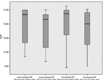
The mean uncorrected and corrected IOP values decreased from 18.06 ± 3.91 and 18.31 ± 4.83 to 16.94 ± 3.87 and 16.95 ± 4.74 mmHg after HD, respectively. This decline was statistically signi-ficant, with a mean reduction of -1.12 ± 3.00 mmHg for the uncorrec-ted IOP and -1.38 ± 3.02 mmHg for the correcuncorrec-ted IOP (p=0.011 and p=0.003, respectively) (Table 2) (Figure 1).
E
FFECTSOFHD
ONCORNEALANDANTERIORCHAMBERMORPHOMETRYTable 1. Mean blood urea nitrogen, body mass, and systolic and diastolic pressure immediately before and after hemodialysis in 50 patients with end-stage renal disease
Before HD After HD Δ* P value†
BUN (mg/dl) 139.54 ± 40.27 044.42 ± 15.46 -95.12 ± 34.98 <0.001
Mean ± SD
Body mass (kg) 066.91 ± 10.09 064.27 ± 10.11 0-2.63 ± 00.66 <0.001
Mean ± SD
Systolic pressure (mmHg) 136.00 ± 20.60 117.20 ± 15.78 -19.60 ± 09.24 <0.001
Mean ± SD
Diastolic pressure (mmHg) 078.00 ± 10.30 067.20 ± 12.62 -10.40 ± 12.28 <0.001
Mean ± SD
HD= hemodialysis; BUN= blood urea nitrogen; SD= standard deviation; Bold face= significant values, p<0.05. *= change in value after HD; †= paired samples t-test.
Figure 1. Boxplot of the uncorrected and corrected intraocular pressure values 30 minutes before and 30 minutes after hemodialysis.
Table 2. Intraocular pressure and corneal and anterior chamber parameters 30 minutes before and 30 minutes after hemodialysis in 50 patients with end-stage renal disease
Before HD After HD Δ* P value
Uncorrected IOP (mmHg) 018.06 ± 03.91 016.94 ± 03.87 -1.12 ± 3.00 0.011† Mean ± SD
Corrected IOP 018.31 ± 04.83 016.95 ± 04.74 -1.38 ± 3.02 0.003†
(mmHg) Mean ± SD
CCT (µm) 536.38 ± 24.73 533.18 ± 27.25 -3.26 ± 7.03 0.002††
Mean ± SD
CV (mm3) 057.52 ± 03.15 055.68 ± 03.55 -0.90 ± 1.23 <0.001††
Mean ± SD
Flattest K (D) 043.93 ± 01.45 043.88 ± 01.50 -0.04 ± 0.30 0.313††
Mean ± SD
Steepest K (D) 044.65 ± 01.46 044.65 ± 01.50 -0.06 ± 0.42 0.829††
Mean ± SD
Maximum K (D) 046.76 ± 01.77 046.41 ± 01.75 -0.37 ± 1.22 0.119††
Mean ± SD
AQD (mm) 002.87 ± 00.46 002.91 ± 00.46 -0.03 ± 0.22 0.250††
Mean ± SD
ACD (mm) 003.41 ± 00.46 003.44 ± 00.48 -0.03 ± 0.13 0.288††
Mean ± SD
ACA (°) 045.90 ± 09.42 046.18 ± 09.02 -1.06 ± 2.66 0.655†
(nasal quadrant) Mean ± SD
ACA (°) 046.38 ± 12.42 045.88 ± 13.91 -0.76 ± 8.05 0.571††
(temporal quadrant) Mean ± SD
HD= hemodialysis; IOP= Intraocular pressure; CCT= central corneal thickness; CV= corneal volume; D= diopter; K= keratometry; AQD= aqueous depth; ACD= anterior chamber depth; ACV= anterior chamber volume; ACA= anterior chamber angle; SD= standard deviation; Bold face= significant values, p<0.05.
*= change in value after HD; †= Wilcoxon signed-rank test; ††= paired samples t-test.
The flattest, steepest, and maximum keratometric values were similar before and after HD (p=0.313, p=0.829, and p=0.119, respectively) (Table 2). The mean values of the anterior chamber parameters before and after HD are summarized in table 2. As shown in the table, ACD, AQD, ACV, and ACA in the nasal and temporal quadrants were similar before and after HD (p>0.05 for all variables).
C
ORRELATIONSBETWEENTHECHANGESINOCULARANDSYSTEMICPARAMETERS
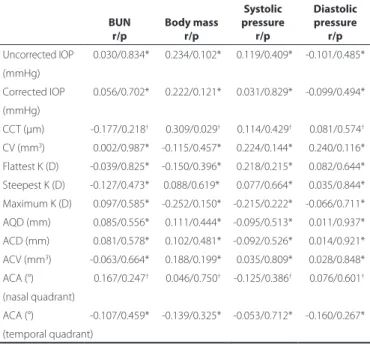
The changes in the uncorrected and corrected IOP and corneal and anterior chamber morphometry values were not significantly correlated with the changes in the systemic hemodynamic parame-ters (p>0.004) (Table 3).
DISCUSSION
In this study, we investigated the short-term effects of HD on corneal and anterior chamber morphometry and IOP, as well as the relationships between changes in ocular and systemic parameters in patients with ESRD. Uncorrected and corrected IOP, CCT, and CV values significantly decreased after HD, whereas anterior chamber
morphometry values did not significantly change. No significant corre lations were found between the changes in the ocular and sys-temic parameters.
Table 3. Correlation analyses between changes in intraocular pressu-re, cornea and anterior chamber parameters, and systemic parame-ters in 50 patients with end-stage renal disease after hemodialysis
BUN Body mass
Systolic pressure
Diastolic pressure
r/p r/p r/p r/p
Uncorrected IOP -0.030/0.834* -0.234/0.102* -0.119/0.409* -0.101/0.485*
(mmHg)
Corrected IOP -0.056/0.702* -0.222/0.121* -0.031/0.829* -0.099/0.494*
(mmHg)
CCT (µm) -0.177/0.218† -0.309/0.029† -0.114/0.429† -0.081/0.574†
CV (mm3) -0.002/0.987* -0.115/0.457* -0.224/0.144* -0.240/0.116*
Flattest K (D) -0.039/0.825* -0.150/0.396* -0.218/0.215* -0.082/0.644* Steepest K (D) -0.127/0.473* 0.088/0.619* -0.077/0.664* -0.035/0.844*
Maximum K (D) -0.097/0.585* -0.252/0.150* -0.215/0.222* -0.066/0.711*
AQD (mm) -0.085/0.556* -0.111/0.444* -0.095/0.513* -0.011/0.937*
ACD (mm) -0.081/0.578* -0.102/0.481* -0.092/0.526* -0.014/0.921*
ACV (mm3) -0.063/0.664* -0.188/0.199* -0.035/0.809* -0.028/0.848*
ACA (°) -0.167/0.247† -0.046/0.750† -0.125/0.386† -0.076/0.601†
(nasal quadrant)
ACA (°) -0.107/0.459* -0.139/0.325* -0.053/0.712* -0.160/0.267* (temporal quadrant)
BUN= blood urea nitrogen; IOP= intraocular pressure; CCT= central corneal thickness; CV= corneal volume; D= diopter; K= Keratometry; AQD= aqueous depth; ACD= anterior chamber depth; ACV= anterior chamber volume; ACA= anterior chamber angle; Bold face= significant values, p<0.004; r= correlation coefficient.
*= Pearson correlation test; †= Spearman’s correlation test.
change in IOP (16). Recent studies have noted that IOP values either
do not change or decrease after HD, and these studies explained
these results as being due to improved HD techniques(5,8,25,26). A few
studies reported significant decreases in IOP and CCT values, and suggested that total body fluid loss and increased plasma colloid osmotic pressure after HD cause an efflux of water from the eye to the plasma, resulting in a decrease in the CCT and IOP values(5,8). Jung
et al.(5) reported a correlation between the changes in IOP and those
of body mass, whereas Dinc et al.(8) did not find this correlation. In
the present study, the mean uncorrected IOP decreased significantly after HD, with a mean reduction of -1.12 ± 3.00 mmHg. Similar to Dinc et al.’s study, no correlations were found between IOP chan-ges and systemic chanchan-ges. Two of the studies reported a positive
correlation between the change in CCT and total body fluid loss(5,8).
In the present study, CCT and CV decreased significantly after HD, but these changes were not correlated with any systemic changes including body fluid loss. In terms of the relationship between IOP
and CCT, Jung et al.(5) found a positive correlation between IOP and
CCT changes, whereas Dinc et al.(8) did not find such a relationship.
In the present study, we did not evaluate the correlation between IOP and CCT; however, we investigated the effect of CCT on IOP using Ehlers’ formula, and corrected the IOP values according to CCT. The corrected IOP values also decreased after HD, meaning the IOP values decreased separately from the corneal thickness. However, the data did not reveal any factors that were related to the decrease in IOP after HD.
Several studies have attempted to characterize the effects of
pos-tural changes and water intake on IOP(27-32). The postural change test
and the water-drinking test are two classic provocative tests used to change IOP. Research shows that IOP is lower in the sitting position
than in the supine position(27-29). In our study, the patients generally
stayed in the supine position during HD, and we measured IOP after
the patients had been in the sitting position for a while. IOP increases with water ingestion and decreases with fasting, so the total body
fluid volume is important in IOP measurement(30-33). HD patients
ex-perience increased water load before HD, restricted water ingestion during HD, and decreased water load after HD. Although we did not find a significant correlation between IOP and body fluid loss, we believe that changes in body fluid volume and the positioning of the patients may have affected our IOP results.
The concentration of aqueous humor, a kind of extracellular fluid, can be affected by blood fluid volume changes after HD. Evaluating anterior chamber parameters is important when defining ocular pharmacokinetics, aqueous humor dynamics, and the pathophysio-logies of primary open-angle glaucoma, angle-closure glaucoma, and pigmentary glaucoma. Anterior chamber parameters are also cru cial in intraocular lens measurement, piggyback and phakic
in-traocular lens implantation, and cataract surgeries(34,35). The timing
of preoperative measurements, phakic intraocular lens implantation, and cataract surgeries in HD patients is important. Therefore, it is crucial to define anterior segment morphometry changes in these patients and, if there are changes, to evaluate the most appropriate time for preoperative measurements and surgeries.
ACD changes during HD have also been investigated in previous
studies(24,25). In a study researching the ocular effects of acetate HD,
ACD was found to decrease after acetate HD, whereas it did not
chan-ge after bicarbonate HD(24). The authors of the study suggested that
the explanation for these findings may be similar to that for dialysis disequilibrium syndrome: As urea is removed from the intracellular compartment, idiogenic osmoles may be generated, resulting in a lower intraocular pH, which may in turn have a local effect on the
formation of aqueous humor(24). Acetate HD may result in a longer
period of acidosis. On the other hand, bicarbonate HD, which is associated with a steady rise in blood pH levels, may more rapidly correct the intraocular acidosis, thus permitting normal aqueous dy-namics to proceed(24). In another study, Gracitelli et al.(25) investigated
ACD and axial length changes after bicarbonate HD, reporting that ACD decreased and axial length did not change after HD. However, an ultrasonic biometer was used in both of these studies, and mea-surements taken using these devices can be inaccurate because of numerous factors, such as corneal impression, excess fluid on the probe, and a thick layer of tear film(36,37). ACD is the distance between
the corneal epithelium and the lens, and may be misinterpreted in HD patients as resulting from CCT changes. Therefore, in the present study, we measured ACD with a 3D rotating Schleimpflug topo-graphy system, which allows rapid, non-contact anterior segment evaluation. Despite changes in CCT, the ACD values did not change after bicarbonate HD. AQD was also measured, and is defined as the distance between the corneal endothelium and the lens. After eli-minating the effects of the CCT changes, the AQD values did not show a significant change after HD. The other anterior chamber parameters, ACV and ACA in the nasal and temporal quadrants, were also similar to the control measurements after HD.
ocular parameters were not measured during HD, OPP was not mea-sured, systemic medication use was allowed, and we did not include a control group.
In conclusion, we found that HD treatment has significant effects on uncorrected and corrected IOP, CCT, and CV. HD likely causes underestimation of IOP and corneal morphometry changes, and these changes may be important when planning corneal or lenticular surgeries in HD patients.
REFERENCES
1. Himmelfarb J, Ikizler TA. Hemodialysis. N Engl J Med. 2010;363(19):1833-45. Comment in: N Engl J Med. 2011;364(6):584; author reply 585.
2. Tomazzoli L, De Natale R, Lupo A, Parolini B. Visual acuity disturbances in chronic renal failure. Ophthalmologica. 2000;214(6):403-5.
3. Mullaem G, Rosner MH. Ocular problems in the patient with end-stage renal disease. Semin Dial. 2012;25(4):403-7.
4. Aktas Z, Ozdek S, Asli Dinc U, Akyürek N, Atalay V, Güz G, et al. Alterations in ocular surface and corneal thickness in relation to metabolic control in patients with chronic renal failure. Nephrology (Carlton). 2007;12(4):380-5.
5. Jung JW, Yoon MH, Lee SW, Chin HS. Effect of hemodialysis (HD) on intraocular pres-sure, ocular surface, and macular change in patients with chronic renal failure. Effect of hemodialysis on the ophthalmologic findings. Graefes Arch Clin Exp Ophthalmol. 2013;251(1):153-62.
6. Nagaoka T, Takeyama Y, Kanagawa S, Sakagami K, Mori F, Yoshida A. Effect of haemo-dialysis on retinal circulation in patients with end stage renal disease. Br J Ophthalmol. 2004;88(8):1026-9.
7. Yang SJ, Han YH, Song GI, Lee CH, Sohn SW. Changes of choroidal thickness, in trao cular pressure and other optical coherence tomographic parameters after haemo dialysis. Clin Exp Optom. 2013; 96(5):494-9.
8. Dinc UA, Ozdek S, Aktas Z, Guz G, Onol M. Changes in intraocular pressure, and cor-neal and retinal nerve fiber layer thickness during hemodialysis. Int Ophthalmol. 2010; 30(4):337-40.
9. Gass JD. Bullous retinal detachment and multiple retinal pigment epithelial deta-chments in patients receiving hemodialysis. Graefes Arch Clin Exp Ophthalmol. 1992; 230(5):454-8.
10. Sitprija V, Holmes JH, Ellis PP. Changes in intraocular pressure during hemodialysis. Invest Ophthalmol. 1964;3:273-83.
11. Sitprija V, Holmes JH, Ellis PP. Intraocular pressure changes during artificial kidney the rapy. Arch Ophthalmol. 1964;72(5):626-31.
12. Gafter U, Pinkas M, Hirsch J, Levi J, Savir H. Intraocular pressure in uremic patients on chronic hemodialysis. Nephron. 1985;40(1):74-5.
13. Cecchin E, De Marchi S, Tesio F. Intraocular pressure and hemodialysis. Nephron. 1986;43(1):73-4.
14. Leiba H, Oliver M, Shimshoni M, Bar-Khayim Y. Intraocular pressure fluctuations during regular hemodialysis and ultrafiltration. Acta Ophthalmol (Copenh). 1990;68(3):320-2. 15. Ulas F, Dogan U, Keles A, Ertilav M, Tekçe H, Celebi S. Evaluation of choroidal and re-tinal thickness measurements using optical coherence tomography in non-diabetic haemodialysis patients. Int Ophthalmol. 2013;33(5):533-9.
16. Levy J, Tovbin D, Lifshitz T, Zlotnik M, Tessler Z. Intraocular pressure during haemodia-lysis: a review. Eye (Lond). 2005;19(12):1249-56.
17. Brandt JD, Beiser JA, Gordon MO, Kass MA; Ocular Hypertension Treatment Study (OHTS) Group. Central corneal thickness and measured IOP response to topical ocular hypotensive medication in the Ocular Hypertension Treatment Study. Am J Ophthal-mol. 2004;138(5):717-22. Comment in: Am J OphthalOphthal-mol. 2004;138(5):847-8; Am J Ophthalmol. 2005;139(6):1148; author reply 1148-9.
18. Tielsch JM, Katz J, Sommer A, Quigley HA, Javitt JC. Hypertension, perfusion pressure, and primary open-angle glaucoma. A population-based assessment. Arch Ophthal-mol. 1995;113(2):216-21.
19. Leske MC, Connell AM, Wu SY, Hyman LG, Schachat AP. Risk factors for open-angle glaucoma. The Barbados Eye Study. Arch Ophthalmol. 1995;113(7):918-924. Comment in: Arch Ophthalmol. 1996;114(2):235.
20. Hu J, Bui KM, Patel KH, Kim H, Arruda JA, Wilensky JT, et al. Effect of hemodialysis on intraocular pressure and ocular perfusion pressure.JAMA Ophthalmol. 2013;131(12): 1525-31.
21. Barbosa CP, Stefanini FR, Penha F, Góes MA, Draibe SA, Canziani ME, et al. Intraocular pressure and ocular perfusion during hemodialysis. Arq Bras Oftalmol. 2011;74(2): 106-9.
22. De Marchi S, Cechin E, Tesio F. Intraocular pressure changes during hemodialysis: prevention of excessive dialytic rise and development of severe metabolic acidosis following acetazolamide therapy. Renal Fail. 1989;11(2-3):117-24.
23. Jaeger P, Morisod L, Wauters JP, Faggioni R. Prevention of glaucoma during hemo-dialysis by mannitol and acetazolamide. N Engl J Med 1980;303(12):702.
24. Rever B, Fox L, Christensen R, Bar-Khayim Y, Nissenson AR. Adverse ocular effects of acetate hemodialysis. Am J Nephrol. 1983;3(4):199-204.
25. Gracitelli CP, Stefanini FR, Penha F, Góes MÂ, Draibe SA, Canziani ME, et al. Anterior chamber depth during hemodialysis. Clin Ophthalmol. 2013;7:1635-9.
26. Samsudin A, Mimiwati Z, Soong T, Fauzi MS, Zabri K. Effect of haemodialysis on in-traocular pressure. Eye (Lond) 2010;24(1):70-3.
27. Weber AK, Price J. Pressure differential of intraocular pressure measured between supine and sitting position. Ann Ophthalmol. 1981;13(3):323-6.
28. Kothe AC. The effect of posture on intraocular pressure and pulsatile ocular blood flow in normal and glaucomatous eyes. Surv Ophthalmol. 1994;38:191-7. 29. Prata TS, De Moraes CG, Kanadani FN, Ritch R, Paranhos A Jr. Posture-induced
in-traocular pressure changes: considerations regarding body position in glaucoma patients. Surv Ophthalmol. 2010;55(5):445-53.
30. Kronfeld PC. Water drinking and outflow facility. Invest Ophthalmol. 1975;14(1):49-52. 31. Brucculeri M, Hammel T, Harris A, Malinovsky V, Martin B. Regulation of intraocular
pressure after water drinking. J Glaucoma. 1999;8(2):111-6.
32. Read SA, Collins MJ. Water drinking influences eye length and IOP in young healthy subjects. Exp Eye Res. 2010; 91(2):180-5.
33. Oltulu R, Satirtav G, Ersan I, Soylu E, Okka M, Zengin N. The effect of dehydration and fasting on corneal biomechanical properties and ıntraocular pressure. Eye Contact Lens. 2016;42(6):392-4.
34. Palamar M, Egrilmez S, Uretmen O, Yagci A, Kose S. Influences of cyclopentolate hydrochloride on anterior segment parameters with pentacam in children. Acta Ophthalmol. 2011;89(5):461-5.
35. Palamar M, Egrilmez S, Uretmen O, Kose S. Evaluation of cornea and anterior chamber using pentacam in pediatric cases. Turk J Ophthalmol. 2011;41(3):133-7.
36. Stainert RF. A-scan biometry and intraocular lens power calculation. In: Alber DM, Jacobiec FA, editors. Principles and practice of ophthalmology. Philedelphia: WB Saunders; 1994. p. 603-6.