ABSTRACT | Purpose: Obesity is associated with eye diseases, but the underlying structural changes and pathogenic mecha-nisms have not been examined in detail. Here, we assessed the effects of morbid obesity on the morphometric indices of eye disease. Methods: Morbidly obese volunteers (n=101, body mass index [BMI] ≥40) and healthy individuals (n=95, BMI: 18.50-24.99) were examined by Goldman applanation tonometry, pachymetry, and spectral domain optical coherence tomography. Intraocular pressure, anterior chamber depth, axial length, central corneal thickness, retinal nerve fiber layer thickness, central foveal thickness, and choroidal thickness were compared between groups. Results: Uncorrected intraocular pressure was significantly greater in the morbidly obese group than in the healthy control group (15.5 ± 2.5 vs. 14.5 ± 2.6 mmHg, p=0.009), whereas axial length, anterior chamber depth, and central corneal thickness did not differ between the groups. The mean retinal nerve fiber layer thickness at the temporal quadrant was reduced in the morbidly obese group (72.7 ± 13.6 vs. 85.05 ± 52.6 µm, p=0.024). Similarly, the mean retinal thicknesses at nasal and temporal 1500-µm locations were lower in the morbidly obese group (346.6 ± 18.2 vs. 353.7 ± 18.8 µm, p=0.008; 323.1 ± 20.3 vs. 330.0 ± 18.9 µm, p=0.001). The mean choroidal thickness was also reduced in almost all measurement locations (fovea, temporal 500 and 1000 µm, and nasal 500, 1000, and 1500 µm) of the obese group (p<0.05). Weight and BMI were negatively correlated with subfoveal choroidal thickness (r=-0.186, p=0.009; r=-0.173, p=0.015). Conclusion: Morbid obesity is associated with elevated uncorrected intraocular pressure and signs of neuropathy and retinopathy. Obesity may thus increase the risks of glaucoma and glaucomatous optic neuropathy.
Keywords: Morbid obesity; Intraocular pressure; Nerve fibers;
Corneal pachymetry; Anterior eye segment; Posterior eye segment
RESUMO | Objetivo: A obesidade está associada a doenças
oulares, mas as mudanças estruturais subjacentes e os meca-nismos patogênicos não foram examinados detalhadamente. Aqui avaliamos os efeitos da obesidade mórbida nos índices morfométricos da doença ocular. Métodos: Voluntários obesos mórbidos (n=101, índice de massa corporal ≥40) e indivíduos saudáveis (n=95, índice de massa corporal 18,50 a 24,99) foram examinados por tonometria de aplanação de Goldman, paquimetria e tomografia de coerência óptica de domício espectral. A pressão intraocular, profundidade da câmara anterior, comprimento axial, espessura central da córnea, espessura da camada de fibras nervosas da retina, espessura foveal central e espessura da coroide foram comparadas entre os grupos. Resultados: A pressão intraocular não corrigida foi significativamente maior no grupo com obesidade mórbida do que no grupo controle saudável (15,5 ± 2,5 vs. 14,5 ± 2,6 mmHg, p=0,009), enquanto que o comprimento axial, profundidade da câmara anterior e espessura central da córnea não diferiram entre os grupos. A espessura média da camada de fibras nervosas da retina no quadrante temporal foi reduzida no grupo com obesidade mórbida (72,7 ± 13,6 vs. 85,05 ± 52,6 µm, p=0,024). Da mesma forma, a média das espesuras da retinianas nas localizações nasal e temporal de 1500 µ foi menor no grupo com obesidade mórbida (346,6 ± 18,2 µm vs. 353,7 ± 18,8 µm, p=0,008; 323,1 ± 20,3 µm vs. 330,0 ± 18,9 µm, p=0,001). A espessura média da coroide também foi reduzida em quase todos os locais de mensuração (fóvea, temporal 500 e 1000 µm, nasal 500, 1000 e 1500 µm) do grupo obeso (p<0,05). Peso e índice de massa corporal foram negativamente correlacionados com a espessura da coroide subfoveal (r=-0,186, p=0,009; r=-0,173, p=0,015). Conclusão: A obesidade mórbida está associada à elevada pressão intraocular não corrigida e a sinais de neuropatia e retinopatia. A obesidade pode, assim, aumentar os riscos de glaucoma e neuropatia óptica glaucomatosa.
Descritores: Obesidade morbida; Pressão intraocular; Fibras
nervosas; Paquimetria corneana; Segmento anterior do olho; Segmento posterior do olho
Ocular abnormalities in morbid obesity
Anormalidades oculares na obesidade mórbida
Kuddusi Teberik1, M. Tahir Eski2, Sami Doğan3, Mevlüt Pehlivan3, Murat Kaya1 1. Department of Ophthalmology, Faculty of Medicine, Duzce University, Duzce, Turkey. 2. Ministry of Health Bingöl State Hospital, Bingöl, Turkey.
3. Department of General Surgery, Faculty of Medicine, Duzce University, Duzce, Turkey.
Submitted for publication: February 7, 2018 Accepted for publication: June 3, 2018
Funding: No specific financial support was available for this study.
Disclosure of potential conflicts of interest: None of the authors have any potential conflicts of interest to disclose.
Corresponding author: Kuddusi Teberik. E-mail: [email protected]
INTRODUCTION
Excessive accumulation of adipose tissue (obesity) can impair physical, psychosocial, and general health(1).
Obesity afflicts all age groups, from youth to old age, but there has been a particularly rapid increase in preva-lence among youth and young adults(2,3). Body mass
index (BMI), calculated as weight (kg)/height (m)2, is the
most widely adopted metric for defining underweight, overweight, and obesity. The World Health Organization defines underweight as a BMI ≤18.50, normal weight as a BMI in the range 18.50-24.99, overweight as 25.00-29.99, obese class I as 30.00-34.99, obese class II as 35-39.99, and obese class III as BMI ≥40. Morbid obesity is defined as BMI ≥35 kg/m2 along with accompanying
illnesses or ≥40 kg/m2 without weight-related illness.
Obesity is strongly related to increased morbidity and early mortality due to associated diseases such as dia-betes mellitus (DM), hypertension, and cardiovascular disease(4). Obesity is also related to various eye disea ses,
such as cataract, age-related maculopathy, diabetic re tinopathy, and glaucoma. However, relatively few studies have examined the potential effects of obesity without associated diseases such as DM on visual health(5).
Obesity is negatively correlated with visual acuity, but the underlying eye-related problems and pathogenic mechanisms are uncertain. Ocular problems in obesity may be associated with dysfunction of both mechanical and vascular elements of the eye, possibly due to chro-nic oxidative stress(6). Thus, obesity may impair visual
function in the absence of associated diseases or in the preclinical stages.
To examine the effects of obesity on ocular function in the absence of obesity-related diseases, we compa-red intraocular pressure (IOP), anterior chamber depth (ACD), central corneal thickness (CCT), axial length (AL), retinal nerve fiber layer (RNFL) thickness, retinal thickness (RT), and choroidal thickness (CT) between morbidly obese subjects without associated diseases (≥40 kg/m2)
and matched healthy subjects with normal BMI.
METHODS
This study was performed at the Department of Oph-thalmology and General Surgery, Düzce University Medical Faculty, and conducted in accordance with the principles of the Declaration of Helsinki. The research sample consisted of two groups. From 353 morbidly obese individuals scheduled for sleeve gastrectomy at Düzce University Medical Faculty Department of Surgery,
101 with BMI ≥40 without weight-related illness were recruited as study volunteers. The control group was composed of 95 non-obese adults matched for age and sex ratio with BMI values ranging from 18.50 to 24.99. The study was approved by the institutional ethics com-mittee and all participants provided informed consent following explanations of research protocols and goals. BMI was assessed as bodyweight in kilograms divided by body height in meters squared (kg/m2). Subject height was
measured in the standing position and body weight using a digital scale. An automatic blood pressure monitor was used to assess systolic and diastolic blood pressures.
Candidates with histories of retinal disease, smoking, high alcohol consumption, IOP above 21 mmHg, systemic diseases (such as diabetes and hypertension), glaucoma, intraocular surgery, laser treatment, contact lens use, strabismus, ocular trauma, amblyopia, or cataract, as well as those with any ocular inflammation, currently recei-ving medical treatment for glaucoma, or harecei-ving taken any prescription drugs over the past 3 months were excluded from the study.
All statistical analyses were conducted using the sta-tistical package SPSS v 22.0 (SPSS Inc., Chicago, IL, USA). Group means were compared by Mann-Whitney U-test or Student’s t-test as appropriate. Correlations between various clinical examination parameters and BMI were analyzed by Spearman’s rank correlation test. A P<0.05 (two-tailed) was considered statistically significant for all tests.
RESULTS
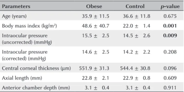
The morbid obesity group (n=101) and healthy control group (n=95) were well matched for age (35.9 ± 11.5 years, range 14-63 years vs. 36.6 ± 11.8 years, range 14-63 years; p>0.05) and sex ratio (68 females and 33 males vs. 67 females and 28 males; p>0.05). The mean uncor-rected IOP was significantly greater in the obese group (15.5 ± 2.5 mmHg vs. 14.5 ± 2.6 mmHg; p=0.009), whereas AL, ACD, and CCT did not differ. Demographic and clinical characteristics of the study groups are sum-marized in table 1.
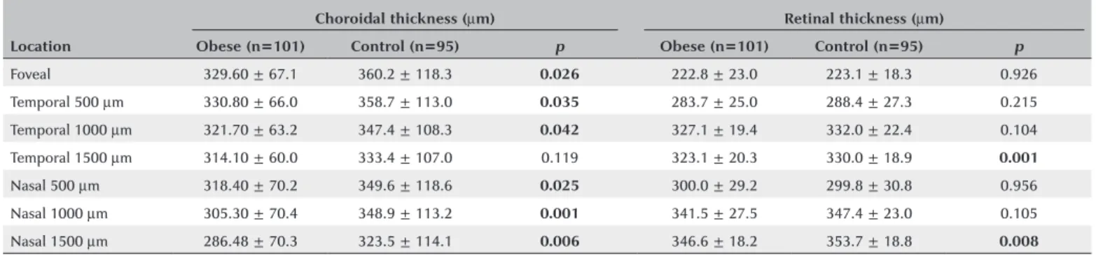
Among morphological measures (Table 2), RNFL thickness in the temporal quadrant was significantly re-duced in the morbidly obese group compared to the heal-thy control group (72.7 ± 13.6 µm vs. 85.05 ± 52.6 µm; p=0.024), but no significant differences were found for the other quadrants. RT was also reduced in the obese group in both the nasal and temporal 1500 µm regions (346.6 ± 18.2 µm vs. 353.7 ± 18.8 µm, p=0.008; 323.1 ± 20.3 µm vs. 330.0 ± 18.9 µm, p=0.001). Mean CT was also re-duced in all measurement locations of the obese group (p<0.05) except for temporal 1500 mm (Table 3).
There was no significant correlation between BMI and any anterior segment parameter except for uncor-rected IOP (Table 4). Alternatively, weight and BMI were strongly and negatively correlated with subfoveal CT (r=-0.186, p=0.009; r=-0.173, p=0.015). Weight and BMI were also negatively correlated with CT at nasal 500, 1000, and 1500 µm locations as well as temporal 500, 1000, and 1500 µm locations. Height was not asso-ciated with CT at any location (Table 5).
DISCUSSION
Uncorrected IOP was significantly elevated in morbidly obese adults without weight-related diseases, whereas corrected IOP, CCT, AL, and ACD measurements did not differ from those of healthy controls. However, the obese group demonstrated signs of retinopathy and vascular abnormalities. Thus, morbid obesity in the absence or
in the preclinical stages of associated diseases may pose risks for glaucoma and other eye diseases.
Risk factors for high IOP have been extensively stu-died due to the strong association with glaucomatous optic nerve damage(7,8). In The Beaver Dam Eye Study, IOP
was positively correlated with BMI among other factors(9).
Furthermore, epidemiologic studies have reported that the increase in IOP associated with obesity is indepen-dent of age, systolic blood pressure, and diastolic blood pressure (DBP)(10,11). Stojanov and colleagues reported
that obese individuals have significantly higher IOP than that of non-obese individuals(12), and Gunes et al.
found elevated IOP in Turkish patients with BMI ≥39(13).
Furthermore, both Gunes et al. and Jang et al. found a positive correlation between IOP and BMI(13,14). In our
study, this correlation was found to be weak. In contrast to IOP, only a small number of studies have analyzed the correlation between BMI and glaucomatous optic neuropathy. One study found that high BMI increases the likelihood of clinical glaucoma diagnosis at hospital ad-mission(15). However, a case-control study found no
statis-tically significant difference in BMI between glaucomatous and non-glaucomatous subjects(16). Further investigations
are needed to determine associations between BMI and glaucoma.
Table 1. Demographic and clinical characteristics of the study groups
Parameters Obese Control p-value
Age (years) 035.9 ± 11.5 036.6 ± 11.8 0.675
Body mass index (kg/m2) 048.6 ± 40.7 022.0 ± 01.4 0.001
Intraocular pressure (uncorrected) (mmHg)
015.5 ± 02.5 014.5 ± 02.6 0.009
Intraocular pressure (corrected) (mmHg)
014.6 ± 02.5 014.2 ± 02.2 0.208
Central corneal thickness (µm) 551.9 ± 31.3 544.4 ± 30.8 0.096
Axial length (mm) 022.8 ±0 2.1 022.9 ± 00.8 0.609
Anterior chamber depth (mm) 003.1 ± 00.4 003.1 ± 00.4 0.911
Table 2. Retinal nerve fiber layer (RNFL) thickness comparisons
Quadrant (µm) Obese Control p-value
Temporal 072.7 ± 13.6 085.05 ± 52.6 0.024
Temporal-superior 143.9 ± 24.4 138.30 ± 23.4 0.108
Temporal-inferior 148.7 ± 23.5 142.80 ± 22.8 0.078
Nasal 079.9 ± 17.3 081.90 ± 18.1 0.434
Nasal-superior 113.7 ± 25.6 117.10 ± 20.9 0.309
Nasal-inferior 115.0 ± 26.8 111.20 ± 23.9 0.300
Table 3. Distribution of mean choroidal and retinal thickness at different locations in morbidly obese and control groups
Location
Choroidal thickness (µm) Retinal thickness (µm)
Obese (n=101) Control (n=95) p Obese (n=101) Control (n=95) p
Foveal 329.60 ± 67.1 360.2 ± 118.3 0.026 222.8 ± 23.0 223.1 ± 18.3 0.926
Temporal 500 µm 330.80 ± 66.0 358.7 ± 113.0 0.035 283.7 ± 25.0 288.4 ± 27.3 0.215
Temporal 1000 µm 321.70 ± 63.2 347.4 ± 108.3 0.042 327.1 ± 19.4 332.0 ± 22.4 0.104
Temporal 1500 µm 314.10 ± 60.0 333.4 ± 107.0 0.119 323.1 ± 20.3 330.0 ± 18.9 0.001
Nasal 500 µm 318.40 ± 70.2 349.6 ± 118.6 0.025 300.0 ± 29.2 299.8 ± 30.8 0.956
Nasal 1000 µm 305.30 ± 70.4 348.9 ± 113.2 0.001 341.5 ± 27.5 347.4 ± 23.0 0.105
Nasal 1500 µm 286.48 ± 70.3 323.5 ± 114.1 0.006 346.6 ± 18.2 353.7 ± 18.8 0.008
Table 4. Correlation of body mass index with clinical parameters
r p
Uncorrected intraocular pressure -0.167 0.020
Corrected intraocular pressure -0.107 0.135
Central corneal thickness -0.071 0.322
Axial length -0.030 0.672
Anterior chamber depth -0.025 0.728
r= correlation coefficient.
Table 5. Correlations of choroidal thickness with weight, height, and body mass index (BMI)
Subfoveal T500 T1000 T1500 N500 N1000 N1500
Weight
r -0.186* -0.189* -0.176* -0.156* -0.181* -0.249* -0.231*
p -0.009† 0.008† -0.014* -0.029* -0.011* -0.001† 0.001†
Height
r -0.069* -0.071* -0.037* -0.056* -0.037* -0.073* -0.116
p -0.340* -0.320* -0.604* -0.435* -0.603* -0.311* -0.107*
BMI
r -0.173* -0.176* -0.172* -0.150* -0.180* -0.237* -0.208*
p -0.015* -0.014* -0.016* -0.036* -0.011* -0.001† -0.003†
†= correlation is significant at the 0.01 level (2-tailed); *= correlation is significant at the 0.05 level (2-tailed); r= correlation coefficient.
Mean corneal thickness ranges from about 537 µm to 544 µm in normal adults, but is thicker in ocular hypertension cases and thinner in normotension glau-coma(17). Bron and colleagues found that CCT correlates
positively with IOP, with each 10-mm increase in CCT associated with a 0.15-1 mmHg increase in the IOP(18),
whereas a manometric study found that a 10-µm increase in CCT predicts a 0.7 mmHg increase in IOP(19). In this
study, we found no significant effect of obesity on the corrected IOP.
There is currently little understanding of the patho-physiology linking obesity, IOP, and glaucoma. Both ‘me chanical’ and ‘vascular’ etiologies of IOP and glau-coma may be driven by obesity. Proposed mechanical pathomechanisms include accumulation of intraorbital fat, elevated blood thickness, elevated episcleral venous pressure, and deterioration of aqueous discharge(20,21).
Additionally, eyes with naturally weak vascular flow to the optic nerve head are more prone to injury under increa-sed or even normal IOP(22,23). Disruption of the arterial
blood supply, changes in autonomic vasoregulation, and endothelial damage associated with obesity activity may also lead to aberrant ophthalmic blood flow(24). Another
possibility is that hyperleptinemia-induced obesity can lead to oxidative injury in the trabecular meshwork(25).
Gunes et al. reported significantly lower ACD in obe-se subjects compared to a control group, but there were no significant difference in AL and CCT values(15).
Moreo-ver, Yilmaz et al. reported that AL and CCT values did not differ significantly among healthy obese individuals with BMI of class 1(26). In our study, these parameters
also did not differ between control and obese groups. Su et al.(27) reported that the cornea is thicker in high-BMI
subjects and that this leads to higher IOP. In contrast, we also found no difference in corrected IOP between groups, although in accord with a previous study we did find a significant increase in unadjusted IOP values(28).
Glaucoma is less likely to develop in individuals with high CCT(15). However, we believe that glaucomatous
optic neuropathy may develop due to ischemic and me-chanical effects caused by obesity, even though there is no difference in corrected IOP.
In our study, the morbidly obese group exhibited sig-nificant thinning of the RNFL in the temporal quadrant . Similarly, Dogan et al.(29) also found that mean RNFL
redu-ced in morbid obesity. In the same study, no significant difference was found in central macular thickness, but CT was reduced. Using a caliper system, we measured significantly lower foveal RT was at both temporal and nasal 1500 mm locations in the morbidly obese group. In addition, CT was significantly reduced in most mea-sured areas (all except temporal 1500 µm), in accord with Dogan et al.(29). Yilmaz et al. also found reduced CT
at the subfoveal area and up to 2000 µm in both nasal and temporal directions from the fovea(26). Unlike retina
and optic nerve head vasculature, the choroidal blood supply is regulated autonomically, with noradrenaline from sympathetic efferent nerve activation reducing and NO from parasympathetic efferents increasing choroidal blood flow(30). The effects of obesity on NO
signaling in particular may explain the impact of obesity on CT because low NO values are observed in morbidly obese individuals. Further, there is a positive correlation between BMI and levels of the vasoconstrictors ET-1 and Ang-II, suggesting that obesity-induced shifting of the vasodilator/vasoconstrictor balance (reduced NO and elevated ET-1 and Ang-II) can affect CT. Spearman’s correlation analysis revealed negative correlations of BMI and weight (but not height). Yilmaz et al. also found negative correlations of between CT and both BMI and weight(26). However, Yumusak et al.(31) reported a positive
correlation between BMI and CT in class two obese women, suggest that factors such as sex and severity of obesity may influence the relationship between BMI and choroidal blood flow.
The major strength of our study is the sample size. To our best knowledge, this is the largest -scale study conducted to date on the ocular effects of obesity in the absence of related diseases. Moreover, both anterior and posterior segment findings were evaluated in detail. Alternatively, the present study is limited by the lack of biochemical blood parameter measures. Another limita-tion is that we did not evaluate obstructive sleep apnea syndrome or obesity hypoventilation syndrome, and these disorders may also impact ocular function.
In conclusion, uncorrected IOP was significantly higher in the morbidly obese compared to matched con-trols with normal BMI. Corrected IOP, CCT, AL, and ACD did not differ significantly between groups, but regional decreases in RNFL thickness and RT were observed as well as more pervasive decreases in CT. IOP elevation and RNFL thinning in morbidly obese adults may increa-se the risks of glaucoma and optic neuropathy.
REFERENCES
1. James PT. Obesity: the worldwide epidemic. Clin Dermatol. 2004; 22(4):276-80.
2. Lobstein T, Baur L, Uauy R; IASO International Obesity Task Force. Obesity in children and young people: a crisis in public health. Obes Rev. 2004;5(Suppl 1):4-104.
3. Kennedy RL, Chokkalingham K, Srinivasan R. Obesity in the elderly: who should we be treating, and why, and how? Curr Opin Clin Nutr Metab Care. 2004;7(1):3-9.
4. Flegal KM, Graubard BI, Williamson DF, Gail MH. Excess deaths associated with underweight, overweight, and obesity. JAMA. 2005;293(15):1861-7.
5. Bergman B, Nilsson-Ehle H, Sjöstrand J. Ocular changes, risk markers for eye disorders and effects of cataract surgery in elderly people: a study of an urban Swedish population followed from 70 to 97 years of age. Acta Ophthalmol Scand. 2004;82(2):166-74. 6. Cheung N, Wong TY. Obesity and eye diseases. Surv Ophthalmol.
2007;52(2):180-95.
7. Wilson MR, Hertzmark E, Walker AM, Childs-Shaw K, Epstein DL. A case-control study of risk factors in open angle glaucoma. Arch Ophthalmol. 1987;105(8):1066-71.
8. Carel RS, Korczyn AD, Rock M, Goya I. Association between ocular pressure and certain health parameters. Ophthalmology. 1984; 91(4):311-4.
9. Klein BE, Klein R, Linton KL. Intraocular pressure in an American community. The Beaver Dam Eye Study. Invest Ophthalmol Vis Sci. 1992;33(7):2224-8.
10. Wu SY, Leske MC. Associations with intraocular pressure in the Barbados Eye Study. Arch Ophthalmol. 1997;115(12):1572-6. 11. Shiose Y. The aging effect on intraocular pressure in an apparently
normal population. Arch Ophthalmol. 1984;102(6):883-7. 12. Stojanov O, Stokić E, Sveljo O, Naumović N. The influence of
retrobulbar adipose tissue volume upon intraocular pressure in obesity. Vojnosanit Pregl. 2013;70(5):469-76.
13. Gunes A, Uzun F, Karaca EE, Kalaycı M. Evaluation of anterior
segment parameters in obesity. Korean J Ophthalmol. 2015;29(4): 220-5.
14. Jang HD, Kim DH, Han K, Ha SG, Kim YH, Kim JW, et al. Rela-tionship between intraocular pressure and parameters of obesity in Korean adults: the 2008-2010 Korea National Health and Nu-trition Examination Survey. Curr Eye Res. 2015;40(10):1008-17. 15. Zang EA, Wynder EL. The association between body mass index
and the relative frequencies of diseases in a sample of hospitalized patients. Nutr Cancer. 1994;21(3):247-61.
16. Gasser P, Stümpfig D, Schötzau A, Ackermann-Liebrich U, Flammer J. Body mass index in glaucoma. J Glaucoma. 1999;8(1):8-11. 17. Wolfs RC, Klaver CC, Vingerling JR, Grobbee DE, Hofman A, de
Jong PT. Distribution of central corneal thickness and its asso ciation with intraocular pressure: the Rotterdam Study. Am J Ophthalmol. 1997;123(6):767-72.
18. Bron AM, Creuzot-Garcher C, Goudeau-Boutillon S, d’Athis P. Fal-sely elevated intraocular pressure due to increased central corneal thickness. Graefes Arch Clin Exp Ophthalmol. 1999;237(3):220-4. 19. Feltgen N, Leifert D, Funk J. Correlation between central corneal
thickness, applanation tonometry, and direct intracameral IOP readings. Br J Ophthalmol. 2001;85(1):85-7.
20. Shiose Y, Kawase Y. A new approach to stratified normal intraocular pressure in a general population. Am J Ophthalmol. 1986;101(6): 714-21.
22. Dielemans I, Vingerling JR, Wolfs RC, Hofman A, Grobbee DE, de Jong PT. The prevalence of primary open-angle glaucoma in a population-based study in The Netherlands. The Rotterdam Study. Ophthalmology. 1994;101(11):1851-5.
23. Maier PC, Funk J, Schwarzer G, Antes G, Falck-Ytter YT. Treatment of ocular hypertension and open angle glaucoma: meta-analysis of randomised controlled trials. BMJ. 2005;331(7509):134.
24. Grieshaber MC, Flammer J. Blood flow in glaucoma. Curr Opin Ophthalmol. 2005;16(2):79-83.
25. Newman-Casey PA, Talwar N, Nan B, Musch DC, Stein JD. The relationship between components of metabolic syndrome and open-angle glaucoma. Ophthalmology. 2011;118(7):1318-26. 26. Yilmaz I, Ozkaya A, Kocamaz M, Ahmet S, Ozkaya HM, Yasa D, et
al. Correlation of choroidal thickness and body mass index. Retina. 2015;35(10):2085-90.
27. Su DH, Wong TY, Foster PJ, Tay WT, Saw SM, Aung T. Central
cor-neal thickness and its associations with ocular and systemic factors: the Singapore Malay Eye Study. Am J Ophthalmol. 2009;147(4): 709-716.e1.
28. Spaide RF, Koizumi H, Pozzoni MC. Enhanced depth imaging spectral-domain optical coherence tomography. Am J Ophthalmol. 2008;146(4):496-500.
29. Dogan B, Kazim Erol M, Dogan U, Habibi M, Bulbuller N, Turgut Coban D, et al. The retinal nerve fiber layer, choroidal thickness, and central macular thickness in morbid obesity: an evaluation using spectral-domain optical coherence tomography. Eur Rev Med Pharmacol Sci. 2016;20(5):886-91.
30. Nilsson SF. Nitric oxide as a mediator of parasympathetic vaso-dilation in ocular and extraocular tissues in the rabbit. Invest Ophthalmol Vis Sci. 1996;37(10):2110-9.