Clinicoradiological Session
Case 3/2008 – Fourteen Month Old Female Infant with Pulmonary
Arteriovenous Fistula
Edmar Atik
Instituto do Coração do Hospital das Clínicas – FMUSP – São Paulo, SP - Brazil
Clinical data
Cyanosis as an isolated sign appeared at 5 months of age, having recently progressed accompanied by an infectious process.
Physical examination
Patient was eupneic, with marked cyanosis, and pulses were palpated normally on all 4 limbs. Weight: 10 Kg, Height: 81 cm, BP: 90/60 mm Hg, HR: 120 bpm. O2 saturation, at rest: 50%. Aorta was not palpable at the supraesternal notch.
There were no deformities or impulsions in the precordium. Ictus cordis was not palpated. Heart sounds were normal and no heart murmurs were identified.
Lung auscultation evidenced a slight difference in characteristics [timbre] of vesicular breath sounds from one side to the other of the chest. The abdomen showed no abnormalities.
The electrocardiogram showed sinus rhythm and signs of left ventricular chamber overload with deep S waves in V2 and V3 (Sokolof Index: 40 mm) with ÂQRS: +100o, ÂP: +60o, and ÂT: +30o.
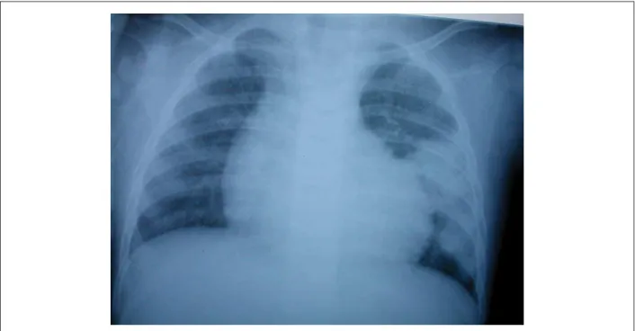
Radiographic image
cardiac area with slightly increased dimensions, elongated and rounded left ventricular arch, and normal pulmonary vascular network (rete). A hypotransparent image with
Correspondence: Edmar Atik •
InCor - Av. Dr. Enéas Carvalho de Aguiar, 44 - 05403-000 - São Paulo, SP - Brazil E-mail: [email protected]
Key words
Pulmonary arteriovenous fistula, congenital cardiopathy, interventional treatment, surgery.
Fig. 1 - &KHVW;UD\VKRZLQJVOLJKWO\LQFUHDVHGFDUGLDFDUHDZLWKHORQJDWHGDQGURXQGHGOHIWYHQWULFXODUDUFK3XOPRQDU\YDVFXODUQHWZRUNLVQRUPDO7KHUHLVDFOHDU
LPDJHRIFRQGHQVDWLRQRIWKHSXOPRQDU\SDUHQFK\PDLQWKHPLGWROHIWSRUWLRQFRQVLVWHQWZLWKDQDUWHULRYHQRXV¿VWXOD
Clinico Radiological Session
Edmar Atik
Arq Bras Cardiol 2008; 90(6): 407-408
distinct borders in the middle left pulmonary field was noted (Fig. 1).
Diagnostic impression
this image is consistent with the diagnosis of possible significant pulmonary arteriovenous fistulae.
Differential diagnosis
image of condensation in pulmonary parenchyma demands differential diagnosis with an infectious and/or tumoral pulmonary process.
Diagnostic confirmation
in light of the absence of signs of cardiopathy, the cyanosis observed may be explained by an arteriovenous shift of blood through the supposed pulmonary fistulae. The magnitude
of this shift may justify the left ventricular increase seen on the chest X-ray and the left ventricular overload observed on the electrocardiogram. The echocardiogram confirmed cardiovascular normality and the hemodynamic study showed the presence of giant arteriovenous fistulae limited to the middle portion of the left lung corresponding to the lingula of the lobe and possibly part of the upper lobe because of the presence of 3 arterial trunks. The left lower lobe was preserved. In the right middle lobe there was also a moderate pulmonary arteriovenous fistula (Fig. 2).
Treatment
During the same diagnostic hemodynamic procedure, coil embolization was performed of the middle right lobe and oxygen saturation increased to around 65%. Since the fistula on the left was pronounced, a lobectomy of the lobe-lingula was considered in order to avoid progression of the hypoxia along with its deleterious consequences.
Fig. 2 - $QJLRJUDSK\RIWKHOREHOLQJXODVKRZVODUJHFDOLEHUDUWHULRYHQRXVYHVVHOVDQGUDSLG¿OOLQJRIWKHOHIWDWULXPFKDUDFWHUL]LQJWKHSXOPRQDU\¿VWXODHLQ$QRUPDO
RSDFL¿HGOHIWORZHUOREHLQ%JUHDWHUGLODWLRQRIWKHOHIWSXOPRQDU\DUWHU\LQ&DUWHULRYHQRXV¿VWXODRIWKHULJKWPLGGOHOREHEHIRUHHPEROL]DWLRQLQ'DQGDIWHUHPEROL]DWLRQLQ ($EEUHYLDWLRQV/$OHIWDWULXP53$ULJKWSXOPRQDU\DUWHU\/3$OHIWSXOPRQDU\DUWHU\$9)3SXOPRQDU\DUWHULRYHQRXV¿VWXOD///ORZHUOHIWOREH
![Fig. 2 - $QJLRJUDSK\RIWKHOREHOLQJXODVKRZVODUJHFDOLEHUDUWHULRYHQRXVYHVVHOVDQGUDSLG¿OOLQJRIWKHOHIWDWULXPFKDUDFWHUL]LQJWKHSXOPRQDU\¿VWXODHLQ$QRUPDO RSDFL¿HGOHIWORZHUOREHLQ%JUHDWHUGLODWLRQRIWKHOHIWSXOPRQDU\DUWHU\LQ&DUWHULRYHQRXV¿VWXODRIWKHULJKWPLGGOHOREHEH](https://thumb-eu.123doks.com/thumbv2/123dok_br/15444921.600097/2.892.127.829.515.884/qjlrjudsk-riwkhoreholqjxodvkrzvodujhfdolehuduwhulryhqrxvyhvvhovdqgudslg-oolqjriwkhohiwdwulxpfkdudfwhul-lqjwkhsxoprqdu-hgohiworzhuorehlq-juhdwhuglodwlrqriwkhohiwsxoprqdu-duwhulryhqrxv-vwxodriwkhuljkwplggohoreheh.webp)
