Left Ventricular Ejection Fraction and Volumes as Measured by 3D
Echocardiography and Ultrafast Computed Tomography
Marcelo Luiz Campos Vieira, César H. Nomura, Bernardino Tranchesi Junior, Wercules A. de Oliveira, Gustavo
Naccarato, Bruna S. Serpa, Rodrigo B. D. Passos, Marcelo B. G. Funari, Cláudio H. Fischer, Samira S. Morhy
Hospital Israelita Albert Einstein, São Paulo, SP - BrazilSummary
Background: Real-time three-dimensional echocargiography (RT-3D-Echo) and ultrafast computed tomography (CT) are two novel methods for the analysis of LV ejection fraction and volumes.
Objective: To compare LVEF and volume measurements as obtained using RT-3D-Echo and ultrafast CT.
Methods: Thirty nine consecutive patients (27 men, mean age of 57±12 years) were studied using RT-3D-Echo and 64-slice ultrafast CT. LVEF and LV volumes were analyzed. Statistical analysis: coefficient of correlation (r: Pearson), Bland-Altman analysis, linear regression analysis, 95% CI, p<0.05.
Results: RT-3D-Echo measurements: LVEF ranged from 56.1 to 78.6 (65.5±5.58)%; end-diastolic volume ranged from 49.6 to 178.2 (87±27.8) ml; end-systolic volume ranged from 11.4 to 78 (33.1±13.6) ml. CT scan measurements: LVEF ranged from 53 to 86% (67.8±7.78); end-diastolic volume ranged from 51 to 186 (106.5±30.3) ml; end-systolic volume ranged from 7 to 72 (35.5±13.4)ml. Correlations between RT-3D-Echo and CT were: LVEF (r: 0.7888, p<0.0001, 95% CI 0.6301 to 0.8843); end-diastolic volume (r: 0.7695, p<0.0001, 95% CI 0.5995 to 0.8730); end-systolic volume (r: 0.8119, p<0.0001, 95% CI 0.6673 to 0.8975).
Conclusion: Good correlation between LVEF and ventricular volume parameters as measured by RT-3D-Echo and 64-slice ultrafast CT was found in the present case series. (Arq Bras Cardiol 2009;92(4):278-284)
Key words: Echocardiography, three-dimensional; tomography, emission-computed; heart/anatomy & histology; stroke volume.
Mailling address: Marcelo Luiz Campos Vieira •
Rua Cardoso de Melo, 463, apto. 21, Vila Olímpia - 04548-002, São Paulo, SP - Brazil
E-mail: [email protected], [email protected]
Manuscript received May 23, 2008; revised manuscript received June 26, 2008; accepted July 17, 2008.
Validation of the analysis of left ventricular ejection fraction and volumes using RT-3D-Echo has been demonstrated in previous studies that used magnetic resonance imaging (MRI)8-11. However, MRI has limitations regarding patients
with mechanical prosthesis, pacemakers, metallic devices, or those who are obese or have difficulty to stay in small physical spaces. In this context, another investigational imaging method - the 64-slice ultrafast computed tomography (CT), has been used for the structural analysis of the heart, especially of the coronary anatomy. The 64-slice CT provides a better anatomical analysis in relation to echocardiography, whereas the echocardiographic analysis allows better temporal identification in relation to CT. Few studies in the literature have compared echocardiography with CT for the assessment of left ventricular ejection fraction and volumes11-14.
Thus, the objective of this study was to compare left ventricular ejection fraction and volumes as measured by RT-3D-Echo and ultrafast 64-slice CT.
Methods
Study population
From October 2006 to July 2007, 39 consecutive patients were prospectively studied. Of these, 17 had a positive and 21
Introduction
Echocardiography has been characterized as the diagnostic method of choice for the morphological and functional study of cardiac structures because it has a good anatomical correspondence and good reproducibility in addition to being a low-cost, easy-to-perform procedure. Nonetheless, two-dimensional echocardiography, which is currently the most frequently used technique for the structural analysis of the heart, has limitations regarding the observation of the cardiac anatomy. This is due to the geometric assumptions for the calculation of cardiac diameters and volumes taken from a limited number of observation planes1-7. Greater
Original Article
Vieira et al LV ejection fraction and volumes on RT-3D-echo and CThad a negative past medical history for coronary artery disease. The individuals underwentechocardiographic and tomographic studies in the echocardiography laboratory and in the computed tomography department of Hospital Israelita Albert Einstein, São Paulo. The mean age of the patients was 57±12 years, and 27 were men. All patients had normal cardiac rhythm (sinus rhythm), and were clinically assessed at baseline by the same investigator (BTJ). The study was approved by the Research Ethics Committee of the Teaching and Research Institute of
Hospital Israelita Albert Einstein, São Paulo, SP.
The individuals underwent two and three-dimensional echocardiographic study as well as a tomographic study.
Echocardiography
The two-dimensional echocardiographic studies were performed according to the American Society of Echocardiography recommendations, and in a commercially available equipment (Philips IE33, Andover, MA, USA), equipped with a 2-5 MHZ transducer and a X3 matrix-array transducer for three-dimensional image acquisition. The following echocardiographic parameters were studied:
I) Two-dimensional echocardiography:
1) Left ventricular end-diastolic volume (LVEDV); 2) Left ventricular end-systolic volume (LVESV); 3) Left ventricular ejection fraction (Simpson’s rule).
II) Three-dimensional echocardiography:
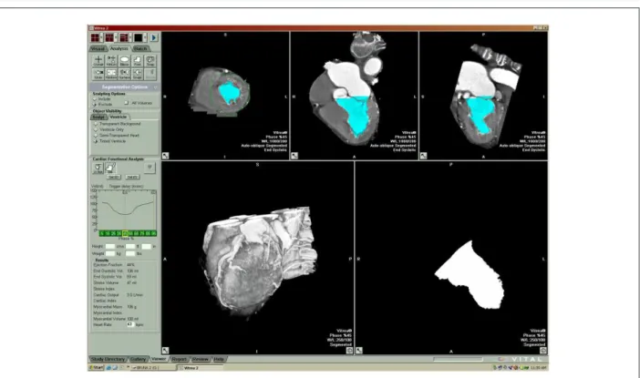
1) Left ventricular end-diastolic volume (LVEDV); 2) Left ventricular end-systolic volume (LVESV); 3) Left ventricular ejection fraction (LVEF). A left ventricular 17-segment model was used for the analysis of left ventricular contractility (Figure 1).
Three-dimensional images were acquired after the two-dimensional echocardiographic study. Images were obtained with the same echocardiography equipment using a
matrix-array transducer with the patient in expiratory breath-hold, and the image was gated to the electrocardiographic recording. The images were stored in the hard disk of the echocardiography equipment and analyzed off-line in a specific software of the equipment (Q Lab, version 5.0, Philips, Andover, MA, USA). The analyses were carried out by two independent observers. Patients whose two-dimensional or three-dimensional echocardiographic images were considered technically inadequate for the analysis of the parameters studied or those presenting significantly irregular heart rhythm were not included in this study. Thus, four patients were excluded from the study: two whose images were considered inadequate for left ventricular analysis, and two who presented significantly irregular heart rhythm.
Computed tomography
Cardiac CT studies were performed in a Toshiba 64-slice multidetector row CT. Nonionic iodinated contrast medium (Henetix- 350 mg/ml) was injected in a peripheral vein at a 5 mL/s speed, followed by infusion of 50 ml of saline solution. The images were acquired with electrocardiographic gating, and an 8-10 second breath-hold period was required. The image parameters included0.4s gantry rotation time, 120kV tube voltage, 400 mA and 64 x 0.5 mm collimation. For the analysis of ventricular function, the images were reconstructed with 1-mm width in ten phases of the cardiac cycle. Intravenous betablocker (metoprolol 5 mg, maximum dose of 20 mg) was used in patients with a higher than 65-bpm heart rate.
Analysis of left ventricular ejection fraction and volumes was carried out off-line in a workstation. Quantitative analysis of the ventricular function was carried out by means of a semi-automated method (area-length method) by two observers using the cardiac short axis in the apical view for the 17-segment model (Figures 2 and 3).
Figure 2 - Tomographic image of the left ventricle (short axis, 4 and 2 chambers) for the determination of reference points for ventricular volume measurement
(in blue).
Figure. 3 - Tomographic image for the three-dimensional determination of ventricular volumes and left ventricular ejection fraction from short-axis images, 4 and 2
Original Article
Vieira et al LV ejection fraction and volumes on RT-3D-echo and CTStatistical analysis
Descriptive, correlation and comparison analyses between the methods were carried out. Descriptive analysis of continuous variables was carried out by observing minimum and maximal values, and calculation of means and standard deviation. The Pearson’s correlation method (r) with 95% confidence interval was used for correlation analysis. Bland & Altman analysis was used for comparison between methods. Linear regression test between the parameters obtained with two and three-dimensional echocardiography was also performed. Measurements were taken by two independent observers. P values <0.05 were considered significant. Data were processed using the MedCalc statistical analysis system, Mariakerke, Belgium.
Results
Descriptive analysis of the echocardiographic and tomographic characteristics (left ventricular ejection fraction and volumes) of the population studied is shown in Table 1. Patients had both ventricular ejection fraction and ventricular volumes (means) within normal limits.
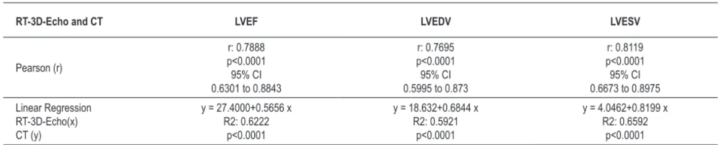
Correlation analysis (Pearson: r) and linear regression equation for the comparison between left ventricular volumes and ejection fraction as measured by three-dimensional echocardiography and computed tomography are shown in Table 2. Good correlation (r) was found for measurement of volumes and ejection fraction with both methods.
Table 2 - Correlation (Pearson:r) and linear regresson equation for the comparison between left ventricular volumes and ejection fraction as
measured by three-dimensional echocardiography and computed tomography in the study population
RT-3D-Echo and CT LVEF LVEDV LVESV
Pearson (r)
r: 0.7888 p<0.0001 95% CI 0.6301 to 0.8843
r: 0.7695 p<0.0001 95% CI 0.5995 to 0.873
r: 0.8119 p<0.0001 95% CI 0.6673 to 0.8975
Linear Regression RT-3D-Echo(x) CT (y)
y = 27.4000+0.5656 x R2: 0.6222
p<0.0001
y = 18.632+0.6844 x R2: 0.5921
p<0.0001
y = 4.0462+0.8199 x R2: 0.6592
p<0.0001
LVEF - left ventricular ejection fraction; LVESV - left ventricular end-systolic volume; LVEDV - left ventricular end-diastolic volume; RT-3D-Echo - real-time three-dimensional
transthoracic echocardiography; CT - computed tomography.
Table 1 - Descriptive analysis of the echocardiographic and tomographic characteristics (left ventricular volumes and ejection
fraction) of the study population
n:39 RT-3D-Echo 64-slice CT
LVEF (%) 56.1 to 78.6(65.5±5.58) (67.8±7.78)53 to 86
LVESV (ml) (33.1±13.6)11.4 to 78 (35.5±13.4)7 to 72
LVEDV (ml) 49.6 to 178.2
(87±27.8)
51 to 186 (106.5±30.3) LVEF - left ventricular ejection fraction; LVESV - left ventricular end-systolic volume; LVEDV - left ventricular end-diastolic volume; RT-3D-Echo - Real-time
three-dimensional transthoracic echocardiography; CT - Computed tomography. Values
are expressed as minimum and maximal values, mean ± standard deviation.
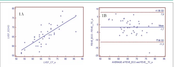
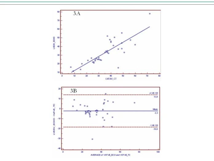
Concordance analysis (Bland & Altman analysis) and linear regression equations for left ventricular ejection fraction and volumes as measured by three-dimensional echocardiography and computed tomography are shown in Graphs 1, 2 and 3.
Good concordance (r) between 3D LVEF and 2D LVEF echocardiographic measurements were observed, with (r): 0.8472, p<0.0001, CI: 0.8254 to 0.9141. The intraobserver concordance for three-dimensional echocardiographic analysis of left ventricular ejection fraction was 0.8634, 95% CI (0.7083 to 0.9436). The interobserver concordance for the three-dimensional echocardiographic analysis of left ventricular ejection fraction was 0.8051, CI (0.5976 to 0.9221).
The intraobserver concordance for the tomographic analysis of left ventricular ejection fraction was 0.8727, 95% CI (0.7437 to 0.9579). The interobserver concordance for the tomographic analysis of left ventricularejection fraction was 0.8347, CI (0.6176 to 0.9291). Concordance (r) between 2D LVEF and LVEF measured by computed tomography was (r): 0.6172, p<0.0001, CI: 0.5654 to 0.8171.
Discussion
Determination of ventricular systolic function and measurement of ventricular volumes can be performed using different noninvasive imaging techniques. Each technique has peculiarities that lead to advantages and limitations to its use. Two-dimensional echocardiography is currently the most widely used method for the anatomical and functional analysis of the heart. It allows anatomical assessment of the different cardiac structures, analysis of cardiac diastole using the different Doppler echocardiography modalities, and can be performed at bedside in intensive care or emergency care environments. However, it has limitations due to suboptimal echocardiographic images in obese patients or those with chronic obstructive pulmonary disease. Also, it depends to a certain extent on the interpretation of the observer and is limited by the use of a small number of anatomical views for the analysis of the heart.
Graphic 1 - Linear regression (Figure 1A) and concordance analysis (Figure 1B) (Bland & Altman plot) for the analysis of left ventricular ejection fraction parameters as
measured by three-dimensional echocardiography (Echo) and computed tomography (CT).
Graphic 2 - Linear regression (Figure 2A) and concordance analysis (Figure 2B) (Bland & Altman plot) for the analysis of left ventricular end-diastolic volume (LVEDV)
parameters as measured by three-dimensional echocardiography (Echo) and computed tomography (CT).
is carried out from new identification planes, without the need for the use of geometric extrapolations for the analysis of complex anatomical shapes (for instance, the right ventricle), which are not always limited to a specific mathematical formulation. However, three-dimensional echocardiography requires specific echocardiography training and a stable heart rhythm for a better analysis, and also depends on the quality of the initial image for best results.
Today, magnetic resonance imaging is considered the reference for the anatomical analysis of most of the structural heart diseases. However, it has limitations regarding costs, or
its use in patients with metallic devices; it requires extensive specific training and is not available in the great majority of the hospitals in Brazil.
64-slice ultrafast computed tomography is a technique that is performed more rapidly than magnetic resonance imaging; it allows imaging of patients with metallic devices as well as the observation of ventricular function together with ventricular volumes and coronary circulation. However, its costs are higher when compared to echocardiographic modalities.
three-Original Article
Vieira et al LV ejection fraction and volumes on RT-3D-echo and CTGraphic 3 - Linear regression (Figure 3A) and concordance analysis (Figure 3B) (Bland & Altman plot) for the analysis of left ventricular end-systolic volume (LVESV)
parameters as measured by three-dimensional echocardiography (Echo) and computed tomography (CT).
dimensional echocardiography and 64-slice computed tomography (r: 0.7888, r: 0.7695, r: 0.8119, p<0.0001, respectively). This finding is consistent with that of a previous study in which a good correlation (r>0.85) was observed between left ventricular volumetric and functional analysis using three-dimensional echocardiography and computed tomography11. For the analysis of ejection
fraction parameters measured by two-dimensional echocardiography and by 64-slice computed tomography, the concordance observed between the methods was lower (r: 0.6172, p<0.0001), but still better than that found in a study with heart transplant patients (r: 0.49)13,and similar
(r: 0.59) to that of a previous study conducted in patients with suspected coronary artery disease in which 16-detector row computed tomography was used12.
Good concordance was observed for ejection fraction parameters measured by three-dimensional and two-dimensional echocardiography (r: 0.8472, p<0.0001). This finding is justified by the observation that left ventricular function and ventricular geometry were preserved in the series of individuals studied. Good reproducibility was also observed regarding ejection fraction measurement by three-dimensional echocardiography (interobserver concordance
of 0.8051) and by computed tomography (interobserver concordance of 0.8347).
The results of the present study show that there was good concordance for the analysis of left ventricular morphology and function between the two noninvasive assessment methods with different possibilities of temporal and spatial observation. The better temporal observation provided by three-dimensional echocardiography allows a more accurate identification of functional cardiac events, whereas the better anatomical definition provided by the tomographic analysis allows a more detailed structural analysis.
In the future, it is important to choose a noninvasive cardiac investigation modality that combines low costs, good reproducibility, diagnostic accuracy, and availability to the low-income population.
Study limitations
within normal limits. These parameters should be further analyzed in patients with ventricular dilatation and contractile dysfunction, as well as in clinical situations accompanied by irregular heart rhythm.
Conclusion
In this case series, a good correlation was observed between ejection fraction measurements, as well as between left ventricular volumes as measured by real-time three-dimensional transthoracic echocardiography and by 64-slice ultrafast computed tomography.
References
1. Roelandt JRT, Yao J, Karsprazak JD. Three-dimensional echocardiography. Curr Opin Cardiol. 1998; 13: 386-98.
2. Kisslo J, Firek B, Ota T, Kang DH, Fleishman CE, Stetten G, et al. Real-time volumetric echocardiography: the technology and the possibilities. Echocardiography. 2000; 17: 773-9.
3. Ahmad M. Real-time three-dimensional echocardiography in assessment of heart disease. Echocardiography. 2001; 18 (1): 73-7.
4. Li J, Sanders SP. Three-dimensional echocardiography in congenital heart disease. Curr Opin Cardiol. 1999; 14: 53-9.
5. Vieira MLC, Pomerantzeff PMA, Leal SB, Mathias Jr W, Andrade JL, Ramires JAF. Ecocardiografia transesofágica tridimensional: acréscimo à informação diagnóstica e à análise anatômica. Rev Bras Ecocardiogr. 2003; 1: 47.
6. Vieira MLC, Ianni BM, Mady C, Encinas J, Pomerantzeff PM, Fernandes PP, et al. Mixoma de átrio esquerdo: avaliação ecocardiográfica tridimensional. Relato de caso. Arq Bras Cardiol. 2004; 82 (3): 281-3.
7. De Simone R, Glombitza G, Vahl CF, Meinzer HP, Hagl S. Three-dimensional Doppler: techniques and clinical applications. Eur Heart J. 1999; 20: 619-27.
8. Bu L, Munns S, Zhang H, Disterhoft M, Dixon M, Stolpen A, et al. Rapid full volume data acquisition by real-time 3-dimensional echocardiography for assessment of left ventricular indexes in children: a validation study compared with magnetic resonance imaging. J Am Soc Echocardiogr. 2005; 18 (4): 299-305.
9. Mannaerts HF, Van Der Heide JA, Kamp O, Papavassilliu T, Marcus JT, Beek
A, et al. Quantification of left ventricular volumes and ejection fraction using freehand transthoracic three-dimensional echocardiography: comparison with magnetic resonance imaging. J Am Soc Echocardiogr. 2003; 16 (2): 101-9.
10. Jenkins C, Bricknell K, Hanekom L, Marwick TH. Reproducibility and accuracy of echocardiographic measurements of left ventricular parameters using real-time three-dimensional echocardiography. J Am Coll Cardiol. 2004; 44 (4): 878-86.
11. Sugeng L, Mor-Avi V, Weinert L, Niel J, Ebner C, Steringer-Mascherbauer R, et al. Quantitative assessment of left ventricular size and function: side-by-side comparison of real-time three-dimensional echocardiography and computed tomography with magnetic resonance refrence. Circulation. 2006; 114 (7): 654-61.
12. Bansal D, Singh RM, Sarkar M, Sureddi R, Mcbreen KC, Griffis T, et al. Assessment of left ventricular function: comparison of cardiac multidetector-row computed tomography with two-dimensional standard echocardiography for assessment of left ventricular function. Int J Cardiovasc Imaging. 2008; 24 (3): 317-25.
13. Ferencik M, Gregory AS, Butler J, Achenbach S, Yeh RW, Hoffmann U, et al. Analysis of cardiac dimensions, mass and function in heart transplant recipients using 64-slice multi-detector computed tomography. J Heart Lung Transplant. 2007; 26 (5): 478-84.
14. Salm LP, Schuijf JD, de Roos A, Lamb HJ, Viegen HW, Jukema JW, et al. Global and regional left ventricular function assessment with 16-detector row CT: comparison with echocardiography and cardiovascular magnetic resonance. Eur J Echocardiogr. 2006; 7 (4): 308-14.
Potential Conflict of Interest
No potential conflict of interest relevant to this article was reported.
Sources of Funding
There were no external funding sources for this study.
Study Association