The role of three-dimensional ultrasound in
pregnancies submitted to cerclage
Thais da Fonseca Borghi1 Mário Henrique Burlacchini de Carvalho1 Antonio Gomes de Amorim Filho1 Silvio Martinelli1 Marcelo Zugaib1 Rossana Pulcineli Vieira Francisco1
1. Department of Obstetrics and Gynaecology, São Paulo University School of Medicine, São Paulo - SP, Brasil
http://dx.doi.org/10.1590/1806-9282.64.07.620
SUMMARY
OBJECTIVE: Cervical cerclage is the standard treatment for cervical incompetence (CI); however, there is still a high risk of preterm birth for women who undergo this treatment. The aim of this study was to longitudinally evaluate findings on two-dimensional transvaginal ultrasonography (2DTVUS) and three-dimensional transvaginal ultrasonography (3DTVUS) that could be related to gestational age at birth.
METHODS: A total of 68 pregnant women who were treated with cerclage were evaluated by 2DTVUS and 3DTVUS in the second and third trimesters of pregnancy. Log-rank tests and Cox regression analyses were used to identify significant findings related to gesta-tional age at delivery.
RESULTS: A cervical length lower than 28.1 mm (p= 0.0083), a proximal cervical length lower than 10 mm (p= 0.0151), a cervical volume lower than 18.17 cm3(p= 0.0152),avascularization index (VI) under 2.153 (p= 0.0044), and a vascularization-flow index (VFI) under 0.961 (p= 0.0059) in the second trimester were all related to earlier delivery. In the third trimester, a cervical length lower than 20.4 mm (p= 0.0009), a VI over 0.54 (p= 0.0327) and a VFI over 2.275 (p= 0.0479) were all related to earlier delivery. Cervical funnelling in the second and third trimesters and proximal cervical length in the third trimester were not related to gestational age at birth. The COX regression analyses showed that cervical volume in the second trimester; FI and VFI in the third trimester were significantly associated with gestational age at birth.
CONCLUSION: In women treated with history-indicated cerclage or ultrasound-indicated cerclage, 2nd trimester cervical volume and 3rd trimester FI and VFI are independent significant sonographic findings associated with time to delivery.
KEYWORDS: Pregnancy. Obstetric labour, premature. Cervix uteri. Cerclage, cervical. Ultrasonography.
INTRODUCTION
Cervical incompetence (CI) is diagnosed based on an obstetric history of second 1,2 or early third
trimester foetal losses 3, following painless cervical
dilation, prolapse or membrane rupture, and expul-sion of a live foetus despite minimal uterine activity. This condition is implicated in 10% to 25% of second trimester pregnancy losses. 4
DATE OF SUBMISSION: 12-Dec-2017
DATE OF ACCEPTANCE: 18-Dec-2017
CORRESPONDING AUTHOR: Mário Henrique Burlacchini de Carvalho
Department of Obstetrics and Gynaecology, São Paulo University School of Medicine 255, Dr. Enéas de Carvalho Aguiar, 255 – São Paulo - SP, Brasil, 05403-000 Tel.: 55 11 26616209, 55 11 983460636
E-mail: [email protected], [email protected]
[email protected] [email protected] [email protected] [email protected] [email protected]
Cervical cerclage is still a standard technique for treating CI, and some reports have estimated that up to 2% of all pregnancies require cerclage procedures
4 Uncontrolled studies have suggested that infant
viability is approximately 25% without cerclage but ranges from 75-90% with cerclage.3
birth rates before 36 weeks vary from 20% – 25% in elective cerclages, 27% – 42% in urgent cerclages, and 53% – 77% in emergent cerclages.2,5
Historically, women treated with cerclage re-ceive digital or speculum follow-up examinations, al-though the usefulness of these procedures has yet to be confirmed by controlled studies.6,7 In contrast, the
introduction of two-dimensional transvaginal ultra-sonography (2DTVUS) for visualizing the cervix and stitches has improved and facilitated both the diag-nosis and follow-up examination of these patients.8
Many studies about the use of 2DTVUS in women treated with cerclage and the prediction of preterm birth been published.
In addition, three-dimensional transvaginal ul-trasonography (3DTVUS) with power Doppler has recently been studied as an option for evaluating the uterine cervix during pregnancy. Previous studies have demonstrated that 3DTVUS can provide more accurate measurements of cervical volume and cer-vical length than 2DTVUS.9-11
There is evidence that angiogenic factors may play a role in cervical ripening and the birth process, and 3DTVUS with power Doppler could provide more in-formation on cervical morphology and vasculariza-tion than 2DTVUS.12 Theoretically, three-dimensional
imaging combined with power Doppler enables the quantitative assessment of volume and Doppler sig-nals of the whole target organ. In contrast, 2DTVUS information on vascularization and blood flow is re-stricted to a single subjectively chosen 2D plane.13
The aim of this study was to longitudinally evalu-ate the findings of 2DTVUS and 3DTVUS with power Doppler that could be related to gestational age at birth in women treated with cerclage.
METHODS
This was a prospective study of women with a singleton pregnancy who underwent elective cer-clage (n= 52) between 12 and 16 weeks of gestation or ultrasound-indicated cerclage (n= 16) between 16 and 24 weeks of gestation. The subjects were fol-lowed at the Recurrent Miscarriage Clinic of the De-partment of Obstetrics and Gynaecology of São Pau-lo University Medical School between June 1, 2012, and October 30, 2015. This study was approved by the University Ethics Committee (CAPPESq 0851/06). All participants in the study provided written informed consent.
The indication for elective cerclage had a history of two or more second trimester pregnancy losses or preterm births with early cervical dilation with-out uterine contractions. Cerclage was indicated by ultrasound when there was a history of one second trimester pregnancy loss or early preterm delivery and a current cervical length of 25 mm or lower up to 24 weeks of gestation.
All women who were treated with elective or ul-trasound-indicated cerclage during this period were invited to participate in this research.
Elective cerclage (n= 52) was placed at 14.06 ± 1.13 weeks, and ultrasound-indicated cerclage (n= 16) was placed at 19.8 ± 2.98 weeks.
Women were included in the study if they met the following criteria: 1) had a singleton gestation of a live foetus; 2) were treated with elective or ul-trasound-indicated cervical cerclage; 3) exhibited no uterine anomalies or cervical conisation; 4) received at least 2 serial transvaginal ultrasound examina-tions for the assessment of cerclage placement and cervical length, one between 20 and 24 weeks (2nd t)
and one between 28 and 32 weeks (3rd t); and 5) had
available delivery records. Patients who were treat-ed with emergency cerclage due to prolapstreat-ed foetal membranes were not included.
All included patients underwent a McDonald cer-clage with a second 5 polyester suture 1 cm below the first stich.
The data obtained included the following: ma-ternal age, race, gestational age at cerclage, cervi-cal length, cervicervi-cal funnelling, proximal cervicervi-cal length, cervical vascular index (i.e., vascularization index (VI), flow index (FI), vascularization-flow in-dex (VFI)), cervical volume, and gestational age at birth. The presence of cervical funnelling was de-fined as a protrusion of membranes into the cervi-cal canal ≥ 5 x 5 mm.
All the exams were performed using a 5 - 9 MHz transvaginal transducer with a 146° field of view (GE Healthcare, Zipf, Austria). The following identi-cal pre-installed settings were used for all patients: a frequency of 3-9 MHz, a pulse repetition frequen-cy of 0.6 kHz, a gain of – 5.0, and a low wall motion filter of 1.
obtained, the probe was gradually advanced again with only enough pressure to restore a satisfactory image. A sagittal view of the cervix where the inter-nal os, the cervical cainter-nal and the exterinter-nal os could all be observed simultaneously was obtained. The cervical length measurements were performed by the first author, who has five years of experience in ultrasounds, as described by Iams et al.14 In cases of
cervical funnelling, the apex of the funnel was con-sidered the beginning of the closed endocervical ca-nal, and the external os was considered the distal end of the endocervical canal. The location of the cerclage was identified as an echo-dense structure in the cer-vical stroma. The system was switched to the power Doppler mode and then into the 3D mode. The cer-vix was centralized within the 3D sector appearing on the ultrasound screen, and data were obtained by holding the transducer stationary while its crystals were mechanically rotated across the sector with a sweep angle of 90°. The fast volume acquisition (i.e., low resolution) setting was always used to minimize periodic flashing artefacts arising from uterine artery pulsation and from foetal movements. The scanned volumes were stored digitally for off-line analysis.
Virtual Organ Computer-Aided Analysis (VO-CALTM) software, which is integrated into the
Volu-son E8 ultrasound system, was used to calculate cer-vical volume (cm3) and power Doppler flow indexes.
The calculated Doppler indexes were the VI, FI, and VFI. The VI is the ratio between the colour voxels
and the total number of voxels in the volume, where a voxel is defined as the smallest unit of each volume; the VI reflects the percentage of the volume consist-ing of blood vessels. The FI is calculated as the sum of weighted colour voxels divided by the number of colour voxels. The FI reflects the average energy per colour voxel reflected from the blood corpuscles in the vessels of the volume; the more blood corpus-cles, the higher the FI values. The VFI is the sum of weighted colour voxels divided by the total number of voxels, and it reflects both the proportion of tissue consisting of vessels and the number of blood corpus-cles in the vessels.
The acquired volumes were manipulated to ob-tain reformatted multi-planar views of the cervix in the mid-sagittal, axial and coronal planes. All cervical measurements were taken on these multi-planar im-ages, and the results were documented on hard cop-ies. The contour mode in the VOCALTM program was
set to manual, and the longitudinal view was used as reference image. The rotation steps were 30°. When drawing the contours, care was taken not to include the lower uterine segment, the vaginal wall, and par-ticularly the large uterine arteries. Once all contours had been drawn, the volume and power Doppler flow indexes of the cervix were computed automatically.
To minimize interobserver variability, a single so-nographer performed all the exams.
The threshold for significance was established at p < 0.05.
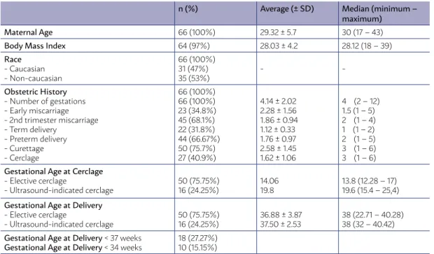
TABLE 1: DEMOGRAPHIC CHARACTERISTICS AND OBSTETRIC DATA.
n (%) Average (± SD) Median (minimum –
maximum)
Maternal Age 66 (100%) 29.32 ± 5.7 30 (17 – 43)
Body Mass Index 64 (97%) 28.03 ± 4.2 28.12 (18 – 39)
Race
- Caucasian - Non-caucasian
66 (100%) 31 (47%) 35 (53%)
-
-Obstetric History
- Number of gestations - Early miscarriage - 2nd trimester miscarriage - Term delivery
- Preterm delivery - Curettage - Cerclage
66 (100%) 66 (100%) 23 (34.8%) 45 (68.1%) 22 (31.8%) 44 (66.67%) 50 (75.7%) 27 (40.9%)
4.14 ± 2.02 2.28 ± 1.56 1.86 ± 0.94 1.12 ± 0.33 1.76 ± 0.97 2.58 ± 1.45 1.62 ± 1.06
4 (2 – 12) 1.5 (1 – 5) 2 (1 – 4) 1 (1 – 2) 2 (1 – 5) 3 (1 – 6) 3 (1 – 6)
Gestational Age at Cerclage
- Elective cerclage
- Ultrasound-indicated cerclage
50 (75.75%) 16 (24.25%)
14.06 19.8
13.8 (12.28 – 17) 19.6 (15.4 – 25,4)
Gestational Age at Delivery
- Elective cerclage
- Ultrasound-indicated cerclage
50 (75.75%) 16 (24.25%)
36.88 ± 3.87 37.50 ± 2.53
38 (22.71 – 40.28) 38 (32 – 40.42)
Gestational Age at Delivery < 37 weeks
Gestational Age at Delivery < 34 weeks
A Kaplan-Meier Curve was developed for each so-nographic characteristic, and the log-rank test was used for continuous variables to form two groups and maximize the differences between them in the survival curves. A Cox regression model was used to identify risk factors related to earlier delivery. The evaluated time periods were analysed separately. The following were considered co-variables for each time periods: funnelling, proximal cervical length, cervical volume, VI, FI, VFI, and cervical length.
RESULTS
68 women referred for cerclage met the study inclusion criteria. Two patients treated with elec-tive cerclage were excluded from the analysis; one was excluded due to spontaneous miscarriage after cerclage at 19 weeks, and the other was excluded due to foetal death at 20 weeks of gestation. The de-mographic characteristics and obstetric data of the study population are described in table 1.
The average gestational age ± standard deviation at the first and second ultrasound exams was 22.23 weeks ± 1.33 and 29.87 weeks ± 1.46, respectively.
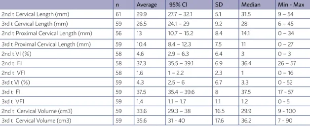
The sonographic findings of 2DTVUS and 3DTVUS
with power Doppler are shown in table 2. Cervical funnelling was present in 16 (26%) patients between 20 and 24 weeks and in 16 (27%) patients between 28 and 32 weeks.
Gestational age at delivery was evaluated as a con-tinuous variable. The Kaplan-Meier curves showed an increased frequency of earlier delivery with a cervical length lower than 28.1 mm (p= 0.0083), a proximal cervical length lower than 10 mm (p= 0.0151), a cervi-cal volume lower than 18.17 cm3 (p= 0.0152),aVI under
2.153 (p= 0.0044), and a VFI under 0.961 (p= 0.0059) in the second trimester (figure 1). In the third trimester, a cervical length lower than 20.4 mm (p= 0.0009), a VI over 0.54 (p= 0.0327) and a VFI over 2.275 (p= 0.0479) were related to earlier delivery (figure 2). Cervical fun-nelling between 20 and 24 weeks, and cervical funnel-ling and proximal cervical length between 28 and 32 weeks were not significant predictors.
The COX regression results showed that for the second trimester ultrasound, a cervical volume ≥ 18.17 was an independent variable that was signifi-cantly associated with later gestational age at birth. Furthermore, in the third trimester, a FI ≥ 44.336 and a VFI ≥ 2.275 were associated with gestational age at birth (table 3).
TABLE 2: ULTRASONOGRAPHIC FINDINGS FROM 2DTVUS AND 3DTVUS IN THE SECOND AND THIRD
n Average 95% CI SD Median Min - Max
2nd t Cervical Length (mm) 61 29.9 27.7 – 32.1 5.1 31.5 9 – 54
3rd t Cervical Length (mm) 59 26.5 24.1 – 29 9.2 28 6 – 45
2nd t Proximal Cervical Length (mm) 56 13 10.7 – 15.2 8.4 14.1 0 – 34
3rd t Proximal Cervical Length (mm) 59 10.4 8.4 – 12.3 7.5 11 0 – 27
2nd t VI (%) 58 4.6 2.9 – 6.3 6.4 3 0 – 3
2nd t FI 58 37.3 35.5 – 39.1 6.9 36.4 26 – 57
2nd t VFI 58 1.6 1 – 2.2 2.3 1 0 – 16
3rd t VI (%) 59 4.3 2.5 – 6 6.7 3.3 0 - 52
3rd t FI 59 37.5 35.4 – 39.6 8 37.5 17 - 57
3rd t VFI 59 1.4 1.1 – 1.7 1.1 1.2 0 - 5
2nd t Cervical Volume (cm3) 59 33.6 29.3 – 38 16.5 29.9 9 - 100
3rd t Cervical Volume (cm3) 59 35.6 31 - 40 17.6 36.2 7 - 90
2nd t: second trimester; 3rd t: third trimester; CI: confidence interval; SD: standard deviation; Min - Max: minimum - maximum.
TABLE 3: COX REGRESSION MODEL FOR ULTRASOUND PARAMETERS IN THE SECOND AND THIRD
TRIMESTERS WITH RESPECT TO GESTATIONAL AGE AT BIRTH.
Variable Estimate Standard
Error
p Value HR 95% CI for HR
Lower Upper
2nd t Cervical Volume ≥ 18.17 cm3 -1.029 0.494 0.037 0.357 0.136 0.941
3rd t FI ≥ 44.336 -1.256 0.423 0.003 0.285 0.124 0.653
mm measured between 18 and 24 weeks and deliv-ery before 34 weeks; however, when these authors considered gestational age as a continuous variable, a cervical length lower than 25 mm was not a signif-icant factor (p= 0.051). In contrast, O’Brien et al.18,
Rust et al.19 and Hedriana et al.20 did not find any
relationship between cervical length measured after cerclage and gestational age at birth. These findings could be explained by factors of cohort heterogene-ity, including a history of second trimester pregnan-cy loss, a history of diethylstilboestrol exposure in utero, cervical conisation, uterine anomalies and treatment with history-indicated, ultrasound-indi-cated and emergency cerclage.
A second trimester proximal cervical length < 10 mm was significantly associated with an earlier deliv-ery. Previous studies have already demonstrated this relationship in pregnant women treated with histo-ry-indicated7, 17, ultrasound-indicated7 and
emergen-cy15 cerclage. However, Hedriana et al.20 found
differ-ent results; they measured proximal cervical length at an average gestational age of 26 ± 4.4 weeks (ges-tational age ± SD) and found that this measurement was not useful for differentiating preterm birth from term birth. The difference between the present study and that study was that we evaluated gestational age as a continuous variable, while Hedriana et al.20
es-tablished the end point as a categorical variable. A few studies have suggested that compared with 2DTVUS, a 3DTVUS examination would allow a more complete assessment of the cervix.9, 11 Nevertheless,
this is the first study regarding 3DTVUS in women treated with cerclage. Our results showed a signifi-cant relationship between a second trimester cervi-cal volume < 18.17 cm3 and an earlier delivery.
Most studies about cervical volume during preg-nancy are related to low-risk patient7, 12, 21 parity and
previous delivery modes22. Rovas et al.12 found no
dif-ferences in the cervical volume of 677 women during low-risk pregnancy; however, their data showed a sig-nificant difference in cervical volume between parous and nulliparous pregnant women. Park et al.23 found
an inverse association between cervical volume mea-sured at 20-24 weeks of gestation and the risk for spontaneous preterm birth before 36 weeks of gesta-tion in pregnant women at a low risk for preterm birth. Similar to our results, they found that the smaller the cervical volume, the higher the likelihood of preterm birth. In a study of 28 pregnant women hospitalized for preterm labour, Rozenberg et al.24 reported that DISCUSSION
This study aimed at evaluating the usefulness of 2DTVUS and 3DTVUS for investigating gestational age at birth in pregnant women treated with cerclage. In this group of patients, a second trimester cer-vical length < 28.1 mm and a third trimester cercer-vical length < 20.4 mm were related to earlier delivery, and these findings are in agreement with current obstet-ric data. Guzman et al.15 evaluated 29 women who
underwent emergency cerclage at 16 to 26 weeks of gestation and found similar results; these authors found significant differences in postoperative endo-cervical canal length between patients who delivered at < 36 versus ≥ 36 weeks. Dijkstra et al. 6 studied 32
women treated with elective or ultrasound-indicat-ed cerclage and found a significant relationship be-tween gestational age at delivery and cervical length between 28 and 32 weeks (r= 1.4, p= 0.002). The av-erage cervical length measured between 28 and 32 weeks was significantly different in women who de-livered preterm compared with those who dede-livered at full term (21.0 ± 5.7 compared with 30.3 ± 9.5 mm, respectively; p= 0.002). Song et al.16 studied a group
of 52 pregnant women treated with elective cerclage and found a significant relationship between cervical length after cerclage and gestational age at delivery before 32 weeks. Miller et al.17 studied 124 women
treated with elective cerclage and found a significant relationship between a cervical length of less than 25
cervical volume increases the positive predictive val-ue of preterm birth. This study reported that the op-timal cervical volume cut-off point for differentiating full term and preterm birth was 20 mm3. In contrast,
Hoesli et al.25 did not find a significant difference in
cervical volume between pregnant women with low and high risk for preterm birth.
When examining gestational age as a continuous variable, the Kaplan-Meier curves showed that the VI and VFI in the 2nd trimester and the VI and VFI in the
3rd trimester were related to gestational age at birth.
We noted that a reduced VI and VFI in the 2nd
trimes-ter were related to earlier delivery, in contrast with an increased VI and VFI in the 3rd trimester. Rovas
et al.12 and Yilmaz et al.21 did not find differences in
these cervical indexes when analysing low-risk preg-nant women in relation to gestational age at delivery.
There are few studies about cervical vascular in-dexes and gestational age at delivery. To our knowl-edge, this is the first study using 3DTVUS with power Doppler to examine women treated with cerclage. De Diego et al.26 studied 29 women with an
asymptom-atic short cervix in the 2nd trimester of pregnancy
and 71 women with threatened preterm labour; they compared these two groups of women in relation to cervical length, cervical volume, VI, FI and VFI. In the group of women admitted for preterm labour, there was a difference in cervical length (18.3 versus 14.9 mm, p= 0.014) between those with at full term and preterm delivery, respectively, but no differenc-es were found in cervical volume, VI, FI or VFI. The authors also found that cervical volume was lower in women with threatened preterm labour than in as-ymptomatic women with the same cervical length; in addition, VI and VFI were higher in women with threatened preterm labour, which reinforced the idea that the cervix increases its vascularization and flow to prepare for labour. The different results we found regarding cervical vascular indexes could be due to the cerclage stitch. Cox regression analyses were used to identify which ultrasound variable could be considered a risk factor for preterm birth; the results showed that a cervical volume ≥ 18.17 cm3 in the 2nd
trimester and a FI ≥ 44.336 in the 3rd trimester
re-duced the risk for preterm birth, whereas a VFI ≥ 2.275 in the 3rd trimester was associated with earlier
delivery. While this behaviour was unexpected for FI, it could be explained by the fact that FI is not an indicator of perfusion and cannot provide informa-tion on the volume of blood being pumped through
a vessel during a particular period. The real meaning of the FI is unclear, and the FI is less predictable than the VI and VFI.27 Furthermore, Park et al.23 have
al-ready reported an association between small cervical volume (≤ 20 cm3) and preterm birth
In our study, standardized equipment settings were used to avoid significant effects on our results, and the use of a transvaginal probe to evaluate the cervix theoretically reduces the influence of attenua-tion on the vascular index.
A methodological difficulty when estimating cer-vical volume and vascularity using 3D ultrasound is defining landmarks when drawing the contours of the cervix. Rovas et al.12 and Hoesli et al.25 have also
noted this difficulty. The delineation between the cervix and the lower uterine segment is particular-ly difficult, especialparticular-ly during earparticular-ly pregnancy and at mid-gestation, when the lower uterine segment is thick and the cervix is often curved. It may also be difficult to clearly distinguish the cervix from the surrounding vaginal tissue.
While we found that according to Kaplan-Meyer curves, the VI and VFI were significantly associated with earlier delivery in women treated with cerclage, the clinical importance of these findings for the vascu-larization of the cervix has yet to be fully understood.
CONCLUSION
In women treated with history-indicated cerclage or ultrasound-indicated cerclage, 2nd trimester
cer-vical volume and 3rd trimester FI and VFI are
inde-pendent significant sonographic findings associated with time to delivery.
COMPLIANCE WITH ETHICAL STANDARDS:
The authors declare that they have no conflict of interest.
All procedures performed in this study involving human participants were in accordance with the ethical standards of the institutional and/or nation-al research committee and with the 1964 Helsinki declaration and its later amendments or comparable ethical standards.
This article does not contain any studies with an-imals performed by any of the author
RESUMO
OBJETIVOS: Determinar quais características ultrassonográficas obtidas por meio da ultrassonografia transvaginal bidimensional (USG TV 2D) e tridimensional (USG TV 3D) associam-se ao parto prematuro em gestantes submetidas à cerclagem profilática e terapêutica.
MÉTODOS: Sessenta e seis gestantes com feto único submetidas à cerclagem profilática ou terapêutica e acompanhadas no ambu-latório de Aborto Habitual da Clínica Obstétrica do Hospital das Clínicas da Faculdade de Medicina da USP, entre 10 de juho de 2012 e 30 de outubro de 2015, foram avaliadas longitudinalmente, por meio das US TV 2D e US TV 3D associadas ao power Doppler para avaliação do VI, FI e VFI , nos três trimestres da gestação. Os resultados foram avaliados em relação ao parto em idade gestacional (IG) menor que 34 semanas e maior ou igual a 34 semanas, assim como em relação à idade do parto como variável contínua.
RESULTADOS: O comprimento do colo uterino (CC) e a distância do ponto de cerclagem ao orifício interno do colo uterino (POI) di-minuíram de forma significativa entre o segundo e terceiro trimestres da gestação. O CC, o POI e o afunilamento cervical no terceiro trimestre da gestação tiveram relação com a ocorrência de parto em IG<34 semanas. Na análise de regressão de COX, em que a variável de interesse foi o tempo até o parto, o volume do colo uterino no segundo trimestre e o FI e VFI no terceiro trimestre foram significativos.
CONCLUSÃO: Foi possível identificar parâmetros ultrassonográficos do colo uterino bi e tridimensionais que se correlacionam com a idade gestacional do parto.
PALAVRAS-CHAVE: Gravidez. Trabalho de parto prematuro. Colo do útero. Cerclagem cervical. Ultrassonografia.
REFERENCES
1. American College of Obstetricians and Gynecologists. ACOG Practice Bulletin No.142: Cerclage for the management of cervical insufficiency. Obstet Gynecol. 2014;123(2 Pt 1):372-9.
2. Nelson L, Dola T, Tran T, Carter M, Luu H, Dola C. Pregnancy outcomes following placement of elective, urgent and emergent cerclage. J Matern Fetal Neonatal Med. 2009;22(3):269-73.
3. Lotgering FK. Clinical aspects of cervical insufficiency. BMC Pregnancy Childbirth. 2007;7(Suppl 1):S17.
4. Ouzounian JG, Korst LM, Lee RH. Cervical length and delivery out-comes in patients with prophylactic cervical cerclage. Am J Perinatol. 2011;28(4):273-6.
5. Khan MJ, Ali G, Al Tajir G, Sulieman H. Evaluation of outcomes associated with placement of elective, urgent, and emergency cerclage. J Obstet Gy-naecol India. 2012;62(6):660-4.
6. Dijkstra K, Funai EF, O’Neill L, Rebarber A, Paidas MJ, Young BK. Change in cervical length after cerclage as a predictor of preterm delivery. Obstet Gynecol. 2000;96(3):346-50.
7. Andersen HF, Karimi A, Sakala EP, Kalugdan R. Prediction of cervical cer-clage outcome by endovaginal ultrasonography. Am J Obstet Gynecol. 1994;171(4):1102-6.
8. Kurup M, Goldkrand JW. Cervical incompetence: elective, emergent, or urgent cerclage. Am J Obstet Gynecol. 1999;181(2):240-6.
9. Bega G, Lev-Toaff A, Kuhlman K, Berghella V, Parker L, Goldberg B, et al. Three-dimensional multiplanar transvaginal ultrasound of the cervix in pregnancy. Ultrasound Obstet Gynecol. 2000;16(4):351-8.
10. Chou CY, Hsu KF, Wang ST, Huang SC, Tzeng CC, Huang KE. Accuracy of three-dimensional ultrasonography in volume estimation of cervical car-cinoma. Gynecol Oncol. 1997;66(1):89-93.
11. Severi FM, Bocchi C, Florio P, Picciolini E, D’Aniello G, Petraglia F. Com-parison of two-dimensional and three-dimensional ultrasound in the as-sessment of the cervix to predict preterm delivery. Ultrasound Med Biol. 2003;29(9):1261-5.
12. Rovas L, Sladkevicius P, Strobel E, Valentin L. Reference data representa-tive of normal findings at three-dimensional power Doppler ultrasound examination of the cervix from 17 to 41 gestational weeks. Ultrasound Ob-stet Gynecol. 2006;28(6):761-7.
13. Yigiter AB, Kavak ZN. Cervical volume and flow indices during pregnancy by transvaginal 3-dimensional ultrasonography and doppler angiography. Timisoara Med J. 2009;58(2):137-42.
14. Iams JD, Goldenberg RL, Meis PJ, Mercer BM, Moawad A, Das A, et al. The length of the cervix and the risk of spontaneous premature delivery. Na-tional Institute of Child Health and Human Development Maternal Fetal Medicine Unit Network. N Engl J Med. 1996;334(9):567-72.
15. Guzman ER, Houlihan C, Vintzileos A, Ivan J, Benito , Kappy K. The significance of transvaginal ultrasonographic evaluation of the cer-vix in women treated with emergency cerclage. Am J Obstet Gynecol. 1996;175(2):471-6.
16. Song RK, Cha HH, Shin MY, Choi SJ, Oh SY, Kim JH, et al. Post-cerclage ultrasonographic cervical length can predict preterm delivery in elective cervical cerclage patients. Obstet Gynecol Sci. 2016;59(1):17-23.
17. Miller ES, Gerber SE. Association between sonographic cervical appear-ance and preterm delivery after a history-indicated cerclage. J Ultrasound Med. 2014;33(12):2181-6.
18. O’Brien JM, Hill AL, Barton JR. Funneling to the stitch: an informative ul-trasonographic finding after cervical cerclage. Ultrasound Obstet Gynecol. 2002;20(3):252-5.
19. Rust OA, Atlas RO, Meyn J, Wells M, Kimmel S. Does cerclage location influence perinatal outcome? Am J Obstet Gynecol. 2003;189(6):1688-91. 20. Hedriana HL, Lanouette JM, Haesslein HC, McLean LK. Is there value for
serial ultrasonographic assessment of cervical lengths after a cerclage? Am J Obstet Gynecol. 2008;198(6):705.e1-6.
21. Yilmaz NC, Yiğiter AB, Kavak ZN, Durukan B, Gokaslan H. Longitudinal examination of cervical volume and vascularization changes during the antepartum and postpartum period using three-dimensional and power Doppler ultrasound. J Perinat Med. 2010;38(5):461-5.
22. Jo YS, Jang DG, Kim N, Kim SJ, Lee G. Comparison of cervical parameters by three-dimensional ultrasound according to parity and previous delivery mode. Int J Med Sci. 2011;8(8):673-8.
23. Park IY, Kwon JY, Kwon JY, Hong SC, Choi HM, Kwon HS, et al. Usefulness of cervical volume by three-dimensional ultrasound in identifying the risk for preterm birth. Ultrasound Med Biol. 2011;37(7):1039-45.
24. Rozenberg P, Rafii A, Sénat MV, Dujardin A, Rapon J, Ville Y. Predictive value of two-dimensional and three-dimensional multiplanar ultrasound evaluation of the cervix in preterm labour. J Matern Fetal Neonatal Med. 2003;13(4):237-41.
25. Hoesli IM, Surbek DV, Tercanli S, Holzgreve W. Three dimensional volume measurement of the cervix during pregnancy compared to conventional 2D-sonography. Int J Gynaecol Obstet. 1999;64(2):115-9.
26. De Diego R, Sabrià J, Vela A, Rodríguez D, Gómez MD. Role of 3-dimen-sional power Doppler sonography in differentiating pregnant women with threatened preterm labour from those with an asymptomatic short cervix. J Ultrasound Med. 2014;33(4):673-9.
27. Pairleitner H, Steiner H, Hasenoehrl G, Staudach A. Three-dimensional power Doppler sonography: imaging and quantifying blood flow and vas-cularization. Ultrasound Obstet Gynecol. 1999;14(2):139-43.
MHB Carvalho: Protocol/project development; data analysis; Manuscript writing/editing, supervisor AG Amorim Filho: Protocol/project development; data