UNIVERSIDADE FEDERAL DA PARAÍBA CENTRO DE CIÊNCIAS DA SAÚDE
DEPARTAMENTO DE CLÍNICA E ODONTOLOGIA SOCIAL
PROGRAMA DE PÓS-GRADUAÇÃO EM ODONTOLOGIA- NÍVEL MESTRADO COM ÁREA DE CONCENTRAÇÃO EM DIAGNÓSTICO BUCAL
RACHEL REINALDO ARNAUD
DENSIDADE DE MASTÓCITOS EM LESÕES DE QUEILITE ACTÍNICA
RACHEL REINALDO ARNAUD
DENSIDADE DE MASTÓCITOS EM LESÕES DE QUEILITE ACTÍNICA
Dissertação apresentada ao Programa de Pós- Graduação em Odontologia da Universidade Federal da Paraíba para obtenção do título de Mestre em Diagnóstico Bucal.
Orientador(a): Prof.(a) Dr.(a) Maria Sueli Marques Soares Co-Orientador(a): Cláudia Cazal Lira
A744d Arnaud, Rachel Reinaldo.
Densidade de mastócitos em lesões de queilite actínica / Rachel Reinaldo. – João Pessoa: [s.n.], 2011.
70 f. : il.
Orientadora: Maria Sueli Marques Soares. Coorientadora: Cláudia Cazal Lira.
Dissertação (Mestrado) – UFPB/CCS.
1. Mastócitos. 2. Queilite actínica. 3. Carcinoma de células escamosas.
RACHEL REINALDO ARNAUD
DENSIDADE DE MASTÓCITOS EM LESÕES DE QUEILITE ACTÍNICA
Dissertação apresentada ao Programa de Pós- Graduação em Odontologia da Universidade Federal da Paraíba para obtenção do título de Mestre em Diagnóstico Bucal.
Data da Defesa : 05/12/2011
Banca Examinadora
Prof . Dr . Jean Nunes dos Santos (Universidade Federal da Bahia - Examinador)
Prof a. Dr a. Claudia Roberta Leite Vieira de Figueiredo (Universidade Federal da Paraíba - Examinadora)
Prof a. Dr a. Maria Sueli Marques Soares
(Universidade Federal da Paraíba - Orientadora)
DEDICATÓRIA
Dedico este trabalho à Deus, aos meus pais
e aos meus irmãos, razão de tudo que eu faço
AGRADECIMENTOS
À Deus, por ser Presença em minha vida e luz nos meus caminhos. Aos meus pais, Reinaldo e Socorro, pelo amor incondicional.
Aos meus irmãos pela amizade, afeto e companheirismo constantes. Ao meu irmão Reinaldo, um agradecimento especial, pelos ensinamentos de vida e exemplo de honestidade, responsabilidade e perseverança.
Aos meus familiares e cunhados, pelas demonstrações de carinho. Aos meus sobrinhos, pelo simples fato de existirem e sorrirem para mim.
À minha orientadora, mestre e amiga, Prof a. Dr a. Maria Sueli Marques Soares, pela
contribuição, experiência, incentivo e paciência dedicados durante o desenvolvimento deste trabalho.
À Prof a. Claudia Cazal Lira, pelos ensinamentos e valiosa contribuição a este estudo.
Ao Prof. Frederico Barbosa de Sousa, por disponibilizar o Laboratório de Microscopia e Imagem Biológica – CCS/UFPB para análise dos dados.
Aos professores da Disciplina de Estomatologia, por ampliarem meus conhecimentos e dispertarem em mim o interesse pela Estomatologia.
Ao Hospital Napoleão Laureano, pelo fornecimento dos casos de Queilite Actinica usados no presente estudo.
Ao laboratório de Patologia da UFPB, pelo fornecimento de material usado no presente estudo.
Aos funcionários do laboratório de Histologia e Patologia da UFPB, pela contribuição nos cortes e colorações deste trabalho.
A todos que direta ou indiretamente contribuíram para que esse trabalho fosse realizado.
“A mente que se abre a uma nova idéia jamais voltará ao seu
tamanho original.”
RESUMO
O objetivo do estudo foi avaliar a densidade de mastócitos nas lesões de queilite actínica e sua correlação com os processos de inflamação e displasia epitelial, comparando a controle normal. Para o grupo queilite actínica, selecionaram-se 33 casos de registrados no Serviço de Cirurgia de Cabeça e Pescoço do Hospital do Napoleão Laureano, PB. E para o controle, 9 blocos com diagnóstico de mucocele Os blocos parafinados foram cortados e corados em Hematoxilina e Eosina e por Azul de toluidina. A contagem dos mastócitos foi realizada em 8 campos por espécimes. Realizou-se análise estatística descritiva e aplicaram-se testes U de Mann-Whitney, Kuskall-Wallis, Qui-Quadrado e o coeficiente de Spearman, considerando p<0,05. No grupo queilite actínica, 57,6% apresentaram algum grau de displasia epitelial, sendo 39,4% displasia leve, 15,2% moderada e 3% severa. Em 21,2% da amostra foi observado carcinoma de células escamosas de lábio. A presença de elastose solar foi observada em 81,8% dos casos e algum grau de inflamação em 84,9%, sendo 39,4% leve, 15,2% moderado e 30,3% intenso. Os mastócitos estavam presentes em 87,8% dos casos. A média de mastócitos no grupo queilite actínica foi de 17,42±10,43 células/μm² e no controle 1,78±1,64 células/μm², com diferença estatisticamente significante (p<0,001). A densidade dos mastócitos foi significativamente maior nos casos de carcinoma com p=0,001. Houve correlação estatisticamente significante entre densidade de mastócitos e os processos de displasia (p=0,004) e infiltrado inflamatório(p=0,000); e entre displasia epitelial e infiltrado inflamatório (p=0,002). O aumento da densidade dos mastócitos sugere uma possível participação dessas células no processo de transformação maligna da lesão.
ABSTRACT
The objective of this study was to evaluate the density of mast cells in the lesions of actinic cheilitis and its correlation with the processes of inflammation and epithelial dysplasia. We selected 33 cases of actinic cheilitis recorded in the Department of Head and Neck Surgery, Hospital Dr. Laureano Napoleon, PB. The paraffin blocks were cut and stained with hematoxylin and eosin and toluidine blue. The count of mast cells was performed in 8 fields per case and final reading with the average value expressed. Analysis was descriptive and inferential statistics, and tests were applied to the Mann-Whitney U, Kuskall-Wallis test, chi-square and Spearman coefficient, considering p <0.05. Of the total sample, 57.6% had some degree of epithelial dysplasia, mild dysplasia was 39.4%, 15.2% moderate and 3% severe. In 21.2% of the sample was observed squamous cell carcinoma of the lip. The presence of solar elastosis was observed in 81.8% and some degree of inflammation in 84.9%, as well as 39.4% mild, 15.2% moderate and 30.3% intense. Mast cells were present in 87,8% of cases. The average number of mast cells in specimens of actinic cheilitis was 17.42 ± 10.43 cells / μm² and normal mucosa 1.78 ± 1.64 cells / μm², and this difference was statistically significant (p <0.001). The density of mast cells was significantly higher in cancer cases with p = 0.001. There was a statistically significant correlation between the density of mast cells and the processes of dysplasia (p = 0.004) and inflammatory cell infiltration (p = 0.000) and between epithelial dysplasia and inflammatory cell infiltration (p = 0.002). The increased density of mast cells suggests a possible role of these cells in the malignant transformation of the lesion.
SUMÁRIO
1 INTRODUÇÃO GERAL... 9
2 OBJETIVOS... 12
2.1 OBJETIVO GERAL ... 13
2.2 OBJETIVOS ESPECÍFICOS... 13
2 CAPÍTULOS... 14
CAPÍTULO 1... 16
CAPÍTULO 2... 31
CAPÍTULO 3... 43
3 DISCUSSÃO GERAL... 55
4 CONCLUSÃO GERAL... 60
REFERÊNCIAS GERAIS... 62
APÊNDICE... 64
10
A queilite actínica (QA) é uma importante lesão bucal com potencial de transformação maligna podendo originar carcinoma de células escamosas (CCE) de lábio (VAN DER WALL, 2009); e que afeta grande número de pessoas que se expõem excessivamente à radiação solar (PERUZETTO, 2006).
A radiação ultravioleta (UV) tem propriedade imunomoduladora e inflamatória. Estudos mostram que os mastócitos estão significantemente aumentados na pele exposta a radiação UV (GONZALEZ, MORAN, KOCHEVER, 1999). O mesmo fasto foi evidenciado em lesões de Q.A (ROJAS et al., 2004) e em carcinoma de lábio (ROJAS et al., 2005) . Autores como Rojas et al. (2004) apontam a necessidade de estudos que possam analisar a contribuição dos mastócitos no processo de malignização da QA.
Os mastócitos são células de grande impacto sobre o microambiente tecidual e reconhecidas como importantes efetoras nas alterações causadas pela radiação ultravioleta (UV) na pele, acredita-se que contribuem no microambiente lesional facilitando a carcinogênese e metástase (HUANG et al., 2008).
Acredita-se que no microambiente tumoral haja fatores capazes de influenciar vários processos celulares tais como o crescimento, morte, diferenciação, migração e invasão, de modo que, o infiltrado inflamatório tumor-associado pode contribuir para a tumorogênese (BALKWILL, MANTOVANI, 2001). E, neste contexto, a participação dos mastócitos tem sido questionada e estudada por vários autores.
maligno da carcinogênese (COUSSENS et al., 1999). A presença de mastócitos e sua distribuição em lesões com potencial de transformação maligna e em lesões malignas indicam possível associação dessas células com a severidade das lesões, bem como com o aumento da imunossupressão provocada pela radiação UV (GOMES et al., 2008; ARAÚJO et al., 2010). Por outro lado, outros estudos sugerem que os mastócitos exercem efeito tumoral antagônico e apresentam efeito inibitório sobre a proliferação de ceratinócitos (HUTTUNEN et al., 2001; THEOHARIDES; CONTI, 2004).
2.1 OBJETIVO GERAL
O objetivo do presente estudo foi verificar a participação dos mastócitos no processo de transformação maligna da QA para CCE através da determinação da sua presença e densidade nos diversos graus de displasia, carcinoma e outros achados histológicos como a presença do processo inflamatório.
2.2 OBJETIVOS ESPECÍFICOS
Correlacionar à densidade dos mastócitos em QA e mucosa normal.
Correlacionar à densidade dos mastócitos com os diversos graus de displasia
epitelial e CCE.
Correlacionar à densidade dos mastócitos com a presença de infiltrado
inflamatório.
Estes capítulos são compostos por 3 artigos. O primeiro, descrito no Capítulo 1, é uma revisão sistemática intitulada “Mast cell participation on malignant
Artigo em processo de envio para periódico International Journal of Dermatology.
Mast cell participation on malignant transformation of Actinic Cheilitis: A systematic review.
SOARES MSM1
ARNAUD RR2
LIRA CC3
Universidade Federal da Paraíba
1 Associated Professor, Department of Clinical and Social Dentistry, School of
Dentistry, Universidade Federal da Paraíba, João Pessoa, Brazil;
2 Postgraduate student, Department of Clinical and Social Dentistry, School of Dentistry, Universidade Federal da Paraíba, João Pessoa, Brazil;
2
Keywords: mast cell, actinic cheilitis, carcinogenesis.
Abstract
Background: No one knows the role of mast cells in the pathogenesis and malignant
transformation of actinic cheilitis (AC) as well as in the carcinogenesis of squamous cell
carcinoma of the lip (SCC).
Objective: Assess the relationship of the presence of mast cells in the pathogenesis and
malignancy of QA through a systematic literature review.
Methods: Electronic search was performed on the primary information basis Medline (via
PubMed), Embase and Cochrane Central Clinical Trials for full-text articles, in English,
published until August 2011 and that cited the level of association between mast cells and
the process of malignancy of AC and / or lip cancer. Terms employed were: mast cell and
actinic cheilitis, and mast cell and lip cancer. The search was limited to the titles of the
articles. The Newcastle-Ottawa scale for qualifying the articles was used.
Results: The assessment was concluded with nine articles on the subject studied, eight of
them of high quality and one of moderate quality. In all studies, the mast cells were
identified both in AC and in SCC of lips and statistically significant differences, as to the
increased density of these cells in AC and SCC, were identified when compared to normal
mucosa. The location of mast cells in tissues was varied, but always related to the region of
the only lamina, solar elastosis and stromal tumor.
Conclusion: There is evidence of the relationship between the presence of mast cells and
Introduction
Actinic cheilitis is a frequent lesion in the lower lip and is characterized by a chronic
inflammatory process caused by excessive exposure of the lips to ultravioleta radiation 1-4. It is considered a potentially malignant lesion in view of its possible transformation to a
squamous cell carcinoma (SCC) of lips3.
Mast cells are connective tissue cells of mesenchymal origin whose chemical
mediators act in the innate and acquired immune response, as well as in the degradation of
extracellular matrix and activate the collagenase providing spaces for the neovascularization
necessary to the recovery process5-6. The mast cell degranulation results in the release of specific substances, among them the tryptase, kinase, MMP, heparin, histamnia, fibroblast
growth factor, TNF-α, interleukins, chemokines and lipid mediator5. In the tumor microenvironment, mast cells are important effector cells of the deleterious effects of
ultraviolet radiation, which can facilitate the process of carcinogenesis and metástase7. Studies show that the density of mast cells is increased in actinic cheilitis, and SCC of the
lip8-9. Some authors claim that the accumulation of these cells is associated with the progression and prognosis of the tumor10-12.
Although several studies investigate the involvement of mast cells in
photocarcinogenesis particularly skin and lip lesions 9, the data are controversial and the real
role of mast cells in the tumor microenvironment of these lesions is still unknown. The issue
has aroused interest and requires more attention, therefore, it is proposed in this study to
perform a systematic review aiming to analyze the literature concerning the presence of mast
cells and their role in the pathogenesis of actinic cheilitis and its transformation to SCC of the
4
Methodology
Literature research
To identify relevant literature on the role of mast cells in actinic cheilitis electronic
search was conducted in the primary information bases Medline, Embase and Cochrane
Central Clinical Trials, and it was limited to articles published retrospectively until August
2011. In the search strategy performed in MEDLINE (via PubMed) were used the following
terms: mast cell and and actinic cheilitis and mast cell and lip cancer. We used the same
keywords in English and in Portuguese for further research in Central and Embase. The
search was limited to the titles of articles. From the results of initial screening, which was
performed by independent evaluators, 29 journals that published relevant articles were
identified (Fig. 1). Disagreements between reviewers were resolved by consensus.
Primary Outcome
The primary outcome of interest in this study was the participation of mast cells in the
process of progression of actinic cheilitis for squamous cell carcinoma of the lip. The
Aspects: density, location and degranulation of mast cells were added to the primary outcome
as indicators of change and cellular activity.
Eligibility criteria and screening process of the studies
The articles identified by the initial search strategy were evaluated according to the
following inclusion criteria: articles published in English, full text which were associated
with the level of concentration of mast cells with epithelial changes produced in actinic
cheilitis and / or cancers of the lip. Articles were excluded from the study of literature
(mastocytoma). The final screening assessment reports consisted of the full-text of selected
articles.
Methodological quality assessment
Two independent reviewers performed a comparative analysis of methodological
quality using the scale NEWCASTLE-OTTAWA13 for observational studies. This scale performs critical assessment where they are considered high-quality studies reach those who
score ≥ 6, a total of 9 points. We conducted an adaptation of the score being as follows:
studies with score ≥ 6 (high quality) studies with a score of 3 to 5 (moderate quality) and
studies with score <3 (low quality). Following the sum of the number of points in the items
assessed in each article.
Elements for the abstractsof the studiessurveyed
The basic elements of the abstract of each study were defined by one investigator
and independently verified by another reviewer, is evaluated and resolved disagreements by
consensus. In Table 1 are shown the elements of interest the articles evaluated in this study
employed.
Results
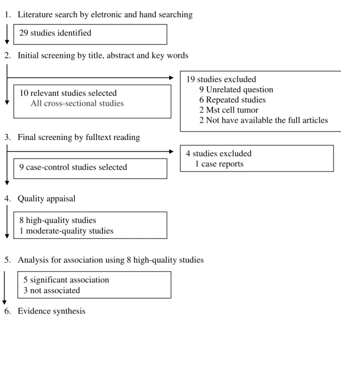
In the initial search by title the electronic search revealed 29 citations of which
20 articles were excluded because they are not related to the question, because they
are repeated, referring to mast cell tumors, do not present the full text available in
English or as they were reported case. Thus, after applying the eligibility criteria were
selected for review nine relevant studies, which were qualified by the
Newcastle-Ottawa scale, and obtained the following result: eight articles of high quality and one
6
In nine studies evaluated the samples were composed of paraffin embedded
specimens of AC and / or lip SCC and controls consisting of normal labial mucosa.
Most of the lesions studied in the samples was obtained from studies of men with
white skin and with a mean age of 54.5 years.
In the study of the density of mast cells was observed that some authors
compared lesions of actinic cheilitis and tissue normal14-16, while in others the comparison was between lesions of actinic cheilitis, lip cancer and tissue normal8 ,10,17-18 or between injuries SCC the lip and tissue normal9,11 (Table 2).
All studies identified mast cells both in AC lesions and SCC of the lip. The
presence of inflammation was recorded in seven of these articles8 ,10,14-18 and the presence of solar elastosis in six 8,10,14,16-18. Moreover, the presence of mast cells was associated with neovascularization in tissue local 9-11,14,16-18.
The location of mast cells in tissues was varied, but always related to the
region for AC or SCC. In samples AC14-16 mast cells were mainly located in the region of the lamina propria, along the surface epithelium and in the areas of solar elastosis.
However, were also identified in the vicinity of areas of epithelial dysplasia, bordering
the neoplastic islands 8 ,10,17-18, or in the vicinity of neoplastic vessels 9,11.
Some of the articles studied reported that mast cells when present were active and
degranulated 11,14,16. In cases related to AC 14,16 degranulation were associated with inflammation and areas of solar elastosis and in the case of squamous cell degranulation was
related to increased number of blood vessels (neovascularization) around the area
tumoral11.
In all studies of actinic cheilitis 8,10,14-18 , mild, moderate, and severe epithelial
dysplasia was identified , always associated with mast cells in these areas. In the cases of
All studies reported a statistically significant difference between the increased density
of mast cells in cases of lesions of actinic cheilitis and / or squamous cell carcinoma
compared with normal mucosa; on the other hand there was no significant association
between the presence of mast cells and differences in the degree of epithelial dysplasia,
although the number of mast cells was increased compared with normal mucosa. Among the
studies that assessed SCC was found in a significant difference between the increased density
of mast cells and the degree of histological differentiation of the tumor 17. The density
of
mast cells was increased in areas of high degradation of collagen10.
Discussion
The activated mast cells release mediators or substances such as histamine, heparin,
and proteoglycan enzymes (tryptase, chymase), growth factors and angiogenesis,
leukotrienes, prostaglandins, among others, who are responsible for promoting
inflammation, matrix destruction, tissue remodeling and angiogenesis19-21. The combined action of these chemical mediators can alter the microenvironment around the damaged
epithelium by UV radiation - initiating agent, secondarily contributing to the process of
malignant transformation of AC lesions and progression to SCC of the lip 22.
In the context of chemical carcinogenesis, a strong intracellular oxidative stress with
the generation of reactive oxygen species such as superoxide radicals and hydrogen peroxide,
which is believed to be responsible for the formation of adducts in DNA 23-25. In AC lesions,
ultraviolet radiation is the physical agent initiating the process of carcinogenesis, being able
to modify the structure of DNA molecules by both oxidative stress and by formation of DNA
8
transformation process allowing the progression of the disease to a clinically detectable
malignancy.
However, it is important to note that not all lesions and conditions with malignant
potential, necessarily undergo transformation into a malignant neoplasia itself28-29. The probabilistic field involves genetic factors (ability to recover the damaged DNA), intermittent
environmental exposures (cumulative effect), time and possibilities for early intervention in
removing the cause.
One of the conditions necessary for tumor growth has something to do to
angiogenesis 30-31, responsible for not only nutrition, but also the metastasis
of malignant
neoplasias 32. Mast cells identified in the peritumoral region, specifically near neoformados11 vessels may be reinforcing the hypothesis that angiogenic chemicals mediators, when
released, provide tumor growth. Remember that the action of metalloproteinases in the
degradation of extracellular matrix favors the process of neoplastic growth.
Conclusion
The concentration of mast cells often increased in areas of dysplastic epithelium and
the tumor area, as well as its location always associated with epithelial most battered areas
(solar elastosis and inflammatory infiltrate) and perivascular region suggest a strong
relationship between mast cells and the process of malignant transformation of AC into SCC
References
1 MARKOPOULOS A, ALBANIDOU-FARMAKI, KAYAVISACTINIC I. Actinic cheilitis: Clinical and pathologic characteristics in 65 cases. Oral Diseases 2004; 10(4):212-216.
2 MARTINS-FILHO PRS, SILVA LCF, PIVA MR. The prevalence of actinic cheilitis in farmers in a semi-arid northeastern region of Brazil. Int J Dermatol 2011; 50(9): 1109-1114.
3 VAN DER WAAL I. Potentially malignant disorders of the oral and oropharyngeal mucosa; terminology, classification and present concepts of management. Oral Oncol 2009; 45(4):317-23.
4 SHAH AY, DOHERTY SD, ROSEN T. Actinic cheilitis: a treatment review. Int J Dermatol. 2010; 49(11):1225-34.
5 WALSH LJ. Mast cells and oral inflammation. Crit Rev Oral Biol Med 2003; 14(3):188-98.
6 HIROMATSU Y, TODA S. Mast cells and angiogenesis. Microsc Res Tech. 2003; 60(1):64-9.
7 HUANG B, LEI Z, ZHANG GM, LI D, SONG C, LI B, LIU Y, YUAN Y, UNKELESS J, XIONG H, FENG ZH. SCF-mediated mast cell infiltration and activation exacerbate the inflammation and immunosuppression in tumor microenvironment. Blood 2008; 112(4):1269-79.
8 GOMES APN, JOHANN JE, LOVATO GG, FERREIRA AM. Comparative Analysis of the Mast Cell Density in Normal Oral Mucosa, Actinic Cheilitis and Lip Squamous Cell Carcinoma. Braz Dent J 2008; 19(3)186-189.
9 PARIZI ACG, BARBOSA RL, PARIZI JLS, NAI GA. A comparison between the concentration of mast cells in squamous cell carcinomas of the skin and oral cavity. An Bras Dermatol 2010; 85(6):811-818.
10 COSTA NL, OTON-LEITE AF, CHEIM-JÚNIOR AP, ALENCAR RDE C, BITTAR GO, SILVA TA, BATISTA AC. Density and migration of mast cells in lip squamous cell carcinoma and actinic cheilitis. Histol Histopathol 2009; 24(4):457-465.
11 ROJAS IG, SPENCER ML, MARTÍNEZ A, MAURELIA MA, RUDOLPH MI. Characterization of mast cell subpopulations in lip cancer. J Oral Pathol Med 2005; 34(5): 268-73.
12 GALINSKY DS, NECHUSHTAN H. Mast cells and cancer--no longer just basic science. Crit Rev Oncol Hematol. 2008; 68(2):115-30.
10
nonrandomized studies in meta-analyses. [http://www.ohri.ca/programs/ clinical_epidemiology/oxford.htm].
14 ROJAS IG, MARTÍNEZ A, PINEDA A, SPENCER ML, JIMÉNEZ M, RUDOLPH MI. Increased mast cell density and protease content in actinic cheilitis. J Oral Pathol Med 2004; 33(9): 567–73.
15 ROJAS IG, MARTÍNEZ A, BRETHAUER U, GREZ P, YEFI R, LUZA S, MARCHESANI FJ. Actinic cheilitis: Epithelial expression of COX-2 and its association with mast cell tryptase and PAR-2. Oral Oncology 2009; 45(3): 284-290. 16 ARAÚJO CP, GURGEL CA, RAMOS EA, FREITAS VS, BARBOSA ADE A JR, RAMALHO LM, DOS SANTOS JN. Accumulation of CD1a-positive Langerhans cells and mast cells in actinic cheilitis. J Mol Hist 2010; 41(6):357-365.
17 SOUZA LR, FONSECA-SILVA T, SANTOS CCO, OLIVEIRA MVM, CORRÊA-OLIVEIRA R, GUIMARÃES ALS, DE PAULA AMB. Association of mast caell, eosinophil leucocyte and microvessel densities in actinic cheilitis and lip squamous cell carcinoma. Histopathology 2010; 57(6): 796-805.
18 FREITAS VS,SANTOS PPAS, FREITAS RA, PINTO LP, SOUZA LB. Mast cells and matrix metalloproteinase 9 expression in actinic cheilitis and lip squamous cell carcinoma. Oral Surg Oral Med Oral Pathol Oral Radiol Endod 2011; 112(3):342-8. 19 BLAIR RJ, MENG H, MARCHESE MJ, REN S, SCHWARTZ LB, TONNESEN MG, GRUBER BL. Human mast cells stimulate vascular tube formation. Tryptase is a novel, potent angiogenic factor. J Clin Invest. 1997; 99(11):2691-700.
20 CAUGHEY GH. Mast cell tryptases and chymases in inflammation and host defense. Immunol Rev 2007; 217:141-54.
21 WASSERMAN SI. The mast cell: its diversity of chemical mediators. Int J Dermatol. 1980;19(1):7-17.
22 KREISLER M, CHRISTOFFERS AB, WILLERSHAUSEN B, D'HOEDT B. Low-level 809 nm GaAlAs laser irradiation increases the proliferation rate of human laryngeal carcinoma cells in vitro. Lasers Med Sci 2003;18(2):100-3.
23 GANNOT G, BUCHNER A, KEISARI Y. Interaction between the immune system and tongue squamous cell carcinoma induced by 4-Nitroquinoline N-oxide in mice. Oral Oncol 2004; 40(3): 287-297.
24 KANOJIA D, VAIDYA MM. 4 – Nitroquinoline – 1- oxide induced experimental oral carcinogenesis. Oral Oncol 2006; 42(7): 655-667.
25 MOGNETTI B, DI CARLO F, BERTA GN. Animal models in oral câncer research. Oral Oncol 2006; 42(5): 448-460.
27 AMES BN, SHIGENAGA MK,HAGEN TM. Oxidants, antioxidants, and the degerative deseases of aging. Proc Natl Acad Sci 1993; 90(17): 7915-22.
28 VAN DER WAAL, I. Potentially malignant disorders of the oral and oropharyngeal mucosa; terminology, classification and present concepts of management. Oral Oncol 2010; 46(6):423-5.
29 GALE N, PILCH BZ, SIDRANSKY D, NAGGAR AEL, WESTRA W, CALIFANO J. Epithelial precursor lesions. In: World Health Organization Classification of Tumors. Head and Neck Tumours 2005; 296-300.
30 RAICA M, CIMPEAN AM, RIBATTI D. Angiogenesis in pre-malignant conditions. Eur J Cancer 2009; 45(11),1924-1934.
31 WEIDNER N. The importance of tumor angiogenesis: the evidence continues to grow. Am J Clin Pathol 2004; 122(5), 675- 677.
12
Figure 1: Number of studies according to the processes of searching, selection and evaluation of literature.
1. Literature search by eletronic and hand searching
2. Initial screening by title, abstract and key words
3. Final screening by fulltext reading
4. Quality appaisal
5. Analysis for association using 8 high-quality studies
6. Evidence synthesis 8 high-quality studies 1 moderate-quality studies 29 studies identified
19 studies excluded 9 Unrelated question 6 Repeated studies 2 Mst cell tumor
2 Not have available the full articles 10 relevant studies selected
All cross-sectional studies
9 case-control studies selected
4 studies excluded
1 case reports
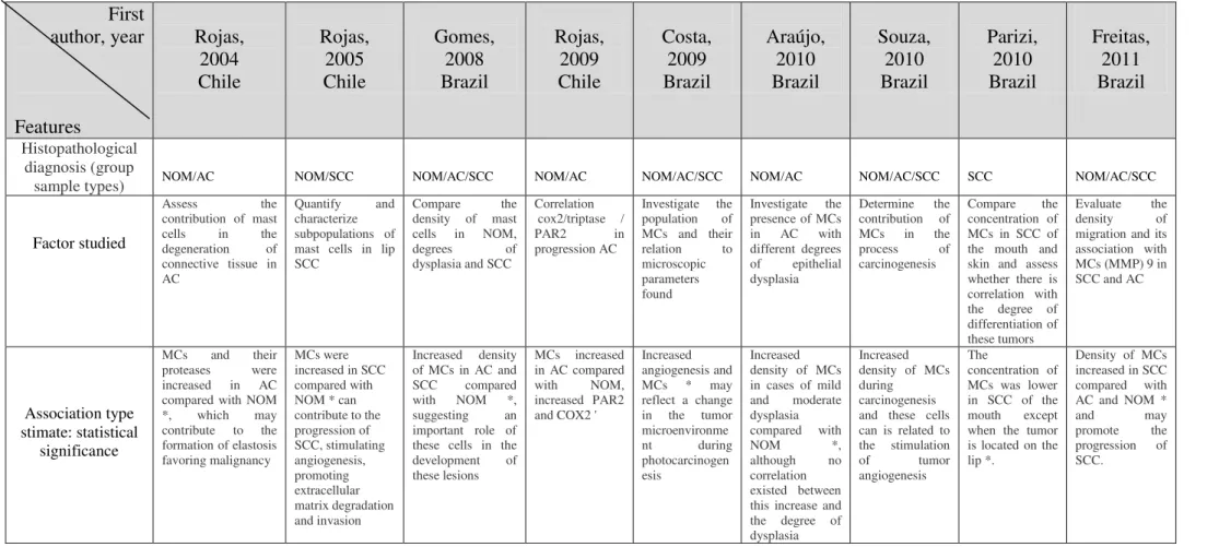
Table 1: Characteristics of the studies summarized researched the type of the study group and factor studied and type of association estimated. First author, year Features Rojas, 2004 Chile Rojas, 2005 Chile Gomes, 2008 Brazil Rojas, 2009 Chile Costa, 2009 Brazil Araújo, 2010 Brazil Souza, 2010 Brazil Parizi, 2010 Brazil Freitas, 2011 Brazil Histopathological diagnosis (group
sample types) NOM/AC NOM/SCC NOM/AC/SCC NOM/AC NOM/AC/SCC NOM/AC NOM/AC/SCC SCC NOM/AC/SCC
Factor studied
Assess the contribution of mast cells in the degeneration of connective tissue in AC
Quantify and characterize subpopulations of mast cells in lip SCC
Compare the density of mast cells in NOM, degrees of dysplasia and SCC
Correlation cox2/triptase / PAR2 in progression AC
Investigate the population of MCs and their relation to microscopic parameters found
Investigate the presence of MCs in AC with different degrees of epithelial dysplasia
Determine the contribution of MCs in the process of carcinogenesis
Compare the concentration of MCs in SCC of the mouth and skin and assess whether there is correlation with the degree of differentiation of these tumors
Evaluate the density of migration and its association with MCs (MMP) 9 in SCC and AC
Association type stimate: statistical
significance
MCs and their proteases were increased in AC compared with NOM *, which may contribute to the formation of elastosis favoring malignancy
MCs were increased in SCC compared with NOM * can contribute to the progression of SCC, stimulating angiogenesis, promoting extracellular matrix degradation and invasion
Increased density of MCs in AC and SCC compared with NOM *, suggesting an important role of these cells in the development of these lesions
MCs increased in AC compared with NOM, increased PAR2 and COX2 '
Increased angiogenesis and MCs * may reflect a change in the tumor microenvironme nt during photocarcinogen esis
Increased density of MCs in cases of mild and moderate dysplasia compared with NOM *, although no correlation existed between this increase and the degree of dysplasia
Increased density of MCs during
carcinogenesis and these cells can is related to the stimulation of tumor angiogenesis
The
concentration of MCs was lower in SCC of the mouth except when the tumor is located on the lip *.
Density of MCs increased in SCC compared with AC and NOM * and may promote the progression of SCC.
Factors studied in the articles and the type of association estimated.
NOM, normal oral mucosa; AC, actinic cheilitis; SCC, Squamous Cell Carcinoma of lip; MCs, mast cells, * statistically significant (p< 0.05).
14
First author,
year and source Tipy of study Sample sizea Mean age
(years)
Increased density of
MCs
Degranulation of MCs
Location of MCs NOS
Coding Score
Rojas, 2004; Chile
Case-control AC/NOM
23 25-74 Y Y Near the area of solar elastosis and
inflammation
10111111 7*
Rojas, 2005; Chile
Case-control SCC/NOM
29 25-84 Y Y Intratumoral and peritumoral stroma,
around vessels
10111111 7*
Gomes, 2008; Brazil
Case-control AC/SCC/NOM
41 --- Y --- --- 10111111 7*
Rojas, 2009; Chile
Case-control SCC/NOM
69 22-73 Y --- Lamina propria 10111111 7*
Costa, 2009; Brazil
Case-control AC/SCC/NOM
58 33-90 Y
---
Scattered in the connective tissue and near the tumor cells
10111111 7*
Araújo, 2010; Brazil
Case-control AC/NOM
38 20-75 Y Y Lamina propria, solar elastosis and around
of vessels
10111111 7*
Souza, 2010; Brazil
Case-control AC/SCC/NOM
71 20 + Y --- Stroma adjacent to dysplastic tissue /
neoplastic, near of vessels and nerves
10111111 7*
Parizi, 2010; Brazil
Case-control SCC
64 50 + Y ---- --- 10001111 5
Freitas, 2011; Brazil
Case-control AC/SCC/NOM
47 --- Y --- Stroma tumor, bordering the
invasion of the epithelium and near the epithelium adjacent to tumor
10111111 7*
AC= actinic cheilitis, SCC= squamous cell carcinoma NOM= normal oral mucosa.
The studies were conducted in Brazil and Chile. The relationship question / answer was evaluated in nine case-control and eight articles were classified as high quality.
Y, Yes; + ages above *evaluated for high-quality methodology by the modified Newcastle-Ottawa Scale (NOS). When a study presented all the criteria of NOS column appears as
1
Artigo em processo de envio para periódico Pathology and Oncology Research.
Actinic cheilitis: histopathological evaluation of 44 cases.
Rachel Reinaldo Arnaud*, Maria Sueli Marques Soares**, Marcos Antônio Farias de Paiva**, Cláudia Roberta Leite Vieira de Figueiredo**, Cláudia Cazal Lira**, Francisco Diogo Carrilho de Oliveira Filho *
* Postgraduate student, Department of Clinical and Social Dentistry, School of Dentistry, Universidade Federal da Paraíba, João Pessoa, Brazil;
**
Abstract
Objective: The purpose of this study was to describe the histopathological findings in lesions
of actinic cheilitis (AC). Methods: We conducted a retrospective study from cases with
clinical diagnosis of AC recorded in the histopathology of the Service of Head and Neck
Napoleon Laureano Hospital, PB, from 2000 to 2007. We selected 44 paraffin blocks that had
histologic revaluation of conditions through the construction of new laminas. These new
sections were stained with hematoxylin and eosin (HE) and histopathological evaluation was
performed by two independent examiners, and the changes classified according to OMS. We
conducted a descriptive statistical analysis in SPSS for Windows version 13. Results: Of the
total sample, 52.3% (23) cases were diagnosed in men and 47.7% (21) in women aged 27 to
92 years old. Most individuals (81.9%) were over 40 years old. Regarding the histologic
features, 68.2% (30) of cases showed some degree of epithelial dysplasia, 36.3% (16)
classified as mild dysplasia, 20.4% (9) moderate dysplasia and 11.3% (5) severe dysplasia. In
15.9% (7) was squamous cell carcinoma of lip (SCC). In epithelial tissue lining lips, the most
frequent histologic findings identified were the presence of hydropic degeneration (79.5%)
and hypergranulosis (56.8%). Inflammatory infiltrate was observed in 88.6% of cases and
86.4% in solar elastosis. Conclusion: Atypical cellular and transformation to SCC are
common features in actinic cheilitis. We stress the importance of early diagnosis and the
proservation patient with this injury.
3
Introduction
Actinic cheilitis (AC) is a disease that affects mainly the lower lip of men over 60
years of age, of fair skin that are exposed chronically and overexposure to ultraviolet (UV)
rays[7.12]. It has great clinical importance because it is a recognized lesion with malignant
potential [25].
Clinically, it has two forms: acute and chronic. The acute form is characterized by
mild edema and erythema, fissures and severe ulcers, and occurs when there is excessive sun
exposure in a short time. Often, there is the resolution of these clinical changes without
irreversible epithelial changes. The chronic form occurs when there is prolonged and
cumulative exposure to ultraviolet rays and usually develops irreversible epithelial changes.
The most common clinical presentation displays dry, cracked lips, slight volume increase and
diffuse, limit loss of semi-labial mucosa and skin, and papules and / or patches leukoplakia
[11.19].
Histologically, AC can make changes in epithelial tissues, including atrophy and
decreased production of keratin, a common occurrence with mild to severe dysplasia [4,22].
In connective tissue there are inflammatory infiltrate and solar elastosis, which are
characterized by the degeneration of collagen fibers, which will then display basophilia and
amorphous appearance [15].
A large number of squamous cell carcinoma (SCC) of the lip is associated with the occurrence of a previous injury of actinic cheilitis [1.19]. The malignant process may not be
visible to the eye at the time of clinical examination, although the histological level, epithelial
dysplastic changes and the presence of inflammatory process in the examined tissue are
predictive of malignant of the lesion [10.20].
Considering that AC is a frequent labial injury which may progress to SCC and it is
important to conduct studies to discuss their histological changes, it is proposed in the present
study to analyze a series of cases of such lesions histologically in order to describe the most
common histological findings and contribute to the diffusion of knowledge among dentists.
Material and methods
We conducted a retrospective laboratory study whose universe was represented by all
cases with clinical diagnosis of AC recorded in the histopathology archive Service of Head
and Neck Napoleon Laureano Hospital, João Pessoa, in the period 2000 to 2007. Eighty-nine
among these, we selected those with pathological conditions for revaluation by making new
cuts. Thus, 44 blocks were cut into sections of 5μm and stained with HE and then evaluated
by two independent observers using light microscopy. Differences between examiners were
resolved by consensus.
To analyze the degree of epithelial atypia, we employed the criteria established by the
World Health Organization, according to Barnes´s considerations [4]. For each specimen, the
grading of epithelial dysplasia was based on observation of the magnitude of changes
involving the extension of the epithelium, considering: Absence of epithelial dysplasia when
there was no change in the epithelium, epithelial mild dysplasia when only the lower 1 / 3 of
the epithelium presented cellular atypia, moderate epithelial dysplasia , when even the middle
1 / 3 of the epithelium had dysplastic changes, severe epithelial dysplasia when changes
reached more than 2 / 3 of the epithelium. It was characterized as carcinoma in situ who
presented the specimen thickness atypia throughout the epithelium and invasive squamous
cell carcinoma when there was disruption in the areas of basement membrane [12].
Other histological changes were also considered, including presence and type of
keratin, hydropic degeneration, spongiosis, epithelial atrophy, epithelial hyperplasia and
hypergranulosis in epithelial tissue. Moreover, the presence of solar elastosis and
inflammatory infiltrate in the connective tissue were considered.
The data were processed using SPSS (Statistical Package for Social Sciences) for
Windows version 13.0, where they performed a descriptive statistical analysis.
This study was approved by the Ethics Committee in Research of Hospital
Universitário Lauro Wanderley CEP/ HULW / UFPB under protocol No. 448/10.
Results
From the total sample it was observed that 52.2% (23) of the cases occurred in men
and 47.7% (21) in women. In relation to the age of patients, it ranged from 27 to 92 , being
81.8% (36) over 40 years old. As for the location of the lesions of actinic cheilitis, it was
observed that 100% were in the lower lip.
Histological evaluation revealed that 68.2% (30) of the cases had some degree of
epithelial dysplasia, and 36.3% (16) of mild dysplasia, 20.4% (9) of moderate dysplasia and
11.3% (5) of severe dysplasia. In 15.9% (7) SCC ocurred, being 4.5% (2) carcinoma in situ
5
Table 1 shows the distribution of the main findings of histological specimens, which
shows that the most frequent were hidropic degeneration with 79.5% and 56.8% with
hypergranulosis.
In the underlying connective tissue, we observed the presence of inflammatory
infiltrate in 88.6% (39) of the cases, and solar elastosis (86%) (38) (Fig. 2).
Discussion
In the Northeast region of Brazil warm climate prevails with high levels of solar
radiation which makes it even more important to conduct studies of lesions such as actinic
cheilitis [24]. According to Miranda, Ferrari, Calandro [17], the longer the exposure time, the
greater the severity of AC lesions in subjects exposed to UV rays. Therefore, early diagnosis
and proactive observation of the patient is of great importance to prevent disease progression.
The reasons for the increased susceptibility of the lips to changes caused by actinic
cheilitis are various and includes a thinner keratin in the area, thin epithelial layer, a small
amount of melanin and decreased secretion of sebaceous glands and sweat glands, ie,
mechanisms normally involved in skin protection against radiation. Moreover, the AC lesions
occur more frequently in the lower lip for this site is more directly exposed to sunlight [5,12].
Our study showed 100% of lesions in the lower lip, and this result is similar to Marcopoulos,
Albanidou-Farmaki, Kayavis [15], however, Kaugars et al [12] and Pimentel et al [20] also
found lesions in the upper lip in 3% of the cases.
The classic demographic profile of patients at high risk for QA reported in the
literature is men over 50 years of age, fair skin and high solar exposure [6]. The results of this
study are consistent with this profile of patients. Most of the patients found having the lesion
was men [2,7,9,12,15,17,26], although the number of women has also been great, probably
due to the nature of work of these patients. Cavalcante, Anbinder, Carvalho [7] claims that
women may be less likely to suffer AC due to the use of lipstick, which can partially protect
lips from the sun. In the present study, patients over 40 years old were most affected,
confirming the study of Cavalcante, Anbinder, Carvalho [7] (75.86%), however, other studies
observed prevalence from the fifth decade of life [2 , 9,21,26].
The histopathological features of actinic cheilitis are important for diagnosis,
prognosis and treatment of injury. The presence of epithelial dysplasia is a common finding
and was observed in most lesions of the present study, this is corroborated by other studies
[12,15], and differs from results of Cavalcante, Anbinder, Carvalho [7] in which all cases of
with dysplasia, it was observed a higher prevalence of mild dysplasia and moderate dysplasia
in agreement with the results of other authors [2-3,12,15,20]. The highest percentage of mild
dysplasia suggests that the degree of epithelial atypia alone does not indicate the malignant
potential of AC [20]. On the other hand, the results of Cavalcante, Anbinder, Carvalho [7]
showed higher percentages of moderate dysplasia or severe dysplasia in cases of AC. Besides
the cases of epithelial dysplasia, it was observed high prevalence of SCC (15.5%) similar to
the findings of Kaugars et al [12] and Markopoulos Albanidou-Farmaki, Kayavis [15]. These
data underscore the potentially malignant lesions such as AC.
Our results showed that most lesions showed changes in epithelial thickness, ranging
from hyperplastic to atrophic, a finding also described by Kaugars et al [12], whose AC
lesions exhibited both hyperplasia and atrophy in 68.4% of their sample. In a smaller
percentage, these same findings were reported by Pimentel et al [20] and Rojas [22], with a
prevalence of 40.6% and 46.6%, respectively. .
The epithelial hyperplasia can occur in up to 100% of the samples of AC [16], as well
as atrophy can occur in relatively high frequency [7,16]. Other epithelial alterations,
frequently observed in this study, were hidropic degeneration, spongiosis and
hypergranulosis. Markopoulos, Albanidou-Farmaki, Kayavis [15] observed a much higher
percentage (75%) of spongiosis in relation to our study. The presence of ortoqueratina was
more prevalent than paraqueratina, being the result similar to those of studies Kaugars et al
[12] and Cavalcante, Anbinder Caravalho and [7].
Some studies indicate the solar elastosis as a histopathological more consistent and
constant finding of actinic cheilitis [2,12,15-16,22-23,]. The solar elastosis reflects the sun
damage to the tissue and may be an important factor in the development of AC without,
however, being predictive of the development of AC to SCC [20]. In the present study, the
solar elastosis was present in 86.36% of cases differing in some studies [7,15,22] that found
solar elastosis in 100% of their samples.
The inflammatory process is recognized as a defensive mechanism of the body.
However, there is evidence that the inflammatory reaction can act in the initiation, promotion
and progression of tumors, since the mediators of inflammation contribute effectively to
changes in the tumor microenvironment [13-14]. Several types of cancers arise at sites of
infection, chronic irritation and inflammation [18]. However, this relationship is not yet fully
known [8]. The results of this study showed that the inflammatory infiltrate occurred in 88.6%
of the cases, being the moderate and severe inflammation the most frequent. Whereas in the
7
100% of the cases. In the study by Pimentel et al [20] this finding was strongly associated
with the presence of epithelial dysplasia.
Conclusion
Based on the results of the sample we can conclude that the cellular atypia and
transformation to CCE are common features in actinic cheilitis. We stress the importance of
early diagnosis and the proservation of the patient with this injury.
References
1 Ackerman AB (2003) Opposing views of 2 academies about the nature of solar keratosis. Cutis 71:391-395.
2 Araújo CP, Barros AC, Lima AAS, Azevedo RA, Ramalho L, Santos JN (2007) Estudo histológico e histoquímico da elastose solar em lesões de queilite actínica. R Ci Med Biol 6:152-159.
3 Araújo CP, Gurgel CA, Ramos EA, Freitas VS, Barbosa A de A Jr, Ramalho LM, dos Santos JN (2010) Accumulation of CD1a-positive Langerhans cells and mast cells in actinic cheilitis. J Mol Hist 41:357-365.
4 Barnes MDL (2005) World Health Organization Classification of tumours. Pathology and genetics of head and neck tumours.Lyon:IARC PRESS.
5 Bentley JM, Barankin B, Lauzon GJ (2003) Paying more than lip service to lip lesions. Candian Family Physician 49:1111-1116.
6 Cataldo E, Doku HC (1981) Solar cheilitis. J Dermatol Surg Oncol 7:989-995.
7 Cavalcante ASR, Anbinder AL, Caravalho YR (2008) Actinic cheilitis: clinical and histological features. J Oral Maxillofac Surg 66:498-503.
8 Coussens LM, Werb Z (2002) Inflammation and cancer. Nature 420:860-867.
9 Corso FM, Wild C, Gouveia LO, Ribas MO (2006) Queilite actínica: prevalência na clínica estomatológica da PUCPR, Curitiba, Brasil. Clin Pesq Odontol 2:227-281.
10 Costa NL, Oton-Leite AF, Cheim-Júnior AP, Alencar RDE C, Bittar GO, Silva TA, Batista AC (2009) Density and migration of mast cells in lip squamous cell carcinoma and actinic cheilitis. Histol Histopathol 24:457-465.
12 Kaugars GE, Pillion T, Svirsky JA, Page DG, Burns JC, Abbey LM (1999) Actinic cheilits: A review of 152 cases. Oral Surg. Oral Med Oral Pathol Oral Radiol Endod 88:181-186.
13 Kinet JP (2007) The essential role of mast cells in orchestrating inflammation. Immunol Rev 217:5-7.
14 Mantovani A, Allavena P, Sica A, Balkwill F (2008) Câncer-related inflammation. Nature 454:436-444.
15 Markopoulos A, Albanidou-Farmaki, Kayavisactinic I (2004) Actinic cheilitis: Clinical and pathologic characteristics in 65 cases. Oral Diseases 10:212-216.
16 Martínez A, Brethauer U, Rojas IG, Spencer M, Mucientes F, Borlando J, Rudolph MI (2005) Expression of apoptotic and cell proliferation regulatory proteins in actinic cheilitis. J Oral Pathol Med 34: 257-262.
17 Miranda AMO, Ferrari TM, Calandro TLL (2011) Queilite actínica: aspectos clínicos e prevalência encontrados em uma população rural no interior do Brasil. Revista Saúde e Pesquisa 4:67-72.
18 Mukherjee S, Bandyopadhyay G, Dutta C, Bhattacharya A, Karmakar R, Barui G (2009) Evaluation of endoscopic biopsy in gastric lesions with a special reference to the significance of mast cell density. Indian Journal of Pathology and Microbiology 52:20-24.
19 Picascia DD, Robinson JK (1987) Actinic cheilitis: a review of etiology, differential diagnosis, and treatment. J Am Acad Dermatol 17:255-264.
20 Neto Pimentel DR, Michalany N, Alchorne M, Abreu M, Borra RC, Weckx L (2006) Actinic cheilitis: Histophatology and p53. J Cutan Pathol 33:539-544.
21 Pontes HAR, Aguiar MCF, Mesquita RA, Pontes FSC, Siveira Júnior JB (2005) Imunoexpressão da proteína de reparo hMSH2 em queilite actínica e mucosa labial normal. Rev Bras de Cancerologia 5:23-30.
22 Rojas IG, Martínez A, Pineda A, Spencer ML, Jiménez M, Rudolph MI (2004) Increased mast cell density and protease content in actinic cheilitis. J Oral Pathol Med 33: 567–573. 23 Santos JN, Sousa SO, Nunes FD, Sotto MN, de Araújo VC (2003) Altered cytokeratin expression in actinic cheilitis. J Cutan Pathol 30:237-241.
9
25 Van der waal I (2009) Potentially malignant disorders of the oral and oropharyngeal mucosa; terminology, classification and present concepts of management. Oral Oncol 45:317-323.
Table 1: Distribution of the main histological characteristics of 44 specimens of actinic cheilitis.
Histological Findings n %
Parakeratosis 14 31,8
Orthokeratosis 35 79,5
Epithelial atrophy 13 29,5
Epithelial hyperplasia 17 38,6
Spongiosis 12 27,2
Hidropic degeneration 35 79,5
Hypergranulosis 25 56,8
Solar Elastosis 38 86,3
Inflammatory infiltrate
Mild 14 31,8
Moderate 9 20,45
Intense 16 36,36
Dysplasia
Mild 16 36,3
Moderate 9 20,4
Severe 5 11,3
Carcinoma in situ 2 4,5
11
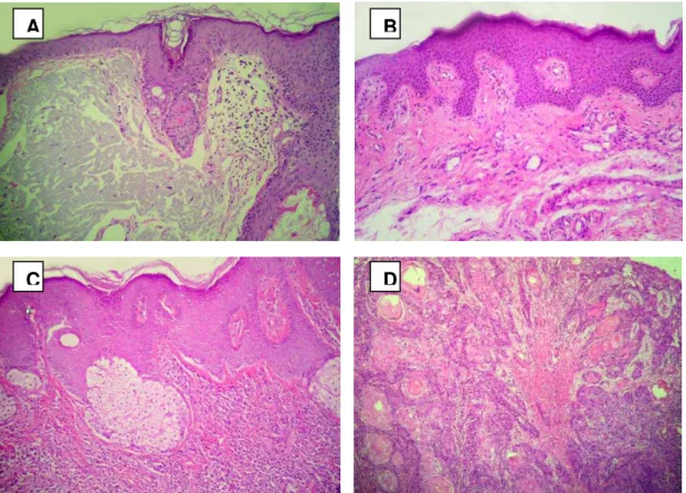
Figure 1: Lesions of actinic cheilitis. A Presence of solar elastosis in injury AC with moderate dysplasia. B
Lesions of AC without epithelial dysplasia. C Epithelium with mild dysplasia.D Squamous cell carcinoma exhibiting well-differentiated neoplastic islets. Hematoxylin-eosin, magnification 100X.
A B
C
A
1
Artigo em processo de envio para periódico Archives of Dermatological Research.
Density of mast cells in lesions of actinic cheilitis.
Rachel Reinaldo Arnaud 1 Maria Sueli Marques Soares 2 Claudia Cazal Lira 2
1
Postgraduate student, Department of Clinical and Social Dentistry, School of Dentistry, Universidade Federal da Paraíba, João Pessoa, Brazil;
2
Abstract
The objective of this study was to analyze the density of mast cells in actinic cheilitis second
the histological features of the lesion compared with normal mucosa. The sample comprised
an experimental group of 33 cases with clinical diagnosed of actinic cheilitis. And a control
group composed of nine blocks with specimens of normal oral mucosa. The paraffin blocks of
the sample were cut and stained with hematoxylin and eosin and blue toluidine. The count of
mast cells was performed 8 fields per case. The final reading was expressed with the average
value of mast per case in cells / μm². In 57.6% of the cases there was some degree of epithelial dysplasia and 21.2% there was squamous cell carcinoma. The presence of
inflammatory infiltrate and solar elastosis was observed in 84.9% and 81.8% of the cases,
respectively. Mast cells were identified in 87.8% of the sample. The density of mast cells in
the cases of actinic cheilitis was 17.4 ± 10.4 cells / μm², and in normal tissue 1.78 ± 1.64 cells / μm², with a significant difference (p <0.001). In addition, there was a statistically significant correlation between the density of mast cells with the processes of dysplasia (p = 0.004) and
inflammatory cell infiltration (p = 0.000). The increased density of mast cells in actinic
cheilitis lesions and its correlation with the processes of dysplasia and inflammation suggest
involvement of these cells in disease progression to squamous cell carcinoma of the lip.
3
Introduction
Actinic cheilitis (AC) is a potentially malignant lesion that can turn into squamous cell
carcinoma (SCC) of the lip. It affects mainly the lower lip of fair-skinned people, who are 50
to 60 years old and exposed to excessive solar radiation [1-5].
Histologically, AC often reveals several epithelial changes, being common the
occurrence of mild to severe dysplasia [6] and carcinoma in situ [7], and in the connective
tissue, it is frequently observed solar elastosis and inflammatory infiltrate of variable degree
[3-4,8].
Some authors claim that the inflammatory infiltrate tumor-associated contributes to
squamous carcinogenesis [9-10]. Others believe that the mechanisms that result in
maliginization of actinic cheilitis may have the participation of components of the
inflammatory process, like the mast cells, which are cells that proliferate after induction by
ultraviolet radiation (UV) and that release potent mediators that act on inflammation,
angiogenesis and degradation of the intercellular matrix [2,11-14]. Mast cells are present in
greater amounts in lesions caused by UV radiation, such as the actinic cheilitis and squamous
cell carcinoma of the lip. Some studies associate higher amount of mast cells to the
development of actinic cheilitis [2,11].
In this context, this study aimed to determine the density of mast cells, the
histopathologic features and qualify the inflammatory process in the lesions of actinic
cheilitis, as well as the possible association between these variables, compared with normal
tissue.
Material and Methods
The study was approved by the Ethics Committee of Hospital Lauro Wanderley - CEP
/ HULW / UFPB under protocol number 448/10.
It was a case-control study where the experimental group was composed of 33 paraffin
blocks of cases with clinical diagnosis of actinic cheilitis in subjects of both sexes, recorded in
the Department of Head and Neck Dr. Napoleon Laureano Hospital, João Pessoa, PB,
obtained in the period between the years 2000 to 2008. The control group consisted of nine
blocks of paraffin embedded specimens of normal oral mucosa. To be included in the sample,
Each selected block was cut into 4 µm thick sections to obtain two histological slides,one of them obtained by a staining routine technique with hematoxylin and eosin (H /
E) and another slide stained with blue toluidine technique for identification of mast cells [11].
A histological evaluation was performed by two independent examiners in an optical
microscope, where microscopic analysis of epithelial histology was conducted according to
the criteria established by the World Health Organization (WHO). The dysplasia was
classified according to WHO in mild dysplasia, moderate dysplasia and severe dysplasia [6].
The presence of solar elastosis and inflammatory infiltrate was also recorded, and the
infiltration classified as mild, moderate or intense and in the selection of areas for analysis
areas of ulceration were disregarded [15].
For the counting of mast cells, we used an optical microscope Motic BA 300 ®,
Canada, with a magnitude of 400x, attached to the computer with program Motic Images Plus
2.0. The reading was performed in 8 fields per slide, using a square reticulum (20x20), where
each side of the field was 12μm, totaling an area of 144 μm² per field, the sum total of the eight fields equals to 1052 μm². The average number of mast cells was obtained by reading the 8 fields and expressed as cells/μm².
Data were tabulated and statistical tests performed in SPSS (Statistical Package for
Social Sciences) for Windows version 15.0. The procedures of statistical inference tests were
performed by Mann-Whitney U, Kuskall-Wallis test, chi-square and Spearman coefficient,
differences were considered statistically significant when p <0.05.
Results
Among the 33 specimens in the experimental group (AC), it was observed that 78.8%
had epithelial changes ranging from mild dysplasia to SCC. In 57.6%(19) there was some
degree of dysplasia, as well as 39.4%(13) mild, 15.2%(5) moderate and 3%(1) severe, and
21.2%(7) of cases occurred CCE. The inflammatory infiltration was observed in 84.9% of the
sample, ranging from 39.4%(13) mild, 15.2%(5) moderate and 30.3%(10) intense. Solar
elastosis was observed in 81.8% of the cases.
Mast cells were identified in 87.8%(29) of the cases (Fig. 1) and were located nearby
the area of dysplastic epithelium (30.3%), in the lamina propria (24.2%), permeating the solar
elastosis (15.5%), in the vicinity of vessels (9%), in the peritumoral region (18.8%) and
submucosa (6%). In 15.2%(5) of the cases, mast cells were active, stage of degranulation,
5
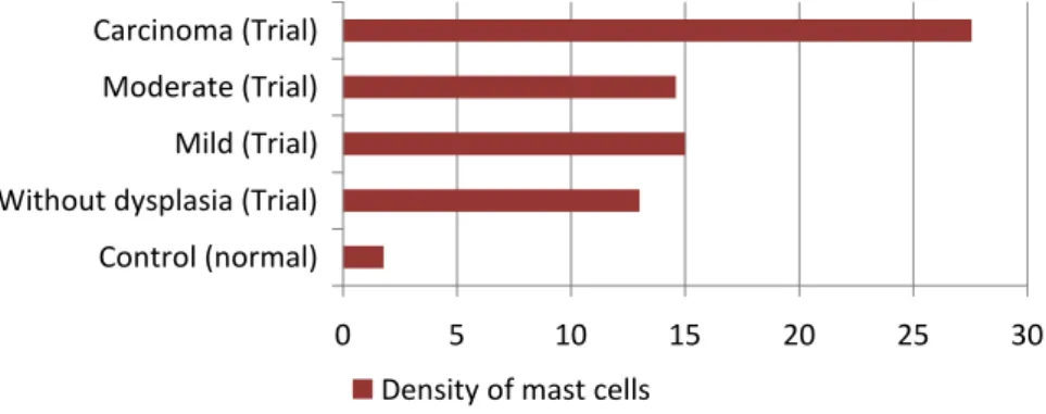
The average number of mast cells in specimens QA (experimental group) was 17.4 ±
10.4 cells/μm² ranging from 5.9 to 33 cells/ μm², while in specimens of normal mucosa (control group) it was 1.78 ± 1.64 cells/ μm², ranging from 0.52 to 3.04 cells / μm². There was a statistically significant difference (Mann-Whitney U, p <0.001) when compared to the
concentration of mast cells between the two groups. The average number of mast cells was
27.57 ± 5.94 cells/μm² in the cases of CCE, which was significantly greater (Kruskal-Wallis test, p = 0.001) when compared with normal tissue and degrees of epithelial dysplasia
(Graphic1).
There was a statistically significant correlation (Spearman coefficient, p = 0.004)
between the density of mast cells and the presence of epithelial dysplasia. Being observed a
significant progressive increase (Kruskal-Wallis test, p = 0.001) in the amount of mast cells,
when there was an increase of of the severity of the degrees of dysplasia.
The inflammatory infiltrate showed a statistically significant correlation with the
density of mast cells (Spearman coefficient, p = 0.000) and the number of mast cells increased
depending on the degree of inflammation (Kruskal-Wallis test, p = 0.003) (Table 1).
It was also observed statistically significant correlation between epithelial dysplasia
and inflammatory infiltration (Spearman coefficient, p = 0.002). However, while checking if
the degree of dysplasia was associated with the degree of inflammation, there was no
significant association (Shi-Square, p = 0.398).
By analyzing the density of mast cells in the presence / absence of solar elastosis, it
was observed that there was no statistically significant difference (Mann-Whitney U, p =
0.05).
Discussion
Mast cells are inflammatory cells that have been recognized as important effecter of
the deleterious effects of solar radiation on the skin [16-17]. Recent studies show that these
cells are significantly increased in lip lesions induced by radiation as actinic cheilitis
[2,18-19] and squamous cell carcinoma of the lip [11,15,20-25]. In the present study we observed
an increase in the number of mast cells in AC lesions compared to normal tissue intraoral,
On the other hand, in the present study, we found a significant association between the
density of mast cells and different degrees of dysplasia, confirming the results of Gomes et al.
[11] and Ch'ng et al. [26]. These authors noted a close correlation between the activation of
mast cells and the different phases of hyperkeratosis, dysplasia, carcinoma in situ and invasive
oral carcinoma. For these authors, during the process of carcinogenesis there is a sequential
infiltration and degranulation of mast cells in CCE. It is noteworthy that the changes in the
cytoskeleton epithelial in potentially malignant lesions may precede structural changes, as
probably observed by Santos et al. [27] who identified changes in the pattern of differentiation
of keratin in AC. Supposedly these changes depend on the stage the process of transformation
is, as well as on the continuity of the action of the etiologic agent on the tissue.
The relationship between the density of mast cells and the different degrees of
dysplasia are not found frequently diverging in our results [15,18-19].
The location of mast cells was related to peritumoral region, around vessels, near the
area of inflammation, epithelial dysplasia and solar elastosis. These findings were similar to
those of other authors [2,11,18-20,22-23] reinforcing the hypothesis that mast cells could
participate in the process of malignant transformation of actinic cheilitis.
As the results of Rojas et al. [20], Rojas et al. [28] and Grimbaldeston, Finlay-Jones,
Hart [29] the density of mast cells was correlated with the presence of local inflammation.
There is evidence that the inflammatory response, in that region tissue may be contributing to
the progression of the benign process to a malignant one [14].
The migration of mast cells to the tumor site and its activation may be a necessary
condition for its effect promoter on the tumor. Since the failure of this process may be
responsible for the decline of these cells in lesions with malignant potential and malignant
lesions of the oral cavity [30].
The consistent presence of mast cells in AC lesions in our sample may have been
mediated by mechanism explained by Huang et al. [31]. In this process, in the presence of
inflammation, the release of chemical mediator SCF (factor derived from tumor stem cells) is
responsible for activation of c-kit receptor on mast cells resulting in differentiation, migration,
maturation and survival of these cells in the tumor microenvironment.
It is worth mentioning that the above experiment involved lesions controlled with
transgenic animals and an artificially controlled tumor microenvironment, which requires
7
Conclusion
The increase of the density of mast cells in AC lesions and its correlation with the
process of dysplasia and inflammation suggests their involvement in the disease progression
to SCC of the lip.
Referências
1. Markopoulos A, Albanidou-Farmaki E, Kayavisactinic I (2004) Actinic cheilitis: Clinical and pathologic characteristics in 65 cases. Oral Dis 10: 212-216.
2. Rojas IG, Martínez A, Pineda A, Spencer ML, Jiménez M, Rudolph MI (2004) Increased mast cell density and protease content in actinic cheilitis. J Oral Pathol Med 33: 567–573. 3. Cavalcante, ASR, Anbinder AL, Caravalho YR (2008) Actinic cheilitis: clinical and histological features. J Oral Maxillofac Surg 66: 498-503.
4. Van Der Waal I (2009) Potentially malignant disorders of the oral and oropharyngeal mucosa; terminology, classification and present concepts of management. Oral Oncol 45: 317-323.
5. Pinera-Marques K, Lorenço SV, Silva LF, Sotto MN, Carneiro PC (2010) Actinic lesions in fishermen's lower lip: clinical, cytopathological and histopathologic analysis. Clinics 65: 363-367.
6. Barnes L, Eveson JW, Reichart P, Sidransky D (2005) World Health Organization Classification of tumours. Pathology and genetics of head and neck tumours. IARC PRESS: 177-179.
7. Silva FD, Daniel FI, Grando LJ, Calvo MC, Rath IBS, Fabro SML (2006) Estudo da prevalência de alterações labiais em Pescadores da ilha de Santa Catarina. Revista Odonto Ciência – Fac. Odonto/PUCRS 21: 37-42.
8. Cury PR, Furuse C, De Araújo NS, De Araújo VC (2007) Signal transducer and activator of transcription-3 expression and activation is dysregulated in actinic cheilitis. J Cutan Pathol 34: 606–611.
9. Mignogna MD, Fedele S, Lo Russo L, Lo Muzio L, Bucci E (2004) Immune activation and chronic inflammation as the cause of malignancy in oral lichen planus: is there any evidence? Oral Oncol 40: 120-130.
10. O’byrne KJ, Dalgleish AG (2001) Chronic immune activation and inflammation as the