The value of diagnostic imaging in the classification
of endoleaks as a complication of endoluminal grafting
of aortic aneurysms*
A importância do diagnóstico por imagem na classificação dos endoleaks como complicação do tratamento endovascular de aneurismas aórticos
Francisco Abaeté das Chagas Neto1, André Rodrigues Façanha Barreto1, Henrique Ferreira dos Reis1, João Paulo Giacomini Bernardes1, Juliana Pinho da Costa Leitão2, Adson Freitas de Lucena3, Valdair Francisco Muglia4, Jorge Elias Junior5
OBJECTIVE: To describe a series of endoleak cases and their respective classification. MATERIALS AND METHODS: The authors developed a retrospective study of endoleaks diagnosed at their institution in the period between 2005 and 2009. Twenty cases were included to illustrate the different types of endoleaks. RESULTS: Seventy percent of the patients were men, and the ages ranged from 43 to 91 years (mean, 76.3 years). Thirteen cases were observed in the infrarenal abdominal aorta, four in the thoracic aorta, two in the iliac artery, and one in the carotid territory. Ultrasonography was the method utilized for diagnosis in three cases, and computed tomography in the other 17 cases. Classification: 60% type I, 25% type II, 15% type III. Other endoleak types were not observed in the present series. CONCLUSION: Early diagnosis and correct classification of endoleaks are crucial for an appropriate management of cases. The knowledge of endoleaks subtypes is fundamental in the education of physicians specialized in radiology and imaging diagnosis as well as for vascular surgeons.
Keywords: Aneurysm; Therapeutics; Complications; Diagnostic imaging.
OBJETIVO: Relatar uma série de casos de endoleaks, com descrição da classificação vigente. MATERIAIS E
MÉTODOS: Realizou-se um estudo retrospectivo dos endoleaks diagnosticados em nossa instituição, entre
2005 e 2009. Foram incluídos 20 casos, utilizados para ilustrar os diferentes tipos de endoleaks.
RESULTA-DOS: Setenta por cento dos pacientes eram do sexo masculino. A idade variou entre 43 e 91 anos, média de 76,3 anos. Treze casos foram observados na aorta abdominal infrarrenal, quatro na aorta torácica, dois nas artérias ilíacas e um no território carotídeo. A ultrassonografia foi o método utilizado para o diagnóstico em 3 casos e a tomografia computadorizada, nos outros 17 casos. Classificação: tipo I, 60%; tipo II, 25%; tipo III, 15%. Não foram observados os demais tipos nesta série. CONCLUSÃO: O diagnóstico precoce e a correta classificação são fundamentais para o manejo adequado dos casos de endoleaks, tornando o
conhe-cimento de seus subtipos conceito fundamental na formação do médico especialista em radiologia e diag-nóstico por imagem e para o cirurgião vascular.
Unitermos: Aneurisma; Terapêutica; Complicações; Diagnóstico por imagem. Abstract
Resumo
* Study developed in the Center of Imaging Sciences and Medical Physics at Hospital das Clínicas da Faculdade de Medi-cina de Ribeirão Preto da Universidade de São Paulo (HCFMRP-USP), Ribeirão Preto, SP, Brazil. Financial support: Fundação de Apoio ao Ensino, Pesquisa e Assistência do Hospital das Clíni-cas da Faculdade de Medicina de Ribeirão Preto da Universida-de Universida-de São Paulo (FAEPA).
1. MDs, Residents, Division of Radiology, Department of Medi-cal Practice – Faculdade de Medicina de Ribeirão Preto da Uni-versidade de São Paulo (FMRP-USP), Ribeirão Preto, SP, Brazil. 2. Graduate Student at Faculdade de Medicina da Universi-dade Federal do Ceará (UFC), Sobral, CE, Brazil.
3. MD, Resident of Neurology at Hospital Geral de Fortaleza (HGF), Fortaleza, CE, Brazil.
4. Professor Doctor, Division of Radiology, Department of Medical Practice at Faculdade de Medicina de Ribeirão Preto da Universidade de São Paulo (FMRP-USP), Ribeirão Preto, SP, Brazil. 5. Professor Doctor, Division of Radiology, Coordinator of the
or heart wall, that is larger than 50% of the presumed normal diameter(1). Among aor-tic aneurysms, 90 % to 95% are located in the abdominal aorta below the renal arter-ies bifurcation(2).
The prevalence of abdominal aortic aneurysms increases with age, affecting approximately 6% of individuals after 65 years of age(3). The mean age at diagnosis is between 65 and 75 years, with male pre-dominance(4). Currently, its incidence is increasing as a consequence of the global population aging(5).
Chagas Neto FA, Barreto ARF, Reis HF, Bernardes JPG, Leitão JPC, Lucena AF, Muglia VF, Elias Junior J. The value of diagnostic imaging in the classification of endoleaks as a complication of endoluminal grafting of aortic aneurysms. Radiol Bras. 2010;43(5):289–294.
INTRODUCTION
Aneurysm is the term utilized to de-scribe a circumde-scribed dilation of a vessel
Center of Imaging Sciences and Medical Physics at Hospital das Clínicas da Faculdade de Medicina de Ribeirão Preto da Universidade de São Paulo (HCFMRP-USP), Ribeirão Preto, SP, Brazil.
Mailing address: Dr. Francisco Abaeté das Chagas Neto. Se-cretaria do Setor de Radiologia (CCIFM), Hospital das Clínicas – FMRPUSP. Avenida Bandeirantes, 3900, Campus Universitário, Monte Alegre. Ribeirão Preto, SP, Brazil, 14048-900. E-mail: [email protected]
Studies indicate that its etiology is mul-tifactorial (atherosclerotic, genetic, trau-matic, infectious, inflammatory and degen-erative) with the actual relevance of each one of these factors remaining controver-sial in the literature(6).
In elderly patients, the conventional surgical treatment of abdominal aortic an-eurysms presents a mortality rate ranging between 2% and 8%(7), being indicated in cases where: a) the abdominal aortic aneu-rysm is symptomatic; b) the aneuaneu-rysm is larger than 5.5 cm, independently of symp-toms; c) the aneurysm is larger than 6.0 cm in patients presenting with high surgical risk(8).
In the early 90’s the experiments devel-oped by Parodi et al.(9) originated the endo-vascular treatment utilizing the percutane-ous placement of intraluminal prosthetic grafts, indicated for patients presenting with high surgical risk and favorable anatomy(8).
The endovascular techniques utilized for the treatment of aortic aneurysms have become an excellent therapeutic option, with the future possibility of becoming the preferential approach for such disease as they are less invasive than conventional surgery, besides presenting satisfactory outcomes(10).
The technological development of pros-thetic grafts with fenestrated and branched systems has allowed the increase of indi-cations in previously unfavorable situa-tions(11).
The purpose of the endovascular treat-ment is to achieve a complete exclusion of the aneurysmal sac by means of the place-ment of intraluminal prosthetic grafts. However, a frequent and feared complica-tion is the persistence of blood flow in the aneurysmal sac after the endovascular re-pair (endoleak), observed in approximately 10% to 25% of cases, with spontaneous resolution in only 40% to 50% of these cases(12).
The objective of the present study is to report a historical series of endoleak cases with emphasis on the diagnostic techniques (CT angiography and ultrasonography [US]), describing the current classification, and contributing to greater knowledge to promote an increase in the suspicion and diagnosis of this complication.
MATERIALS AND METHODS
A retrospective, descriptive study was developed with data collected in the data bank of the system of electronic reports and didactic files of a public university hospi-tal in the São Paulo State, Brazil. Cases of endoleaks diagnosed by means of US and mainly by CT angiography at the institution in the period from 2005 to 2009 were se-lected. Twenty cases were selected and uti-lized to illustrate the different types of endoleaks.
The studies were performed in a Soma-tom Emotion single detector row helical CT equipment (Siemens AG; Erlangen, Germany), adopting the following param-eters: slice thickness: 3.0 mm; pitch: 0.5 and reconstruction of 1.0 mm; mAs: 100; kV: 110; CTDIw: 4.85 mGy; and with a Logic Pro 500 US unit (General Electric; Milwaukee, WI, USA) with a convex 3–5 MHz transducer.
Non-ionic contrast medium (iodine concentration: 300 mg/ml) administered by means of an infusion pump through a pe-ripheral venous access was utilized in the CT angiography, with a flow of 3–4 ml/s and total infused volume of approximately 150 ml, with a delay time of 25 seconds.
The option was made to perform the post-processing of the axial images with multiplanar volumetric and three-dimen-sional reconstructions with multiplanar reconstruction (MPR), volume rendering technique (VRT) and maximum intensity projection (MIP) in workstations for a bet-ter documentation and classification of the endoleaks.
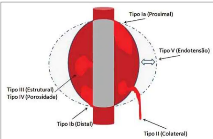
The following parameters were inves-tigated: gender, age, race, imaging method utilized for the diagnosis, affected region, aneurysmal sac dimensions and endoleak type, according to the modified White et al classification, consisting of:
Type I – Blood flow to the endoleak originating at the coupling extremity of the prosthesis. It can be sub-classified as proxi-mal (IA) and distal (IB).
Type II – Blood flow originating from collateral vessels branching from the aorta filling the aneurysmal sac.
Type III – Secondary to structural fail-ure of the endoprosthesis. Fractfail-ures, ori-fices or modular devices separation.
Type IV – Related to the stent porosity, being observed soon after placement of the prosthesis.
Type V – Aneurysmal sac expansion without the identification of an endoleak.
It is also known as endotension.
The images were independently evalu-ated by at least two radiologists with the final consensus prevailing as the result.
Subsequently, data were compiled and tabulated by means of a specific software and submitted to statistical and descriptive analysis.
RESULTS
Amongst the 20 patients diagnosed with endoleak, 14 (70%) were men and 6 (30%) were women. The race with greater preva-lence was white (80%) followed by mixed (10%) and black (10%).
As regards sample distribution by age, the mean value was 76.3 years ranging from 43 to 91 years.
The most frequently involved vascular sites were the infrarenal abdominal aorta with 13 cases (65%), followed by the tho-racic aorta with 4 cases (20%), the iliac territory with 2 cases (10%), and carotid vessels, with 1 case (5%).
CT angiography was the imaging method preferentially utilized for the diag-nosis and classification of the endoleaks in 17 cases (85% of the sample) distributed among types IA, IB, II and III; however US could detect IA type endoleaks in the other three cases (15% of the sample).
The cross diameter of the aneurysmal sacs ranged from 3.8 to 9.8 cm (mean value of 5.8 ± 1.5 cm), the anteroposterior diam-eter ranged between 3.5 and 9.7 cm (mean value of 5.8 ± 1.4 cm), and the longitudi-nal length ranged from 4.6 to 20.0 cm (mean value of 9.8 ± 4.5 cm).
Distribution of the samples according to endoleak types (Figures 1 and 2)
Type II endoleak presents with the blood flow coming from collateral aortic vessels, filling the aneurysmal sac (Figures 6 and 7). Five such cases (25%) were observed in the present sample.
Type III endoleak is characterized by the structural failure of the endoprosthesis, whether such failure is due to partial or total detachment of its components in modular prosthesis or to fracture in such components causing a persistent flow into the aneurysmal sac (Figure 8). Three such cases (15%) were observed in this study.
Type IV endoleak is related to the stent porosity, and can be observed soon after the placement of the prosthesis. It is also re-lated to the anticoagulation status of the patient, and is generally spontaneously corrected, or after the coagulation time is restored to a normal level.
The aneurysmal sac expansion without the identification of an endoleak is known as endotension or endoleak type V. Its cause is uncertain, being possibly related to an-other type of endoleak not detectable at the currently available imaging methods.
Types IV and V endoleaks were not observed in the present sample.
DISCUSSION
The typical patient affected by this com-plication is the male elderly patient with infrarenal abdominal aortic disease(6).
In the present series, type I endoleak was the most prevalent. However, in the literature, type II endoleak is reported as being the most frequently identified type(8).
Figure 1. Scheme illustrating the different types of endoleaks, according to the classification proposed by White et al.
Figure 2. Sample percentage distribution chart as regards endoleak types classification.
Figure 3. Volumetric reconstruction, in the coronal plane of abdominal aorta CT angiography demonstrating endoprosthesis in the aorto-bi-iliac segment and leakage of contrast into the aneurysmal sac in the proximal region of the prosthesis (endoleak type IA – arrow).
Figure 7. Volumetric reconstruction in the sagittal plane of abdominal aorta CT angiography demonstrating endoprosthesis in good conditions and enhanced collateral vessel irrigating the aneurysmal sac (endoleak type II – arrow).
Figure 8. Volumetric reconstruction in the sagittal plane of abdominal aorta CT angiography evidencing structural failure of the endoprosthesis with leak-age of contrast into the aneurysmal sac (endoleak type III – arrow). Figure 5. Three-dimensional reconstruction, in the sagittal plane of CT
an-giography of abdominal aorta demonstrating endoprosthesis in the abdomi-nal segment and contrast leakage into the aneurysmal sac in the distal re-gion of the prosthesis (endoleak type IB – arrow).
Endoleaks may develop during the pro-cedure or sometime after it(13). The early diagnosis and type identification are ex-tremely relevant, as in some cases endo-leaks may be associated with the increased pressure in the aneurysmal sac and in-creased risk for rupture(14–17). Therefore, lifetime follow-up by means of periodic imaging studies is necessary to assure the success of the percutaneous treatment and the timely detection of complications rep-resenting peculiar conditions for the ben-efit of patients’ health(18).
Currently, the classification most rec-ommended by the vascular surgeon’s soci-eties is the one developed by White et al., that was modified in recent years, dividing endoleaks into five types(19-21). This classi-fication is based on the origin of the blood flow into the aneurysmal sac and has direct implication on the treatment required for each case.
Most frequently, type I endoleaks occur after endovascular repair of thoracic aortic aneurysms. Additionally, they are most fre-quently found in patients with a complex arterial anatomy. Short necked, angled, ul-cerated and trapezoid aneurysms contain-ing mural thrombus, pose a challenge for the construction of appropriate seals be-tween the prosthesis and the native aorta(22,23).
Generally, the prognosis for type I endo-leaks is reserved, and an aggressive treat-ment is mandatory. Surgical or endovascular interventions are recommended whenever a type I endoleakis documented between two and four weeks following endopros-thesis implantation. A type I endoleak lo-cated in the proximal neckregion (type IA) is a very dangerous event, as the false lu-men is continuously submitted to high pres-sures, increasing the risk for rupture. In such cases, an immediate intervention is mandatory, with the implantation of one or more prostheses(24).
Type II endoleaks occur as the blood flows through the branches of the aorta segment that did not receive a stent or iliac arteries whose anastomoses communicate directly with the aneurysmal sac. Typical sources include the inferior and lumbar mesenteric arteries(22). This is the most com-mon type of endoleak found after endovas-cular repair of abdominal aortic aneurysms.
The number of patent collateral vessels and the amount of thrombi present in the aneurysmal sac observed in the preopera-tive period seems to be correlated with the risk for development of type II endoleaks.
In cases of increased aneurysmal sac or false lumen patency persistence, percutane-ous selective embolization is suggested, and is easily performed(22).
Type III endoleak is secondary to the disconnection of the endoprosthesis ele-ments, and requires immediate treatment in order to avoid severe complications caused by the continuous flow within the aneu-rysm or the false lumen. In such cases, endovascular therapy may be performed by means of the insertion of a new endopros-thesis inside the previous one. In more complex cases, surgical removal is the most appropriate approach(22).
Repeated stress from arterial pulses on the prosthesis graft may cause such types of leaks. Additionally, as the aneurismal sac decreases along time, additional forces are applied on the graft, and may lead to fail-ure. In spite of being very uncommon, type III endoleaks will probably become most prevalent in the long term follow-up of patients with endoprostheses(22).
Type IV endoleaks are caused by the endoprostheses porosity. Such endoleaks are identified, at the moment of the pros-thesis implantation, as a blurring observed at the immediate post-implantation angio-gram, while the patients are under anti-co-agulant drugs effect. Such endoleaks do not require specific intervention besides the wait for the restoration of normal coagula-tion times. The diagnosis of a type IV endoleak is based on exclusion, as other types of endoleaks may simulate it in the immediate post-implantation evaluation(22). Multidetector CT angiography is the imaging method of choice for the follow-up of these patients, because of the high sensitivity of the method in the identifica-tion of endoleaks and other complicaidentifica-tions associated with the procedure(19,25).
Several authors have demonstrated that CT angiography is the best noninvasive method for the diagnosis of endoleaks, and it is considered as the gold standard and method of choice in such cases(26). With a single breath-hold following the contrast medium injection, the thoracic and
dominal aortas, supra-aortic vessels, ab-dominal branches and the iliaco-femoral axis can be evaluated. The images can be studied in the standard axial plane and can be later be processed through multiplanar reconstructions, utilizing different algo-rithms (VRT, MIP, MPR, shaded surface display [SSD], etc.) providing three-dimen-sional characterizations.
CT angiography can provide the follow-ing data: aorta diameter and morphology, diameter and length of the distal and proxi-mal necks, presence of thrombi or calcifi-cations, abdominal branches patency, size, tortuosity and status of the iliac and femo-ral arteries disease.
At ECG-gated CT angiography, volu-metric three-dimensional data allow the rotation of the aorta, while visualizing it in different phases of the cardiac cycle. This resource may improve the diagnostic accu-racy, as motion artifacts are, many times, the cause of false-positive results(24,27).
Because of variable flow rates, endo-leaks may not be detected at several mo-ments following contrast injection. For this reason, multiphase CT angiography has been recommended: a typical protocol in-cludes images before the administration of contrast medium, images after the admin-istration of contrast in the arterial phase and images in the delayed phase. Pre-contrast images can be useful in the differentiation of calcifications in the endoleak aneurys-mal sac, thus reducing the number of in-conclusive studies(20,28).
Recently, with the development of to-mography apparatuses with 320 detector rows, new techniques for images acquisi-tion have been developed. The literature has already reported the successful utiliza-tion of dynamic volumetric 4D CT angiog-raphy (DV-CTA) in selected cases, for a better characterization of the endoleaks and their appropriate treatment(29).
defined. Additionally, initial studies have demonstrated a limited success of this method(20,30).
The development of sonographic con-trast agents in association with softwares and specific contrast-enhanced imaging techniques (CEUS), have aroused new in-terest in this imaging method and its poten-tial use for the routine follow-up of these patients. Most recently, studies in the litera-ture have demonstrated that sonographic contrast agents seem to increase the sensi-tivity of the method in the post-endovas-cular repair follow-up, by increasing the blood flow echogenicity, allowing a better evaluation of possible complications of the graft, such as the case of endoleaks. On the
other hand, the use of contrast-enhanced US does not replace CT angiography with regards to data related to the integrity of the frat anchoring, changes in the morphology of the aneurysmal sac and visceral vessels patency (30).
Gadolinium-enhanced magnetic reso-nance imaging is capable of demonstrating endoleaks, but its performance depends upon the stent composition. Nitinol stents are generally more appropriate for mag-netic resonance imaging; elgiloy stents may obscure the lumen and stainless steel stents cause extensive artifacts, which leads the study to be inconclusive. It should be high-lighted that in several studies involving a small number of patients with a predomi-nance of nitinol stents, MRI angiography
was at least as sensitive as CT angiography, and in some cases, it demonstrated endo-leaks that were not detected at CT angiog-raphy(24).
The ideal frequency for post-procedural imaging has not been systematically deter-mined. Initial suggestions, based on em-pirical observations of abdominal aortic aneurysms, indicate the follow-up with CT angiography and radiography at one month and at six months after the repair and at every six months thereafter along the patient’s lifetime(23).
CONCLUSION
Early diagnosis and correct classifica-tion of endoleaks are crucial for an
appro-priate management of cases. The knowl-edge of endoleaks subtypes is fundamen-tal in the education of physicians special-ized in radiology and imaging diagnosis as well as for vascular surgeons.
REFERENCES
1. Johnston KW, Rutherford RB, Tilson MD, et al. Suggested standards for reporting on arterial an-eurysms. J Vasc Surg. 1991;13:452–8. 2. Brunkwall J, Hauksson H, Bengtsson H, et al.
Solitary aneurysms of the iliac arterial system: an estimate of their frequency of occurrence. J Vasc Surg. 1989;10:381–4.
3. Bickerstaff LK, Hollier LH, Van Peenen HJ, et al. Abdominal aortic aneurysms: the changing natu-ral history. J Vasc Surg. 1984;1:6–12. 4. Leopold GR, Goldberger LE, Bernstein EF.
Ul-trasonic detection and evaluation of abdominal aortic aneurysms. Surgery. 1972;72:939–45. 5. Law M. Screening for abdominal aortic
aneu-rysms. Br Med Bull. 1998;54:903–13. 6. Thurmond AS, Semler HJ. Abdominal aortic
an-eurysm: incidence in a population at risk. J Cardiovasc Surg (Torino). 1986;27:457–60. 7. Treiman RL, Levine KA, Cohen JL, et al.
Aneu-rysmectomy in the octogenarian. A study of mor-bidity and quality of survival. Am J Surg. 1982; 144:194–7.
8. Albuquerque ELC, Braile DM, Palma JH, et al. Diretrizes para o tratamento cirúrgico das doenças da aorta da Sociedade Brasileira de Cirurgia Car-diovascular. Rev Bras Cir Cardiovasc. 2007;22: 137–59.
9. Parodi JC, Palmaz JC, Barone HD. Transfemoral intraluminal graft implantation for abdominal aortic aneurysms. Ann Vasc Surg. 1991;5:491–9. 10. EVAR trial participants. Endovascular aneurysm repair versus open repair in patients with abdomi-nal aortic aneurysm (EVAR trial 1): randomised controlled trial. Lancet. 2005;365:2179–86. 11. Greenberg RK, Clair D, Srivastava S, et al. Should
patients with challenging anatomy be offered endovascular aneurysm repair? J Vasc Surg. 2003; 38:990–6.
12. Hallett JW Jr. Management of abdominal aortic aneurysms. Mayo Clin Proc. 2000;75:395–9. 13. Sampaio SM, Shin SH, Panneton JM, et al.
Intra-operative endoleak during EVAR: frequency, nature, and significance. Vasc Endovascular Surg. 2009;43:352–9.
14. Lumsden AB, Allen RC, Chaikof EL, et al. De-layed rupture of aortic aneurysms following endo-vascular stent grafting. Am J Surg. 1995;170: 174–8.
15. Alimi YS, Chakfe N, Rivoal E, et al. Rupture of an abdominal aortic aneurysm after endovascular graft placement and aneurysm size reduction. J Vasc Surg. 1998;28:178–83.
16. Torsello GB, Klenk E, Kasprzak B, et al. Rupture of abdominal aortic aneurysm previously treated by endovascular stentgraft. J Vasc Surg. 1998;28: 184–7.
17. Harris PL, Vallabhaneni SR, Desgranges P, et al. Incidence and risk factors of late rupture,
conver-sion, and death after endovascular repair of infra-renal aortic aneurysms: the EUROSTAR experi ence. European Collaborators on Stent/graft tech-niques for aortic aneurysm repair. J Vasc Surg. 2000;32:739–49.
18. Zarins CK, Wolf YG, Lee WA, et al. Will endo-vascular repair replace open surgery for abdomi-nal aortic aneurysm repair? Ann Surg. 2000;232: 501–7.
19. White GH, Yu W, May J, et al. Endoleak as a com-plication of endoluminal grafting of abdominal aortic aneurysms: classification, incidence, diag-nosis, and management. J Endovasc Surg. 1997; 4:152–68.
20. Golzarian J, Dussaussois L, Abada HT, et al. He-lical CT of aorta after endoluminal stent-graft therapy: value of biphasic acquisition. AJR Am J Roentgenol. 1998;171:329–31.
21. Iezzi R, Cotroneo AR, Basilico R, et al. Endoleaks after endovascular repair of abdominal aortic an-eurysm: value of CEUS. Abdom Imaging. 2010; 35:106–14.
22. Fanelli F, Dake MD. Standard of practice for the endovascular treatment of thoracic aortic aneu-rysms and type B dissections. Cardiovasc Intervent Radiol. 2009;32:849–60.
23. Ueda T, Fleischmann D, Dake MD, et al. Incom-plete endograft apposition to the aortic arch: bird-beak configuration increases risk of endoleak for-mation after thoracic endovascular aortic repair. Radiology. 2010;255:645–52.
24. Pannu HK, Jacobs JE, Lai S, et al. Gated cardiac imaging of the aortic valve on 64-slice multi-detector row computed tomography: preliminary observations. J Comput Assist Tomogr. 2006;30: 443–446.
25. White GH, Yu W, May J. Endoleak – a proposed new terminology to describe incomplete aneu-rysm exclusion by an endoluminal graft. J Endovasc Surg. 1996;3:124–5.
26. Thomaz FB, Lopez GE, Marchiori E, et al. Ava-liação pós-operatória do tratamento endovascu-lar de aneurisma
da aorta abdominal por angiotomografia com mu tidetectores. Radiol Bras. 2008;41:213–7. ı 2 7 . Zhang J, Fletcher JG, Vrtiska TJ, et al. L-arge-vessel distensibility measurement with electro cardiographically gated multidetector CT phantom study and initial experience. Radiology. 2
07; 245:258–66.ı28. Bley TA, Chase PJ, Reeder S-B, et al. Endovascular abdominal aortic ane-urysm repair: nonenhanced volumetric CT fo-r follow-up. Radiology. 2009;253:253–62.ı2 . Bent CL, Jaskolka JD, Lindsa
y TF, et al. The use of dynamic volumetric CT angiog-raphy (DV-CTA) for the characterization of endoleaks following fenestrated endovascular aortic aneurysm repair (f-EVAR). J Vasc Surg. 2010;51:203–6.