ISSN 1806-3713 © 2015 Sociedade Brasileira de Pneumologia e Tisiologia
Right lung exclusion in massive pulmonary
thromboembolism
Rodrigo Abensur Athanazio1, Samia Zahi Rached1
Figure 3. Coronal reconstruction demonstrating the complete lack of pulmonary perfusion in the right lung, together with ipsilateral oligemia.
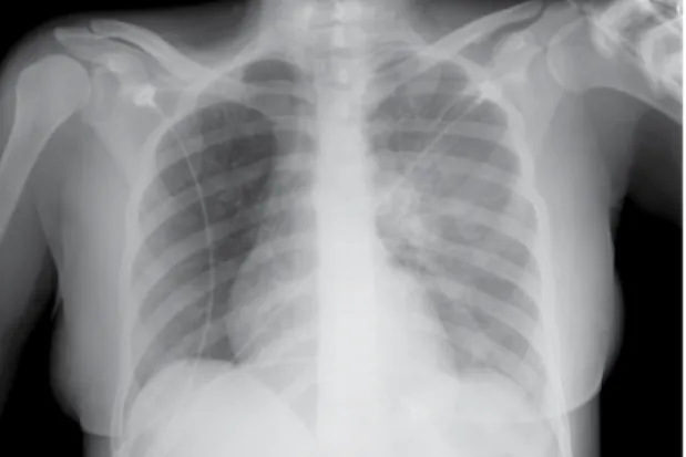
Figure 1. Thoracic X-ray revealing oligemia in the right hemithorax and engorgement of the left pulmonary artery.
Figure 2. CT scan conirming a thrombus in the pulmonary
artery trunk and full occlusion of the right segment (arrow).
1. Divisão de Pneumologia, Instituto do Coração, Hospital das Clínicas, Faculdade de Medicina, Universidade de São Paulo, Brasil. A 37-year-old female patient presented to the emergency
department with a 3-week history of dyspnea, hypoxemia, pleuritic chest pain, and lower limb edema. She had no history of comorbidities and had had two normal pregnancies. There was no family history of thrombosis. An electrocardiogram showed right axis deviation, and blood tests revealed elevated D-dimer levels. A routine chest X-ray showed oligemia in the right hemithorax and engorgement of the left pulmonary artery (Figure 1). Chest CT angiography conirmed the presence of a thrombus in the pulmonary artery trunk and full occlusion of the right segment (Figure 2). The coronal reconstruction shown in Figure 3 elegantly demonstrates the complete
lack of pulmonary perfusion in the right lung, together with ipsilateral oligemia. Echocardiography conirmed pulmonary hypertension (systolic pulmonary artery pressure, 80 mmHg) and right ventricular dysfunction. Because of hemodynamic instability, the patient was submitted to thrombolysis with alteplase and started on anticoagulation therapy. Her dyspnea persisted, and she was categorized as New York Heart Association functional class III. After 6 months, she evolved to chronic pulmonary thromboembolic disease. Positron emission tomography and nuclear magnetic resonance imaging were performed to exclude angiosarcoma. At this writing, the patient is under evaluation for thromboendarterectomy.
486
J Bras Pneumol. 2015;41(5):486-486