Radiol Bras. 2016 Mai/Jun;49(3):190–195 190
Multidetector computed tomography angiography of the renal
arteries: normal anatomy and its variations
*
Angiotomografia computadorizada por multidetectores das artérias renais: anatomia normal e suas variações
Mello Júnior CF, Araujo Neto SA, Carvalho Junior AM, Rebouças RB, Negromonte GRP, Oliveira CD. Multidetector computed tomography angiography of the renal arteries: normal anatomy and its variations. Radiol Bras. 2016 Mai/Jun;49(3):190–195.
Abstract
R e s u m o
Conventional angiography is still considered the gold standard for the study of the anatomy and of vascular diseases of the abdomen. However, the advent of multidetector computed tomography and techniques of digital image reconstruction has provided an alternative means of performing angiography, without the risks inherent to invasive angiographic examinations. Therefore, within the field of radiology, there is an ever-increasing demand for deeper knowledge of the anatomy of the regional vasculature and its variations. Variations in the renal vascular system are relatively prevalent in the venous and arterial vessels. For various conditions in which surgical planning is crucial to the success of the procedure, knowledge of this topic is important. The aim of this study was to familiarize the general radiologist with variations in the renal vascular system. To that end, we prepared a pictorial essay comprising multidetector computed tomography images obtained in a series of cases. We show patterns representative of the most common anatomical variations in the arterial blood supply to the kidneys, calling attention to the nomenclature, as well as to the clinical and surgical implications of such variations.
Keywords: Anatomic variation; Renal artery; Multidetector computed tomography.
A angiografia convencional ainda é considerada o exame padrão ouro no estudo da anatomia e das doenças vasculares do abdome. Entretanto, com o advento da tomografia computadorizada com multidetectores e técnicas de reconstrução de imagens digitais, este exame tem-se tornado uma opção, com a vantagem de não ter os riscos habituais dos exames angiográficos invasivos. Com isso, o aprofundamento do conhecimento dos detalhes anatômicos da vasculatura regional e suas variações é cada vez mais exigido nesta área da radiologia. As variações do sistema vascular renal são relativamente prevalentes, tanto no leito venoso quanto no arterial. O conhe-cimento do tema é de importância nas várias condições em que o prévio planejamento cirúrgico é crucial para o sucesso do procedimento. Para familiarizar o radiologista geral, enriquecendo sua experiência sobre o tópico, os autores elaboraram um ensaio iconográfico a partir de uma série de casos extraídos do banco de imagens de tomografia computadorizada com multidetectores da região abdominal, com os padrões representativos das mais frequentes variações anatômicas da irrigação arterial renal, chamando a atenção para sua desig-nação terminológica e para suas implicações clinicocirúrgicas.
Unitermos: Variação anatômica; Artéria renal; Tomografia computadorizada com multidetectores.
* Study conducted at the Universidade Federal da Paraíba (UFPB), João Pessoa, PB, Brazil.
1. PhD, Adjunct Professor IV of Clinical Radiology, Universidade Federal da Paraíba (UFPB), João Pessoa, PB, Brazil.
2. PhD, Adjunct Professor II of Clinical Radiology, Universidade Federal da Paraíba (UFPB), João Pessoa, PB, Brazil.
3. PhD, Professor in the Department of Surgery, Universidade Federal da Paraíba (UFPB), João Pessoa, PB, Brazil.
4. MD, Urologist, Professor of Urology, Faculdade de Ciências Médicas da Paraíba, João Pessoa, PB, Brazil.
5. Medical Student, Universidade Federal da Paraíba (UFPB), João Pessoa, PB, Brazil.
Mailing address: Dr. Severino Aires Araujo Neto. Avenida Sapé, 1780, ap. 2201, Manaíra. João Pessoa, PB, Brazil, 58038-382. E-mail: [email protected].
Received June 5, 2014. Accepted after revision May 12, 2015.
“normal” pattern. Up until the middle of the last century, the vascular anatomy of the abdomen was a topic of discus-sion restricted to surgeons and anatomists. With the advent and rapid development of imaging tests, radiologists have become indispensible in the diagnostic process and therapy planning for many vascular conditions(1). It is the job of the
radiologist to identify and describe the anatomical patterns of venous and arterial vasculature, particularly when the tests in question are done in preparation for complex kidney sur-geries.
Digital angiography continues to be the gold standard for comparison with any other type of tests for morphologi-cal analysis of renal arterial anatomy(2,3). However, computed
tomography angiography (CTA) studies carry fewer risks and are more accurate than digital angiography studies, with the advantage of evaluating not only the vascular lumina but also the vessel walls and other viscera, and is now used more fre-quently in various scenarios: kidney transplant, Takayasu’s
Carlos Fernando de Mello Júnior1, Severino Aires Araujo Neto2, Arlindo Monteiro de Carvalho Junior3, Rafael Batista Rebouças4, Gustavo Ramalho Pessoa Negromonte5, Carollyne Dantas de Oliveira5
INTRODUCTION
disease, and ureteropelvic junction (UPJ) stenosis due to compression of the inferior polar artery. In addition, CTA has the advantages of allowing a better evaluation of the re-nal collecting system—to identify hydronephrosis—and of the kidneys themselves—to identify tumors, parenchymal atrophy, and congenital disorders, such as horseshoe kidney and duplication of the renal pelvis(4).
Digital image processing and manipulation in diagnos-tic workstations equipped with programs and monitors dedi-cated to this purpose are indispensible for CTA studies. Such studies allow two-dimensional (2D) and three-dimensional (3D) multiple reconstructions to be performed on the basis of raw data extracted from the original axial plane images. The multiplanar reconstruction (MPR), maximum intensity projection (MIP) and volume rendering (VR) methods are widely used and merit a brief explanation. The MPR method provides 2D sectional images in all axial, coronal, and sag-ittal planes, which can be perpendicular to the axial plane, but variations on oblique or even curved planes are particu-larly useful in the study of tortuous structures such as ves-sels. The MIP method selects the higher-density voxels in contiguous axial, coronal, or sagittal sectional images, sum-ming and projecting them in a single image, generally in 3D. After intravenous injection of contrast, when vascular luminal density increases significantly, MIP highlights veins and arteries against the other less dense intra-abdominal structures. Combining consecutive sections allows long tor-tuous vascular segments, which usually enter and exit from an isolated conventional section plane, to be shown in a single image. This effect gives the MIP image the visual sensation of three dimensions. A limitation of this technique is in fact the excess of structures that are added to the image as the slab encompasses more sections, which may visually confuse the examiner. The VR method attributes opacity values rang-ing from 0% (transparent) to 100% (opaque) between vari-ous sections in any plane, in an artificial line of sight projec-tion. By combining these values with luminous effects, the VR 3D image generated reproduces the perspective of depth in a more reliable way than does MIP(1). Therefore, although
MIP and VR have similar resolution and contrast, some au-thors, such as Urban et al.(5), ascribe a certain advantage to
VR, particularly in the visualization of tortuous vessels when it is necessary to determine which are closer or farther from the observer (more superficial or deeper in the examined region, for example).
The objective of this pictorial essay was to familiarize radiologists with the variations found in the renal vascular system, emphasizing prevalence, the most adequate appro-priate terms and the clinical and surgical implications in-volved. To that end, we searched our teaching files, select-ing sample cases in which multidetector computed tomog-raphy (MDCT) studies had produced scans that illustrated the most common anatomical patterns. The studies were carried out with a Brilliance 64-channel scanner (Philips, Eindhoven, the Netherlands). All patients were adults and
were given intravenous iodinated contrast Ultravist®
(Bayer Pharma AG; Leverkusen, Germany), at a concentration of 769 mg/mL. The injection was applied with an injector pump (Envision CT; Medrad, Indianola, PA, USA) with a flow rate of 5 mL/s, and the dosage was approximately 1.5 mL/kg (maximum total dose of 150 mL). Only the post-contrast arterial phase was used for the sample cases. The axial sec-tions (1 mm thick) were acquired at a pitch of 0.8, a recon-struction thickness of 2 mm, and a standard 250 mm field-of-view. During the arterial phase, image acquisition was started with a 6-s delay, after the threshold of 100 Hounsfield units had been reached at the region of interest within the abdominal aorta. The acquisition parameters used for the arterial phase in the abdominal protocol were similar to those used for CTA tests and showed sufficient spatial and tempo-ral resolution to characterize the arterial vessels studied in this paper. The images were processed in a Philips Extended Brilliance workstation using a viewerprogram. Finally, we used the MPR (2D), MIP and VR (3D) methods to acquire images in the axial, coronal, and sagittal planes.
NORMAL ANATOMY OF THE RENAL ARTERIES AND ITS VARIATIONS
The vascularization of the embryonic kidney (proneph-ros, mesoneph(proneph-ros, and metanephros) originates from a group of lateral branches of the abdominal aorta. During the up-ward migration of the kidney to the lumbar region, many arterial branches regress and a main (or hilar) artery sup-plies blood to the renal parenchyma. Despite sequential re-gression of those structures, the caudal arteries, located be-tween the tenth thoracic segment and the third lumbar seg-ment, can persist in the fully formed kidney, evolving into the superior and inferior polar arteries(1,6).
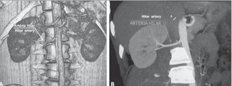
In the so-called “normal” pattern, the kidneys are sup-plied by a single main renal artery, which originates from the abdominal aorta at the L1–L2 level. The main renal ar-tery is also referred to as the hilar arar-tery, because it splits into two, three, or four branches near the hilum (Figure 1), providing the blood supply to various regions of the kidneys. In general, the hilar artery is 4–6 cm long and 5–6 mm in diameter. However, that classic configuration is seen in less than 25% of cases(7).
Given the diverse patterns of renal arterial blood supply, a standardized nomenclature should be adopted, avoiding du-bious terms and contemplating designations that carry an ob-jective anatomical sense and are self-sufficient in transmit-ting an idea of the morphology to which they refer. Uniform usage of these terms among professionals in various fields is fundamental to facilitating communication, avoiding errors, and allowing statistical data from various studies to be syn-thesized. The terms “extra”, “aberrant”, and “supernumerary”, which have been used by some authors(8,9), should be avoided,
suggested, the nomenclature adopted by Sampaio et al.(6)
is, in our opinion, the one that comes closest to the aforemen-tioned precepts and was therefore chosen to classify the find-ings described in this paper. The arterial patterns of the kid-neys (normal and variants) are listed and described below, referenced to the corresponding examples in the figures.
Hilar artery (Figure 1) – Branch of the aorta that en-ters the kidney around the hilum and only at the hilum or renal sinus offers terminal branches. Palmieri et al. reported the prevalence of this pattern to be 62.49% in the right kid-ney and 72.50% in the left kidkid-ney(7).
Upper and lower extrahilar artery (Figures 2 and 3, respectively)– Branch originating from the hilar artery be-fore it reaches the hilum and entering the renal parenchyma outside the hilum (at the upper or lower pole). The reported prevalence of an upper extrahilar artery is 28.6% and 11.6% in the right and left kidney, respectively, compared with and 0% and 1.4%, respectively, for that of a lower extrahilar ar-tery(7).
Superior polar artery (Figure 4) – Branch of the aorta that enters the kidney at the upper pole. The reported preva-lence of a superior polar artery is 7.14% in the right kidney and 11.6% in the left kidney(7).
Inferior polar artery – Branch of the aorta or of the com-mon iliac artery that enters the kidney at the lower pole. The reported prevalence of an inferior polar artery is 3.57% in the right kidney and 2.9% in the left kidney(7).
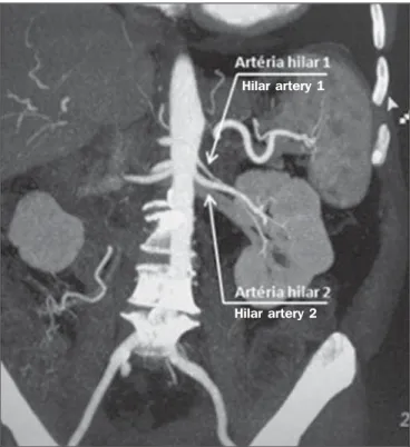
Early bifurcation (Figures 5 and 6) – Right or left re-nal artery with a main trunk less than 1 cm long before branch-ing. This pattern was observed in only one case out of 200 renal pedicles studied(7). In the case of two or more hilar
arteries, the one with the largest caliber will be referred to as the main artery(6).
The prevalence statistics for the morphological patterns mentioned above were restricted to the study by Palmieri et al.(7), because the authors employed a nomenclature closest
to that employed by Sampaio et al.(6). The lack of unifor-mity in the nomenclature employed in many papers published
Figure 1. MDCT, frontal plane VR reconstruction (A) and coronal plane MIP reconstruction (B) showing the right hilar renal artery (arrow), which is a branch of the aorta that enters the kidney near the hilum and has terminal branches only at the hilum or renal sinus.
Hilar artery
Hilar artery
Figure 2. MDCT, coronal plane MIP reconstruction (A) and frontal plane VR reconstruction (B). The arrows indicate the left upper extrahilar polar artery, which branches off the left hilar artery and moves toward the left upper pole.
Upper extrahilar polar artery
on this subject hinders and many times precludes data unifi-cation or cross-study comparisons.
CLINICAL AND SURGICAL IMPLICATIONS OF IMAGING FINDINGS
When planning surgical procedures such as partial ne-phrectomy, pyeloplasty for UPJ stenosis, and kidney trans-plantation, imaging studies are indispensible diagnostic tools(1,3,5). The anatomical information provided can affect
the chosen surgical technique.
Recent studies have shown that partial nephrectomy for tumors produces an oncological result equivalent to that of radical nephrectomy, with a lower rate of progression to chronic kidney disease and cardiovascular events(10–12). When planning partial nephrectomies, previous knowledge
of the vascular anatomy is indispensible. Reducing warm ischemia time is one of the technical measures that can im-prove the functional results of partial nephrectomy(13–16). The
main technique applied is segmental arterial clamping, which potentially improves renal function in the immediate post-operative phase, in comparison with clamping of the main artery(17). The development of high-definition 3D models
of renal vasculature based on imaging studies allows greater precision in the application of the segmental arterial clamp-ing technique(17).
Polar or extrahilar renal arteries are involved in 29–65% of cases of UPJ stenosis(18,19). Prior knowledge of the
pres-ence of these vessels can influpres-ence the surgical approach because they can make endoscopic procedures more difficult and reduce the success rate of conventional treatment(20–22). Figure 3. MDCT, coronal MIP plane reconstruction (A) and frontal plane VR reconstruction (B). The arrows indicate the right lower extrahilar polar artery, which branches off the right hilar artery and moves toward the right lower pole. The key shows the limits of the renal hilum.
Renal hilum
Lower
extrahilar polar artery
Lower extrahilar polar artery
Figure 4. MDCT, coronal plane MIP reconstruction (A) and posterior plane VR reconstruction (B). The arrows indicate the right and left superior polar arteries, which branch off the aorta and move toward the right and left upper poles, respectively.
Therefore, MDCT offers advantages over methods such as ultrasound and intravenous urography in the evaluation of UPJ stenosis, particularly when applied in order to identify polar arteries.
In live-donor kidney transplantation, prior identification of a single renal artery is a favorable factor and lowers the incidence of complications. The presence of variations(6) increases the incidence of vascular thrombosis, warm is-chemia time, blood loss, and the difficulty of carrying out anastomosis, as well as the possibility of urinary fistulas and urethral lesions(6,23). However, the rate of graft rejection in the first year and five-year survival rate do not seem to be
Figure 8. MDCT, axial plane MIP reconstruction, for measuring the length of the right hilar artery from its origin, in the aorta, to its first bifurcation.
Figure 7. MDCT, oblique sagittal plane multiplanar reconstruction, for measuring the caliber of the right hilar artery. Axial and coronal plane images of the same region are shown in the right-hand margin. Note the orientation of the section, perpendicular to the vessel.
Hilar artery
affected by the presence of arterial anatomical variations(23).
As well as mentioning variations in quantity and bifurcation measurements, it is important that the radiology report makes reference to two other aspects: a) the orthogonal diameter of the renal arteries and its variations (Figure 7), given that anastomosis is difficult to perform in arteries with a diam-eter of less than 3 mm and there are greater risks of throm-bosis; and b) the length of the artery from its origin to the first bifurcation (Figure 8), because surgeons recommend that the renal artery be at least 20 mm long to ensure good anastomosis(24).
PRACTICAL RECOMMENDATIONS
IN COMPUTED TOMOGRAPHY ANGIOGRAPHY INTERPRETATION
Axial images are still the basis of diagnosis. However, MPR, MIP, and VR provide important additional informa-tion(3). In CTA, it is recommended that the examiner
oper-ate the workstation directly. In practice, the dynamic use of
Figure 5. MDCT, oblique coronal plane MIP reconstruction. Note the right main hilar artery, designated hilar artery 1, and a right accessory hilar artery, designated hilar artery 2. In this case, both arteries originate from the aorta.
Hilar artery 1
Hilar artery 2
Figure 6. MDCT, coronal plane MIP reconstruction. Note the left main hilar artery (hilar artery 2) entering the hilum, as well as the left accessory hilar artery (hilar artery 1) that follows it. Both originate from the aorta.
Hilar artery 1
reconstruction resources confers greater security and agility in comparison with the isolated use of films.
When interpreting and elaborating the test report, it is recommended that the radiologist follow a basic script. The identification of arterial calcifications is possible in the pre-contrast phase. During the arterial phase, both renal arteries should be followed, from emergence to the renal sinus, in order to identify early bifurcation and extrahilar branches, after which the kidneys should be checked for the presence of polar arteries. Especially in the case of kidney transplanta-tion, it is important to mention the orthogonal diameter of the hilar arteries and occasional variations, as well as the length of the arteries from their origin to the first bifurcation.
CONCLUSIONS
Variations in the arterial vasculature of the kidney are highly prevalent. The low risk and excellent accuracy of MDCT in the evaluation of the arterial anatomy of the kid-ney(5) makes it a practicable alternative to digital
angiogra-phy in many situations. Considering the importance of this anatomy in the treatment planning for various clinical and surgical urological conditions, the radiologist should be prepared to identify and describe findings using standard-ized terminology that is in accordance with the consensus in the literature.
REFERENCES
1. Pérez JA, Torres FG, Toribio AM, et al. Angio CT assessment of anatomical variants in renal vasculature: its importance in the living donor. Insights Imaging. 2013;4:199–211.
2. European Association of Urology. EAU guidelines. Edition pre-sented at the 25th EAU Annual Congress, Barcelona; 2010. 3. American College of Radiology. ACR-SIR-SPR practice parameter
for performance of arteriography. Res. 5 – 2012, Amended 2014 (Res. 39).
4. Türkvatan A, Ozdemir M, Cumhur T, et al. Multidetector CT an-giography of renal vasculature: normal anatomy and variants. Eur Radiol. 2009;19:236–44.
5. Urban BA, Ratner LE, Fishman EK. Three-dimensional volume ren-dered CT angiography of the renal arteries and veins: normal anatomy, variants, and clinical applications. Radiographics. 2001; 21:373–86.
6. Sampaio FJB, Passos MARF. Renal arteries: anatomic study for sur-gical and radiolosur-gical practice. Surg Radiol Anat. 1992;14:113–7. 7. Palmieri BJ, Petroianu A, Silva LC, et al. Study of arterial pattern of 200 renal pedicle through angiotomography. Rev Col Bras Cir. 2011;38:116–21.
8. Chai JW, Lee W, Yin YH, et al. CT angiography for living kidney donors: accuracy, cause of misinterpretation and prevalence of varia-tion. Korean J Radiol. 2008;9:333–9.
9. Guan WH, Han Y, Zhang X, et al. Multiple renal arteries with renal cell carcinoma: preoperative evaluation using computed tomogra-phy angiogratomogra-phy prior to laparoscopic nephrectomy. J Int Med Res. 2013;41:1705–15.
10. Clark MA, Shikanov S, Raman JD, et al. Chronic kidney disease before and after partial nephrectomy. J Urol. 2011;185:43–8. 11. Sun M, Bianchi M, Hansen J, et al. Chronic kidney disease after
nephrectomy in patients with small renal masses: a retrospective ob-servational analysis. Eur Urol. 2012;62:696–703.
12. Huang WC, Elkin EB, Levey AS, et al. Partial nephrectomy versus radical nephrectomy in patients with small renal tumors – is there a difference in mortality and cardiovascular outcomes? J Urol. 2009; 181:55–61.
13. Thompson RH, Lane BR, Lohse CM, et al. Renal function after partial nephrectomy: effect of warm ischemia relative to quantity and quality of preserved kidney. Urology. 2012;79:356–60. 14. Porpiglia F, Fiori C, Bertolo R, et al. Long-term functional
evalu-ation of the treated kidney in a prospective series of patients who underwent laparoscopic partial nephrectomy for small renal tumors. Eur Urol. 2012;62:130–5.
15. Nguyen MM, Gill IS. Halving ischemia time during laparoscopic partial nephrectomy. J Urol. 2008;179:627–32.
16. Gill IS, Patil MB, Abreu AL, et al. Zero ischemia anatomical partial nephrectomy: a novel approach. J Urol. 2012;187:807–14. 17. Shao P, Qin C, Yin C, et al. Laparoscopic partial nephrectomy with
segmental renal artery clamping: technique and clinical outcomes. Eur Urol. 2011;59:849–55.
18. Sampaio FJ, Favorito LA. Ureteropelvic junction stenosis: vascular anatomical background for endopyelotomy. J Urol. 1993;150:1787– 91.
19. Rouvière O, Lyonnet D, Berger P, et al. Ureteropelvic junction ob-struction: use of helical CT for preoperative assessment – compari-son with intraarterial angiography. Radiology. 1999;213:668–73. 20. Faerber GJ, Richardson TD, Farah N, et al. Retrograde treatment of ureteropelvic junction obstruction using the ureteral cutting balloon catheter. J Urol. 1997;157:454–8.
21. Schwartz BF, Stoller ML. Complications of retrograde balloon cau-tery endopyelotomy. J Urol. 1999;162:1594–8.
22. Van Cangh PJ, Wilmart JF, Opsomer RJ, et al. Longterm results and late recurrence after endoureteropyelotomy: a critical analysis of prognostic factors. J Urol. 1994;151:934–7.
23. Kok NFM, Dols LFC, Hunink MGM, et al. Complex vascular anatomy in live kidney donation: imaging and consequences for clinical outcome. Transplantation. 2008;85:1760–5.