INTRAOPERATIVE ULTRASONOGRAPHIC EVALUATION
OF INSULINOMAS: AN UPDATE*
Ana Cláudia Ferreira Rosa1
, Márcio Martins Machado2
, Marcella Stival Lemes3
, Mariana
Caetano Barreto3
, Rodrigo Alvarenga Nunes4
, Nestor de Barros5
, Orlando Milhomem da Mota6
,
Osterno Queiroz da Silva7
, Jales Benevides7
, Paulo Adriano Barreto7
, Giovanni Guido Cerri8
The authors review the literature about intraoperative ultrasonography for evaluation of pancreatic insulinomas. Results of intraoperative ultrasound, preoperative ultrasound and computed tomography are discussed, as well as results of inspection and palpation of the pancreas during surgery, reported in the literature. Keywords: Insulinoma; Ultrasonography; Intraoperative ultrasonography.
Avaliação dos insulinomas pela ultra-sonografia intra-operatória: estado atual do tema.
Os autores fazem uma revisão da literatura sobre a utilização da ultra-sonografia intra-operatória para a avaliação dos insulinomas pancreáticos. São referidos os resultados da ultra-sonografia intra-operatória, ultra-sono-grafia e tomoultra-sono-grafia computadorizada realizadas no pré-operatório, e os resultados da inspeção e palpação do pâncreas realizadas durante procedimentos cirúrgicos referidos na literatura.
Unitermos: Insulinoma; Ultra-sonografia; Ultra-sonografia intra-operatória.
Abstract
Resumo
* Study developed at Department of Radiology, Hospital das Clínicas da Faculdade de Medicina da Universidade de São Paulo, at Hospital Sírio Libanês Diagnosis Center, São Paulo, SP, and at Hospital Araújo Jorge Department of Digestive Tract Diseases – Associação de Combate ao Câncer em Goiás, Goiânia, GO.
1. MD, Radiologist at Hospital das Clínicas da Faculdade de Medicina da Universidade Federal de Goiás.
2. Invited Professor at Department of Radiology of Faculdade de Medicina da Universidade Federal de Goiás.
3. Academic Students of Medicine at Universidade Federal de Goiás.
4. Academic Student of Medicine at Faculdade de Ciências Médicas da Universidade do Vale do Sapucaí, Pouso Alegre, MG. 5. Doctor Professor at Department of Radiology, Faculdade de Medicina da Universidade de São Paulo.
6. Head at Department of Digestive Tract Diseases, Hospital Araújo Jorge da Associação de Combate ao Câncer em Goiás. 7. Surgeons at Department of Digestive Diseases, Hospital Araújo Jorge da Associação de Combate ao Câncer em Goiás. 8. Titular Professor at Department of Radiology, Faculdade de Medicina da Universidade de São Paulo.
Mailing address: Dr. Márcio Martins Machado. Rua Rui Brasil Cavalcante, 496, Ed. Art-1 (Siron Franco), ap. 1101, Setor Oeste. Goiânia, GO, Brazil 74140-140. E-mail: marciommachado@ ibest.com.br
Received October 28, 2004. Accepted March 29, 2006.
In this article, the authors review the role of the intraoperative ultrasonography (IOUS) in the evaluation of pancreatic insulinomas, comparing its applicability with that of other methods like ultrasound (US) and computed tomography (CT), as well as establishing a correlation with sur-gical staging data.
DISCUSSION
Hormone-secreting pancreatic tumors with clinical repercussions are rarely seen. The occurrence of these tumors is esti-mated in about four to five cases per mil-lion inhabitants(3). However, they awake a
great clinical interest because of the differ-ent syndromes they may produce as a result of their excessive hormone production(3).
The treatment of these lesions has caught both clinicians and surgeons´ attention, and, currently, the most appropriate ap-proach is the multidisciplinary one, involv-ing endocrinologists, gastroenterologists, surgeons and oncologists(3). Usually the
therapy is aimed at controlling the in-creased hormone secretion, and the tumor resection afterwards. In case of metastasis, chemotherapy represents an alternative at-tempt to control the disease(3).
In the literature, one may find that en-docrine cells of the digestive system are de-rived from neural crest cells and present
common features in relation to biochemi-cal and ultrastructural aspects related to the synthesis of amines and peptides(3,4). These
cells are considered as pertaining to the APUD (amine precursor uptake and decar-boxilation)(3).
It should be reported that there are ex-perimental evidences discussing the origin of the gastroenteropancreatic APUD cells in the neural crest(5). Notwithstanding, in
spite of this controversy, the application of the APUD system concept explains the di-versity of syndromes associated with endo-crine pancreatic tumors(3).
The term APUD originates from the three initial letters of the words in English designating three important characteristics of these cells: 1) high amines concentra-tion; 2) amine precursoruptake capacity; 3) presence of an amino acid descarboxy-lase(3). They are widely distributed in dif-ferent locations of the organism such us central nervous system, thyroid (parafolli-cular cells), pancreas, bowel, supra-renal medulla and urogenital tract(3).
Tumors originating from APUD cells may be called “apudomas”. Endocrine pan-creatic tumors contain APUD system cells and, therefore, may be considered as apud-omas(3).
At least five types of pancreatic islet cells are found in the normal pancreas. Each type produces a different peptide or INTRODUCTION
The pancreas is a solid organ and should be easily demonstrated by ultrasonogra-phy(1). However, because of its
retroperi-neal localization with anteposition of hol-low viscera, the gas present in the viscera may complicate its study by means of ul-trasound (2).
amine. Beta cells produce insulin, alpha cells produce glucagon; gamma cells, somatostatine; F cells, pancreatic polypep-tide; and enterochromafin cells produce serotonine(6).
Insulinomas are the most frequent neu-roendocrine pancreatic tumors and usually are of benign nature(7). Gastrinoma is the
second more frequent neuroendocrine pan-creatic tumor, and 60% of them, or more, may be of malignant nature(8).
Hormone secreting tumors, like insulinomas, gastrinomas and glucagon-omas, may produce identifiable clinical syndromes as a consequence of the exces-sive hormones production(9). The presence
of these symptoms determines an earlier presentation than the usual presentation of other pancreatic neoplasms. So, usually, these tumors are small, frequently measur-ing less than 1.5 cm.(9,10).
On the other hand, somatostatinomas and neuroendocrine tumors producing pan-creatic polypeptide define the presentation of no or few symptoms, although, at the moment of diagnosis, they may present large dimensions(9).
The insulinomas were the first pancre-atic neuroendocrine tumors to be identi-fied, as reported by Whipple & Frantz(11).
The most important finding in cases of insulinomas is the prevalence of high lev-els of autonomous insulin secretion in the presence of concurrent hypoglycemia. This behavior leads to a diagnosis based on the finding of high fasting (72 hours) levels of plasmatic insulin(12). Consequently, they
cause hypoglycemia and symptoms charac-teristic of the hypoglycemic syndrome(3). A
wide variety of symptoms may occur, in-cluding tremor, flutter, irritability, weak-ness, hunger, sweating, tachycardia and, occasionally, nauseas and emesis. Addi-tionally, behavioral and consciousness dis-turbances may occur(3). More severe cases
may progress to unconsciousness and coma as a result of hypoglycemia(3). As a result
of these behavioral disturbances, about 20% of patients are initially misdiagnosed with psychiatric or neurological disor-ders(13).
Insulinomas may occur at any age range, but the greatest majority occurs be-tween 30 and 60 years of age(14). Similarly,
Machado et al.(15) report a casuistic with
54.2% of patients in this age range, with 94.8% with less than 60 years(15,16).
As regards sex, insulinomas would have a slight incidence in females(16–18). Other
authors, like Norton et al.(19), have
ob-served an even higher (about 83%) inci-dence in females.
Most of insulinomas are benign and small, and in 80%-90% of cases are soli-tary lesions(3). Comi et al.(20) also report that
insulinomas usually are intrapancreatic (about 100%) and small (less than 2 cm). Others report benign insulinomas with less than 1.5 cm in 70% of cases(10).
Approxi-mately 10% of insulinomas are malignant and present with metastasis at the moment of diagnosis(3,21).
Multiple and benign lesions may occur in about 10% of cases, and are more fre-quent in patients with MEN type I syn-drome (multiple endocrine neoplasm)(3).
Some authors, like Welbourn et al.(22),
re-port that the number of patients with MEN type 1 would correspond to 5% of all the patients with insulinomas.
Benign insulinomas have no predilec-tion to a specific site within the pancreas(19),
and have been usually identified with the same frequency in different portions of the pancreas: head (30%), body (35%) and tail (35%)(3).
The tumors may be on the surface of the gland or within the pancreatic paren-chyma(3). The majority of lesions is covered
by a layer of pancreatic parenchyma and is typically firmer than the normal pancreas(3).
As regards patients with MEN-1, De-meure et al.(23) report the significance of a
careful evaluation of these patients, consid-ering the higher possibility of malignancy (about 20%) when compared with non-MEN-1-related sporadic insulinomas (ma-lignancy in about 10% of cases). They have observed that in patients with MEN-1 insulinomas were multiple in 76% of cases. Machado et al.(16) have described cases
where multiple insulinomas in patients with MEN-1 were localized in the pancre-atic body and tail in 87%. In this study, the patients underwent left pancreatectomy (caudal body) associated with enucleation in cases of concurrent cephalic lesions. This surgical alternative had already been previously proposed with excellent re-sults(24–26).
At US, insulinomas, in 90% of cases are small, well-defined, round, homogeneous and hypoechogenic lesions(9,10) (Figures 1,
2, 3, 4 and 5). The pancreatic tail presents a special difficulty for insulinomas local-ization(9). Calcifications, configured as
ar-eas of higher echogenicity may, occasion-ally, be identified (9).
Norton et al.(19) and Lo et al.(7) have
ob-served that insulinomas may be of difficult localization due their small dimensions, and their correct identification, both preop-eratively and during the surgical procedure is of paramount importance for their thera-peutic management(19).
Machado et al.(16) have described the
IOUS as an extremely useful method for insulinomas management, routinely uti-lized by the authors for allowing an ad-equate localization of lesions, with a cor-rect definition of the pancreatic anatomy and for avoiding extensive pancreatic re-sections, besides identifying small-sized lesions usually missed by preoperative ex-aminations.
Machado et al.(16) have also observed
that, notwithstanding the progress of diag-nostic methods during the latest years, the identification of insulinomas remains a difficult clinical problem both for clini-cians and surgeons.
According to Norton et al.(19) the main
advantages of the IOUS for the treatment of benign insulinomas include: precise operative localization; enucleation of nonpalpable, nonvisible tumors; and avoid-ance of ductal and vascular injuries as well as injuries to other vital structures by pre-cise localization of adjacent vital struc-tures. These authors have observed that the IOUS is capable of accurately identifying insulinomas in a higher number than those identified by palpation.
Norton et al.(19) have described a benign
These authors have reaffirmed that the pan-creas evaluation should be precisely per-formed, with perpendicular and oblique insonation of pancreatic regions in order to maximizing the US scanning results, iden-tifying pancreatic insulinomas. This care-ful and systematic evaluation allows the visualization of lesions with high diagnos-tic difficulty, as it can be observed in this case of an isoechogenic insulinoma re-ported by the authors(19).
Norton et al.(19) have also demonstrated
that IOUS has affected the surgical proce-dure, facilitating the excision of occult insulinomas, and in about 41% of cases the IOUS has allowed enucleation of cephalic insulinomas which could not be removed except by means of higher morbidity meth-ods like duodenopancreatectomy. In these 41% of cases reported by the authors, IOUS has defined the incision, determining the
most direct and shorter route to the tumor, without traversing the pancreatic duct or other vascular structures. Also, according these authors, after the surgical incision, the shortest route was continually reconfirmed by IOUS up to the complete enucleation of the lesion.
Doherty et al.(27), in a study with 25
pa-tients, have concluded that IOUS was par-ticularly useful for identifying cephalic pancreatic tumors detected in all the cases, and for definition of the relationship be-tween the tumor and the pancreatic duct in the 25 patients evaluated. Additionally, IOUS was utilized to guide the dissection during enucleation both in cases of pal-pable and nonpalpal-pable lesions. Also, they have affirmed that the higher significance of the IOUS is not in the fact of identify-ing lesions, but rather in the utility of this information for the success of the surgical
procedure. Grant et al.(10) have
corrobo-rated such IOUS relevance, demonstrating its influence on the surgical conduct in 62% of the patients with insulinoma, also for the finding of nonpalpable lesions in the head of the pancreas in four of 29 patients.
In any of the surgical alternatives avail-able for approaching insulinomas, it is de-fined that IOUS is of paramount signifi-cance for the evaluation of the pancreatic parenchyma, since it may guide the char-acterization of the pancreatic ducts and vascular structures(28), resulting in reliable
anatomical information for definition of the surgical strategy.
It is very important to highlight that the major breakthrough in the localization of occult insulinomas is the use of IOUS. Some authors have reported their concern related to the results of the blind resection, a method until recently utilized. According to Norton et al.(19), in cases where
insulin-omas could not be identified, blind (distal, subtotal or even total) pancreatectomy would be performed with high morbidity, possibly resulting in pancreatitis, pancre-atic fistulas, pancrepancre-atic abscesses and even progress to exocrine pancreatic failure and diabetes mellitus. On the other hand, Ka-plan et al.(29) also have observed that blind
distal pancreatic resections (caudal bodies) would have a 33% chance of not removing the insulinoma because some occult insu-linomas will be in the pancreatic head. Norton et al.(19), reported a casuistic with
45% of cephalic insulinomas which would have not been successfully removed by blind distal pancreatectomy.
Figure 5. Hypoechogenic nodule (insulinoma) with 0.5 cm (N, insulinoma; DUOD, duodenum).
Figure 1. Hypoechogenic nodules (insulinomas) with 1.5 cm (black arrow) and 0.3 cm (white ar-row).
Figure 2. Hypoechogenic nodule (insulinoma) with 1.5 cm (arrow).
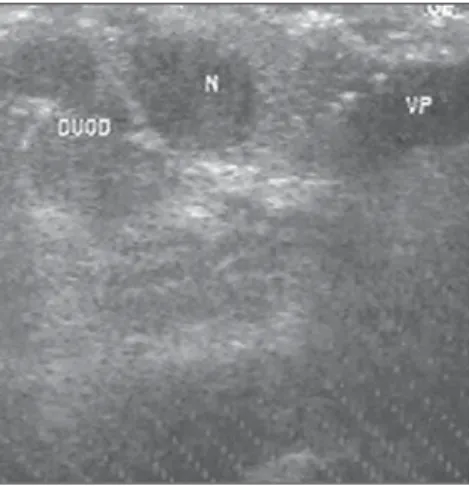
Figure 3. Hypoechogenic nodule (insulinoma) with 0.6 cm. (N, insulinoma; DUOD, duodenum; VP, portal vein).
All of these difficulties were resolved with the utilization of IOUS to identify occult insulinomas, probably in association with a careful palpation of the pancreas.
Demeure et al.(23) reported the
possibil-ity of demonstrating malignant and ectopic extrapancreatic insulinomas. Extrapancre-atic insulinomas would occur in less than 1% cases.
Brazilian and foreign authors concern in searching for a more accurate method for identifying pancreatic insulinomas is a re-sult of the poor diagnostic capacity of pre-operative examinations (US and CT) when compared with IOUS(10,16,27,30–34). Many
series report varied frequencies of preop-erative US detection rates for insulinomas of 0%(32), 26%(27), 28.1%(16), 29.5%(33),
59%(10) and 62%(31). Preoperative CT
de-tection rates also vary, with some authors identifying low rates like those reported by Doherty et al.(27), of 17%. Machado et al.(16)
have reported rates of about 25% and Grant
et al.(10), of 36%. However, other studies
report higher values, like those of Stark et al.(30), ranging between 50% and 60%.
As regards palpation, Kuzin et al.(33)
have reported a 90% detection rate with this procedure. The precise localization of insu-linomas by inspection/palpation depends on the surgeon experience. Doherty et al.(27)
have identified 64% of insulinomas by means of palpation. Machado et al.(16) have
reported the detection of 98.2% of cases by means of inspection/palpation, a result similar to those of other authors(35–37).
The literature review demonstrates that smaller the lesions to be evaluated, the greater will be the limitations of preopera-tive examinations. For insulinomas < 1.00 cm , authors(33) have demonstrated IOUS
detection rate of about 10%, and 20% for preoperative CT. Kuzin et al.(33),
consider-ing only < 1.00 cm insulinomas, have re-ported a 82% detection rate with palpation. In a recent study, Machado et al.(16),
showed that IOUS, in association with pal-pation, allows a precise localization of in 100% of cases, as previously reported in the literature by authors like Van Heerden
et al.(38) and Kisker et al.(37).
The clinicians and surgeons´ confidence in the IOUS capacity of identifying pancre-atic insulinomas has been publicized in several recent publications(16,34).
As a matter of fact, Machado et al.(16)
have affirmed that patients with a con-firmed diagnosis for hyperinsulinism could be evaluated exclusively with conventional abdominal US associated with inspection/ palpation during the surgery for insulin-omas resection in association with IOUS, virtually succeeding in all the cases. Later complications, like diabetes, could be avoided by preserving a larger amount of pancreatic tissue.
Zeiger et al.(39) have confirmed that
IOUS is an unique and safe method for assisting in the localization of insulinomas and is supplementary and additional to the other diagnostic methods. As a result of the practice, the IOUS may reduce the use of other imaging methods, influence the de-cision making and the surgical conduct, al-lowing a complete resection of the primary pancreatic lesion and even the resection of possible associated metastases.
It is important to mention that some of our studies have been developed with the first publications in 1997, evidencing the applicability of the vídeolaparoscopic in-traoperative ultrasound or conventional (non-videolaparoscopic) IOUS (with an open abdominal cavity – laparotomy) in the hepatobiliarypancreatic surgical proce-dures(40–49).
CONCLUSIONS
The authors conclude that IOUS pre-sents higher accuracy for identification of insulinomas when compared with other methods of preoperative staging, proving to be of high value during surgeries, effec-tively supplementing the surgical staging (palpation) itself.
REFERENCES
1. Francisco Neto MJ, Machado MM, Oliveira IRS, Cerri GG. Pâncreas. In: Cerri GG, Oliveira IRS, editores. Ultra-sonografia abdominal. São Paulo: Sarvier, 2002;261–294.
2. Luck AJ, Maddern GJ. Intraoperative abdominal ultrasonography. Br J Surg 1999;86:5–16. 3. Ellison EC, Wise SR, Johnson JA. Endocrine
tu-mors of the pancreas. In: Moody FG, editor. Sur-gical treatment of digestive disease. 2nd ed. Chi-cago: Year Book Med Publ, 1990;545–598. 4. Welbourne RB, Polak JM, Bloom SR. Apudomas
of the pancreas. In: Bloom SR, editor. Gut hor-mones. Edinburgh: Churchill Livingstone, 1978; 561–569.
5. Andrew A, Kramer B, Rawdon BB. Gut and
pan-creatic amine precursor uptake and decarboxyla-tion cells are not neural crest derivatives. Gastro-enterology 1983;84:429–431.
6. Kloppel G, Maillet B. Classification and staging of pancreatic nonendocrine tumors. Radiol Clin North Am 1989;27:105–119.
7. Lo CY, van Heerden JA, Thompson GB, Grant CS, Soreide JA, Harmsen WS. Islet cell carci-noma of the pancreas. World J Surg 1996;20:878– 884.
8. Kisker O, Bastian D, Bartsch D, Nies C, Rothmund M. Localization, malignant potential, and surgi-cal management of gastrinomas. World J Surg 1998;22:651–658.
9. Cosgrove DO. The pancreas. In: Meire H, Cos-grove DO, Dewbury K, Farrant P, editors. Ab-dominal and general ultrasound. 2nd ed. London: Churchill Livingstone, 2001;349–378. 10. Grant CS, van Heerden J, Charboneau JW, James
EM, Reading CC. Insulinoma. The value of in-traoperative ultrasonography. Arch Surg 1988; 123:843–848.
11. Whipple AO, Frantz VK. Adenoma of islet cells with hyperinsulinism. Ann Surg 1935;101:1299– 1335.
12. Fajans SS, Vinik AI. Insulin-producing islet cell tumors. Endocrinol Metab Clin North Am 1980; 18:45–74.
13. Powers RD, Robb JF. Hypoglycemia due to insu-linoma. An unusual cause of altered mental sta-tus in a young man. Minn Med 1983;66:13–15. 14. Galbut DL, Markowitz AM. Insulinoma: diagno-sis, surgical management and long-term follow-up. Am J Surg 1980;139:682–690.
15. Machado MCC, Jukemura J, Monteiro da Cunha JE, et al. Tratamento cirúrgico dos insulinomas. Estudo de 59 casos. Rev Ass Med Brasil 1998;44: 159–166.
16. Machado MCC, Cunha JEM, Jukemura J, et al. Insulinoma: diagnostic strategies and surgical treatment. A 22–year experience. Hepatogastro-enterology 2001;48:854–858.
17. Stefanini P, Carboni M, Patrassi N, Basoli A. Beta-islet cell tumors of the pancreas: results of a study on 1067 cases. Surgery 1974;75:597–609. 18. Service FJ, McMahon MM, O’Brien PC, Ballard DJ. Functioning insulinoma – incidence, recur-rence, and long-term survival of patients: a 60-year study. Mayo Clin Proc 1991;66:711–719. 19. Norton JA, Shawker TH, Doppman JL, et al.
Localization and surgical treatment of occult insulinomas. Ann Surg 1990;212:615–620. 20. Comi RJ, Gorden P, Doppman JL, Norton JA.
Insulinoma. In: Go VLW, editor. The exocrine pancreas: biology, pathobiology and diseases. New York: Raven Press, 1986;745–761. 21. Reber HA, Way LW. Pancreas. In: Way LW,
edi-tor. Current surgical diagnosis & treatment. Con-necticut: Appleton & Lange, 1994;345–359. 22. Welbourne RB, Wood SM, Polak JM, Bloom SR.
Pancreatic endocrine tumors. In: Bloom SR, edi-tor. Gut hormones. New York: Churchill Living-stone, 1981;547–554.
23. Demeure MJ, Klonoff DC, Karam JH, Duh QY, Clark OH. Insulinomas associated with multiple endocrine neoplasia type I: the need for a differ-ent surgical approach. Surgery 1991;110:998– 1005.
neopla-sia type I syndrome. Arch Surg 1985;120:584– 589.
25. Skogseid B, Grama D, Rastad J, et al. Operative tumour yield obviates preoperative pancreatic tumour localization in multiple endocrine neopla-sia type 1. J Intern Med 1995;238:281–288. 26. Thompson NW. The surgical management of
hyperparathyroidism and endocrine disease of the pancreas in the multiple endocrine neoplasia type 1 patient. J Intern Med 1995;238:269–280. 27. Doherty GM, Doppman JL, Shawker TH, et al.
Results of a prospective strategy to diagnose, lo-calize, and resect insulinomas. Surgery 1991;110: 989–997.
28. Deixonne B, Lopez FM. Operative ultrasonogra-phy during hepatobiliary and pancreatic surgery. Berlin: Springer, 1988.
29. Kaplan EL, Arganini M, Kang SJ. Diagnosis and treatment of hypoglycemic disorders. Surg Clin North Am 1987;67:395–410.
30. Stark DD, Moss AA, Goldberg HI, Deveney CW, Way L. Computed tomography and nuclear mag-netic resonance imaging of pancreatic islet cell tumors. Surgery 1983;94:1024–1027. 31. Bottger TC, Weber W, Beyer J, Junginger T. Value
of tumor localization in patients with insulinoma. World J Surg 1990;14:107–114.
32. Vinik AI, Delbridge L, Moattari R, Cho K, Thom-pson N. Transhepatic portal vein catheterization for localization of insulinomas: a ten-year expe-rience. Surgery 1991;109:1–11.
33. Kuzin NM, Egorov AV, Kondrashin SA, Lotov AN, Kuznetzov NS, Majorova JB. Preoperative and intraoperative topographic diagnosis of insulinomas. World J Surg 1998;22:593–598. 34. Machado MM. Ultra-sonografia intra-operatória
(USIO) (Editorial). Radiol Bras 2001;34:V–VI. 35. Galiber AK, Reading CC, Charboneau JW, et al.
Localization of pancreatic insulinoma: compari-son of pre- and intraoperative US with CT and an-giography. Radiology 1988;166:405–408. 36. Menegaux F, Schmitt G, Mercadier M, Chigot JP.
Pancreatic insulinomas. Am J Surg 1993;165: 243–248.
37. Kisker O, Bastian D, Frank M, Rothmund M. Diagnostic localization of insulinomas. Experi-ence with 25 patients with solitary tumors. Med Klin 1996;91:349–354.
38. van Heerden JA, Grant CS, Czako PF, Service FJ, Charboneau JW. Occult functioning insulinomas: which localizing studies are indicated? Surgery 1992;112:1010–1015.
39. Zeiger MA, Shawker TH, Norton JA. Use of in-traoperative ultrasonography to localize islet cell tumors. World J Surg 1993;17:448–454. 40. Paula AL, Machado MM, Mota O, et al.
Detec-ção de metástases hepáticas em tumores de có-lon e reto com a utilização da ultra-sonografia intra-operatória vídeo-laparoscópica (IOUS-LAPA). Rev Bras Coloproc 1997;17 (Supl 1):30. 41. Paula AL, Machado MM, Mota O, et al. Metodi-zação e racionalidade do emprego da ultra-sono-grafia intra-operatória vídeo-laparoscópica (IOUS-LAPA) na avaliação de metástases hepá-ticas de tumores colorretais. Rev Bras Coloproc 1997;17(Supl 1):67.
42. Machado MM, Paula AL, Mota O, et al. Ultra-sonografia intra-operatória vídeo-laparoscópica. Estudo de sua evolução técnica e análise crítica de sua aplicabilidade em coloproctologia. Rev Bras Coloproc 1997;17 (Supl 1):67.
43. Machado MM, Cerri GG. Proposta de
metodiza-ção da ultra-sonografia intra-operatória vídeo-laparoscópica (USIO-LAPA) no estadiamento do fígado em pacientes portadores de neoplasia gas-trointestinal. Radiol Bras 1998;31:375–377. 44. Machado MM, Oliveira IRS, Cerri GG.
Conside-rações sobre a evolução técnica dos transdutores na ultra-sonografia intra-operatória vídeo-lapa-roscópica (USIO-LAPA). Radiol Bras 1999;32: 85–87.
45. Machado MM, Cerri GG, Oliveira IRS, et al. Contribuição da ultra-sonografia intra-operatória (USIO) no estudo de pequenas imagens nodula-res hipoatenuantes identificadas à tomografia computadorizada (TC) no exame pré-operatório de pacientes com adenocarcinoma colorretal e de pâncreas. Comunicação original. Radiol Bras 1999;32:255–258.
46. Machado MM, Rosa ACF, Barros N, Machado MCC, Cerri GG. Ultra-sonografia intra-operató-ria (USIO) do pâncreas e das vias biliares. Radiol Bras 2002;35:105–108.
47. Machado MM, Rosa ACF, Cerri GG. Ultra-sono-grafia intra-operatória (USIO). In: Cerri GG, Oli-veira IRS, editores. Ultra-sonografia abdominal. Rio de Janeiro: Revinter, 2002;573–584. 48. Machado MM. Contribuição da ultra-sonografia
intra-operatória (USIO) no estudo do fígado em pacientes candidatos à ressecção hepática por metástase de adenocarcinoma colorretal. (Tese de Doutorado). São Paulo: Universidade de São Paulo, 2002.