ABSTRACT
Sealability of MTA and calcium
hydroxide-containing sealers
João Eduardo GOMES-FILHO2, Jaqueline Viana MOREIRA5, Simone WATANABE4, Carolina Simonetti LODI4, Luciano
Tavares Angelo CINTRA3, Eloi DEZAN JUNIOR2, Pedro Felício Estrada BERNABÉ1, Mauro Juvenal NERY2, José
Arlindo OTOBONI FILHO2
1- DDS, MSc, PhD, Full Professor, Department of Endodontics, Araçatuba School of Dentistry, UNESP - Univ. Estadual Paulista, Araçatuba, SP, Brazil. 2- DDS, MSc, PhD, Associate Professor, Department of Endodontics, Araçatuba School of Dentistry, UNESP - Univ. Estadual Paulista, Araçatuba, SP, Brazil. 3- DDS, MSc, PhD, Assistant Professor, Department of Endodontics, Araçatuba School of Dentistry, UNESP - Univ. Estadual Paulista, Araçatuba, SP, Brazil. 4- DDS, MSc, Graduate Program in Pediatrics, Araçatuba School of Dentistry, UNESP - Univ. Estadual Paulista, Araçatuba, SP, Brazil.
5- Undergraduate student, Araçatuba School of Dentistry, UNESP - Univ. Estadual Paulista, Araçatuba, SP, Brazil.
Corresponding address: Dr. João Eduardo Gomes-Filho - Faculdade de Odontologia de Araçatuba, Universidade Estadual Paulista - R. José Bonifácio, 1193 - Araçatuba - SP - Brasil - Phone (0055) 18 36363252 - Fax: (0055) 18 36363279 - e-mail: [email protected]
Received: October 2, 2010 - Modiication: August 16, 2011 - Accepted: September 1, 2011
O
bjectives: The aim of this study was to evaluate the apical sealability of Fillapex®,endo-CPM-Sealer® and Sealapex®. Material and Methods: Ninety-four freshly extracted
single-rooted teeth were selected and decoronated. All teeth were radiographed to conirm the existence of a single and straight root canal, which was prepared using Protaper Universal and 2.5% sodium hypochlorite. The teeth were randomly divided in groups of 10 specimens each according to the sealer, and the canals were illed using the single cone technique and one of the sealers. Four additional teeth were used as controls. The teeth were submitted to dye leakage with Rhodamine B for 24 h but using vacuum on the initial 15 min. Thereafter, they were cut longitudinally and the leakage was measured in a linear fashion from apex to crown. Data were analyzed by ANOVA and Tukey’s tests at 5% signiicance level. Results: Fillapex® and Sealapex® showed signiicantly less dye
leakage than endo-CPM-Sealer® (p<0.05). Conclusions: It was concluded that Fillapex®
and Sealapex® were able to prevent apical dye leakage differently from endo-CPM-Sealer®.
Key words: Root canal illing material. Leakage. Fillapex.
INTRODUCTION
One of the of root canal obturation goals is to obtain hermetic sealing of the root canal system favoring the process of apical and periapical repair after endodontic therapy17. Inadequate illing can
result in luid movements into the illing defects favoring a periapical chronic inlammatory reaction
and compromising the treatment success29.
Root canal ramifications, such as lateral, secondary and accessory canals can establish connection between the main root canal and periodontal ligament, as well as the apical foramen3,9. Several authors described that localized periodontal problems might be associated with necrotic and
infected root canal ramiications highlighting the
importance of the capacity of the endodontic sealer to flow into these irregularities3,4. Despite the
signiicance of this physical property, the relationship
between low and its ability to penetrate narrow root canal ramiications has not been investigated3,30.
Root canal sealers used clinically have several bases including zinc oxide-eugenol, epoxy resin, glass ionomer, and calcium hydroxide. Sealapex® (Sybron endo Glendora, CA, USA) is an endodontic sealer that contains calcium oxide, which, in contact with water, forms calcium hydroxide and it was used in the present article as a reference.
A new formulation of MTA-labeled endo-CPM-Sealer® (eGeO S.R.L., Buenos Aires, Buenos Aires, Argentina) was created to be used as root canal sealer. The composition of CPM Sealer is MTA with the addition of calcium carbonate to reduce
the pH from 12.5 to 10.0 after set. This way, the
surface necrosis in contact with the material is restricted, which allows the action of the alkaline phosphatase14.
The aim of this study was to evaluate the apical sealability of Fillapex®, endo-CPM-Sealer® and Sealapex® endodontic sealers.
MATERIAL AND METhODS
Tooth selection and preparation
Ninety-four single-rooted teeth recently extracted for several reasons were selected and stored in neutral formaldehyde for at least 72 h from the tooth bank of the Araçatuba School of Dentistry, UNeSP – Univ. estadual Paulista, Brazil. The teeth
were radiographed to conirm the existence of a
single and straight canal and were decoronated at a mean distance of 11 mm from the apex.
Root canal instrumentation and obturation
Instrumentation was held by a single operator. A #10 K file (Dentsply Maillefer, Ballaigues, Switzerland) was introduced into the root canal until it was visible at the apical foramen. The working length was determined by subtracting 1 mm from this length. Root canal instrumentation was performed using the ProTaper Universal rotary instruments (S1, S2, F1, F2 and F3) (Dentsply Maillefer) activated by an electric motor with controlled speed of 300 rpm (endo-Plus, Driller, Jaguaré, SP, Brazil). Originally a #10 or #15 K
ile was irst introduced to the middle third of the
canal. Instruments S1 and S2 were advanced until resistance was encountered, but not more than two thirds of the depth of the canal. It was carried out
to introduce a #15 K ile up to the working length
followed by reintroduction of the instrument S1 to this length. The other instruments were then inserted into full-length work in sequence S2, F1, F2 and F3. The canals were irrigated with 3 mL of 2.5% sodium hypochlorite (Apothicário Compounding
Pharmacy, Araçatuba, SP, Brazil) after each ile. The luid content was aspirated and the canals were
dried with sterile absorbent paper points.
All root canals were illed by the single cone
technique with the use of master gutta-percha F3 cone (Dentsply Maillefer). The teeth were
condenser followed by vertical condensation. No sealer was used in the positive control group and
the negative control teeth were illed in the same
way as in Group 1.
Dye leakage assay
After instrumentation, the teeth were sealed with nail polish leaving only the apex free for penetration of 0.2% Rhodamine B during 7 days, using vacuum
in the irst 15 min. The teeth in the negative control
group were sealed, including apex. For analysis of the sealing ability of the tested materials, leakage of the dye was linearly measured on the photographs that were taken under stereomicroscopy using
the SigmaScan® Pro Image Analysis Software
(Systat Software, San Jose, CA, USA). Leakage measurement considered the line with longer length of dye, on the material/dentin wall interface, from the most apical to the most cervical portion. If the leakage length were not similar on both sides of the root canal, only the longer length was considered. Leakage was independently measured by three calibrated examiners. The results (mean of leakage values obtained by each examiner) obtained in millimeters were tabulated and analyzed by ANOVA to investigate possible differences
between materials, and Tukey’s test to conirm the signiicance of difference between groups, using Pacotico statistical software. The signiicance level
was set at 5%.
RESULTS
Kappa statistics showed that agreement between the three examiners was higher than 90%. The positive control showed total leakage in all specimens and the negative control did not show leakage (Figure 1). Analysis of the data for the 3 experimental groups revealed that
endo-CPM-Sealer® allowed more leakage than the other
materials (p=0.001258). Fillapex® was similar (p>0.05) to Sealapex® and both materials showed
signiicantly less (p<0.05) leakage when compared
DISCUSSION
The goal of this study was to evaluate the degree of apical leakage of Fillapex® compared with Sealapex® and endo-CPM-Sealer® after illing of root canals prepared with the Protaper Universal system
using the single-cone illing technique.
All procedures were performed by the same operator to avoid intra-operator discrepancies. Only teeth with single straight root canals were used because they can offer a more standardized method for evaluation of apical leakage. In addition,
assessing the dificulties of managing curved canals
was not within the study’s scope.
The Protaper Universal system is the new version of the Protaper™ NiTi rotary system, which includes
shaping, inishing and retreatment instruments1,2. Protaper Universal system was used in this study for reducing the time required for biomechanical preparation and improving the standardization of instrumentation1,2. Moreover, using the single cone
technique for root canal illing allowed observing the
sealability of the sealers in a more critical situation than that offered by the lateral condensation technique, as it is possible to speculate that the single master cone needs a greater interaction with
the sealer to promote the sealing.
There is a lot of skepticism about the value of in vitro leakage studies with regard to clinical
signiicance and limitations of the results13,31. even so, they are widely used to evaluate and compare
the sealing eficiency of materials, which must be
assessed prior to their use in patients. Although in the present study the intensity of the coloration produced after the leakage was not evaluated, Rhodamine B was used because it does not suffer discoloration by calcium hydroxide-based materials, as occurs with methylene blue12,30.Vacuum was used according to studies that showed higher levels of leakage when it was employed compared with groups without its utilization, probably because the
presence of air bubbles in the illing mass makes it dificult the penetration of dye, though they do not
prevent the penetration of bacteria11,13.
The results of this study showed lower leakage
of dye in the groups illed with Sealapex® and
Fillapex®, and higher leakage with
endo-CPM-Sealer®.
Sealapex® is a material speciically developed to be used as a sealer. It contains calcium hydroxide that will only be biologically active if calcium and hydroxyl ions are released over time10,12,26. An
increase of pH has been shown to be bactericidal,
interfering with the osteoclastic activity and promoting an alkalinization in the adjacent tissues. Calcium ions are also important in the activation of calcium-dependant adenosine triphosphatase, cell migration and differentiation, and reaction with carbonic gas to form calcium carbonate crystals (birefringent to polarized light), which serve as
a nucleus for calciication and make possible the
observation of mineralization with Von Kossa technique12,24. These biologic activities can explain the good clinical results observed with the use of
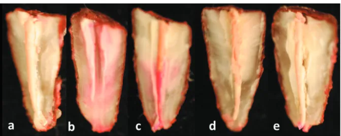
Figure 1- Leakage pattern according to the groups. a) Negative Control, observe absence of leakage (reddish staining); b) Positive Control, observe leakage along the walls from apical to the cervical root third; c) Endo CPM-Sealer®, observe leakage along the walls from the apical to the middle root third; d) MTA Fillapex®, observe absence of staining; e) Sealapex®, observe leakage only on the apical portion of the mater cone
Table 1- Comparison of mean dye leakage values (mm) and standard deviations (SD) of the endodontic sealers
Materials n Mean±SD (mm)
Fillapex® 30 0.80±0.61a
Endo-CPM-Sealer® 30 2.44±1.00b
Sealapex® 30 1.22±0.53a
based on MTA with additional substances to obtain a consistency suitable to be used in root canal treatment. MTA can form calcium ions and hydroxyl ions important for stimulation of hard tissue deposition11. The presence of MTA also suggests a possibility of setting expansion, which might have favored the sealability. Most dental materials have a tendency to shrink away from their interfacial margins, exposing a gap through which contaminating elements can penetrate. MTA setting results in the hydration of anhydrous mineral oxide compounds to produce calcium silicate hydrate and calcium hydroxide phases6,7,22, which produces
expansion against its conining margins, enhancing
the seal and minimizing leakage5,16,25.
According to its manufacturer, endo-CPM-Sealer® has similar or better physical, chemical and biological characteristics compared with MTA, and have the same clinical indications. endo-CPM-Sealer® has similar composition to that of MTA, but with the addition of calcium carbonate to reduce
the pH after setting to 10, thus limiting the surface
necrosis of the adjacent tissue and allowing the alkaline phosphatase action14. endo-CPM-Sealer® has been shown to have adequate radiopacity, hydroxyl and calcium ion release, antimicrobial activity, biocompatibility (including stimulation of
mineralization), and no cytotoxicity to ibroblast
culture14,16,23,27,28. However, in the present study, this material allowed the highest leakage level, demonstrating that it was not able to prevent dye apical leakage, which can hazard the success of the endodontic treatment. Maybe the physical
properties of the material can explain this inding.
CONCLUSIONS
Based on the results of the present study, it may be concluded that Sealapex® and Fillapex®
were able to signiicantly prevent apical leakage
differently from endo-CPM-Sealer®, which showed the highest levels of leakage.
CJ. Inluence of cement particle-size distribution on early age
autogenous strains and stresses in cement-based materials. J Am Ceram Soc. 2001;84:129-35.
6- Camilleri J. Characterization of hydration products of mineral trioxide aggregate. Int endod J. 2008;41:408-17.
7- Camilleri J. Hydration mechanisms of mineral trioxide
aggregate. Int endod J. 2007;40:462-70.
8- Cobankara FK, Orucoglu H, Sengun A, Belli S. The quantitative
evaluation of apical sealing of four endodontic sealers. J endod. 2006;32:66-8.
9- De Deus QD. Frequency, location, and direction of the lateral, secondary, and accessory canals. J endod. 1975;1:361-6. 10- estrela C, Sydney GB, Bammann LL, Felippe O Jr. Mechanism of action of calcium and hydroxyl ions of calcium hydroxide on tissue and bacteria. Braz Dent J. 1995;6:85-90.
11- Goldman M, Simmonds S, Rush R. The usefulness of dye-penetration studies reexamined. Oral Surg Oral Med Oral Pathol. 1989;67:327-32.
12- Gomes-Filho Je, Bernabé PFe, Nery MJ, Otoboni-Filho JA, Dezan-Júnior e, Costa MMTM, et al. Reaction of rat connective tissue to a new calcium hydroxide-based sealer. Oral Surg Oral Med Oral Pathol Oral Radiol endod. 2008;106:71-6.
13- Gomes-Filho JE, Hopp RN, Bernabé PFE, Nery MJ, Otoboni Filho JA, Dezan Júnior E. Evaluation of the apical iniltration after root
canal disruption and obturation. J Appl Oral Sci. 2008;16:345-9. 14- Gomes-Filho Je, Watanabe S, Bernabé PFe, Costa MTM. A mineral trioxide aggregate sealer stimulated mineralization. J endod. 2009;35:256-60.
15- Gomes-Filho Je, Watanabe S, Gomes AC, Faria MD, Lodi
CS, Penha Oliveira SH. Evaluation of the effects of endodontic materials on ibroblast viability and cytokine production. J Endod.
2009;35:1577-9.
16- Hawley M, Webb TD, Goodell GG. Effect of varying
water-to-powder ratios on the setting expansion of white and gray mineral trioxide aggregate. J endod. 2010;36:1377-9.
17- Holland R, Murata SS, Barbosa HG, Garlipp O, Souza V, Dezan
Júnior e. Apical seal of root canals with gutta-percha calcium hydroxide. Braz Dent J. 2004;15:26-9.
18- Holland R, Otoboni Filho JA, Souza V, Nery MJ, Bernabé PF,
Dezan Junior e. Calcium hydroxide and a corticosteroid-antibiotic association as dressings in cases of biopulpectomy. A comparative study in dogs’ teeth. Braz Dent J. 1998;9:67-76.
19- Hosoya N, Kurayama H, Iino F, Arai T. Effects of calcium
hydroxide on physical and sealing properties of canal sealers. Int endod J. 2004;37:178-84.
20- Ishimura H, Yoshioka T, Suda H. Sealing ability of new adhesive root canal illing materials measured by new dye penetration
method. Dent Mater J. 2007;26:290-5.
21- Kielbassa AM, Uchtmann H, Wrbas KT, Bitter K. In vitro study
assessing apical leakage of sealer-only backills in root canals of
22- Lee YL, Lee BS, Lin FH, Yun Lin A, Lan WH, Lin CP. Effects of
physiological environments on the hydration behavior of mineral trioxide aggregate. Biomaterials. 2004;25:787-93.
23- Scarparo RK, Haddad D, Acasigua GA, Fossati AC, Fachin
eV, Grecca FS. Mineral trioxide aggregate-based sealer: analysis of tissue reactions to a new endodontic material. J endod. 2010;36:1174-8.
24- Seux D, Couble ML, Hartmann DJ, Gauthier JP, Magloire H.
Odontoblast-like cytodifferentiation of human dental pulp cells in vitro in the presence of a calcium hydroxide-containing cement.
Arch Oral Biol. 1991;36:117-28.
25- Storm B, eichmiller FC, Tordik PA, Goodell GG. Setting expansion of gray and white mineral trioxide aggregate and Portland cement. J endod. 2008;34:80-2.
26- Tagger M, Tagger E, Kir A. Release of calcium and hydroxyl
ions from set endodontic sealers containing calcium hydroxide. J endod. 1988;14:588-91.
27- Tanomaru JM, Tanomaru-Filho M, Hotta J, Watanabe E, Ito
IY. Antimicrobial activity of endodontic sealers based on calcium hydroxide and MTA. Acta Odontol Latinoam. 2008;21:147-51.
28- Tanomaru-Filho M, Chaves Faleiros FB, Saçaki JN, Duarte
MAH, Guerreiro-Tanomaru JM. Evaluation of pH and calcium ion release of root-end illing materials containing calcium hydroxide
or mineral trioxide aggregate. J endod. 2009;35:1418-21.
29- Valera MC, Camargo CH, Carvalho AS, Gama ERP. In vitro
evaluation of apical microleakage using different root-end illing
materials. J Appl Oral Sci. 2006;14:49-52.
30- Venturi M, Prati C, Capelli G, Falconi M, Breschi L. A preliminary
analysis of the morphology of lateral canals after root canal illing
using a tooth-clearing technique. Int endod J. 2003;36:54-63. 31- Wu MK, Van Der Sluis LW, Ardila CN, Wesselink PR. Fluid
movement along the coronal two-thirds of root illings placed
by three different gutta-percha techniques. Int endod J. 2003;36:533-40.
32- Yang SE, Baek SH, Lee W, Kum KY, Bae KS. In vitro evaluation