ABSTRACT
Prevalence of enamel defects and associated risk
factors in both dentitions in preterm and full term
born children
Vanessa Resende Nogueira CRUVINEL1, Danuze Batista Lamas GRAVINA1, Tatiana Degani Paes Leme AZEVEDO1,
Catharina Siqueira de REZENDE2, Ana Cristina Barreto BEZERRA3, Orlando Ayrton de TOLEDO4
1- DDS, MSc, Professor, Department of Pediatric Dentistry, School of Dentistry, Catholic University of Brasília, Brasília, DF, Brazil.
2- Private Practice, Catholic University of Brasília, Brasília, DF, Brazil.
3- DDS, MSc, PhD, Associate Professor, Department of Pediatric Dentistry, School of Dentistry, University of Brasília, Brasília, DF, Brazil.
4- DDS, PhD, Associate Professor, Department of Pediatric Dentistry, School of Dentistry, University of Brasília, Brasília, DF, Brazil.
Corresponding address: Vanessa Resende Nogueira Cruvinel - Universidade Católica de Brasília - Curso de Odontologia Colônia Agrícola Samambaia, Rua 01 - Chácara 99 - Casa C - 72110600 - Taguatinga - DF - Brasil - Fone: 55 61 35615376 - Fax: 55 61 3352-4130 - e-mail: [email protected]
Received: September 09, 2010 - Modiication: August 01, 2011 - Accepted: August 25, 2011
O
bjectives: The aim of this study was to evaluate the prevalence of enamel defects andtheir risk factors on primary and permanent dentitions of prematurely born children
and full-term born children born at Regional Hospital of Asa Sul, Brasília, DF, Brazil. Material
and Methods: eighty 5-10-year-old children of both genders were examined, being 40 born prematurely (G1) and 40 born full term (G2). The demographic variables, medical history and oral health behaviors were retrieved using a questionnaire and data obtained from clinical examination were recorded. The teeth were examined and the presence of enamel defects was diagnosed according to the DDe Index and registered in odontograms. Subsequently, the defects were categorized in four groups according to one of the criteria
proposed in 1992 by the FDI Commission on Oral Health, Research and Epidemiology.
Kruskal-Wallis, Chi-square, Kappa, Mann-Whitney tests and logistic regression were used for statistical analysis. Results: 75% of total sample had enamel defects. There was a
major prevalence of hypoplasia of the enamel in G1 (p<0.001). There was a signiicant
relationship between low weight and presence of the imperfections on the enamel in G1 on the primary dentition. The logistic regression model showed that the other risk factors
such as monthly per capita family income, educational level, dietary and hygiene habits,
luoride exposure, trauma, and diseases were not associated with enamel defects and
caries. Conclusions: Pre-term labor can be a predisposing factor for the presence of the enamel hypoplasia in the primary dentition.
INTRODUCTION
According to the World Health Organization (WHO)21, a newborn of less than 37 weeks gestation or born within fewer than 259 days after the last menstrual period is considered premature or pre-term. Prematurity can be classiied as mild, when the baby is born between the 35th and 36th weeks of gestation; moderate, if the birth occurs between the 31st and 34th weeks; or extreme, if the gestational age is less than or equal to 30 weeks.
The birth of pre-term and/or low birth weight newborns represents a public health problem with increased economic, social, family, and individual costs10. Preventive and health promoting measures become necessary in order to improve the quality of life of these children. As such, knowing the risk factors to which they are subject is of fundamental importance for the adoption of such measures.
Low gestational age and low birth weight are the main factors that determine the incidence of neonatal complications. Among the most frequent complications are neonatal rickets, hypocalcemia, perinatal anoxia, anemia, infections, and metabolic, renal, respiratory, cardiovascular and hematological diseases. In these circumstances the use of various drug therapies and, frequently, orotracheal or laryngoscopic intubation to overcome respiratory difficulties, are necessary. These pathologies, whether or not associated with ventilatory support, may cause anomalies in the oral structures of these babies. In a previous study, low birth weight preterm infants presented a higher prevalence of hypoplasia than normal birth weight controls, and the most affected primary teeth by hypoplasia were maxillary incisors7.
Among the most prevalent oral alterations in these children are hypoplasias and opacities of the dental enamel6,8. The formation of these defects is related to disorders during amelogenesis6. The term during which the attack on the ameloblasts occurs is very important to the location and the appearance of enamel defects. The inal enamel shows a record of injuries received during the development of the teeth, which may appear as hypoplasia of the enamel, diffuse or marked opacity, and hypomineralization of the enamel20. enamel hypoplasia is the most common of the changes in human tooth development, with relevant clinical implications due to esthetic reasons, symptoms involved, susceptibility to caries, and also to the dificulty of treatment in many cases. This condition occurs as a direct result of disorders of metabolism of the ameloblast layers of the enamel organ19.
Hypoplastic areas are reported as being highly susceptible to dental caries3,9,15 because, through ultrastructural analysis, they have shown enamel with less mineralization, more porosity, with
irregular surfaces allowing the accumulation of bacteria which is favorable to the development of colonies of Streptococcus mutans, resulting in carious lesions3,11,13.
Knowing that children born prematurely present alterations in the dental structures, and that these can be related to the development of carious lesions, we must focus our attention on certain risk groups within which they fall. Given this evidence, this study aimed at evaluating the prevalence of enamel defects in the dentition of children born prematurely compared with those born full-term, and the main risk factors associated with those defects.
MATERIAL AND METhODS
Sample
This was a cross-sectional study involving children born in the Regional Hospital of Asa Sul – HRAS-DF, a public hospital from the Brazilian Uniied National Health System (SUS), considered to be a reference for high-risk births.
The sample for this study comprised 80 children of both genders, born in this hospital between 2000 and 2004, between 5 and 10 years of age, with birth weights that ranged from 605 g to 4300 g. The children were taken from a case-control study done in 2004, aimed at comparing the oral alterations present in 96 premature babies and 96 full-term babies born in this maternity hospital, making a total of 192 children. In calculating the initial sample (1st study), a conidence interval of 95%, with a margin of error of 10% (more or less)8, was considered. Of the children analyzed in the previous study, 98 were selected by having their charts updated with address and telephone number. Of these, 18 chose not to participate in the research. To calculate this sample, we took into account a 10 to 15% annual loss, which can be expected in longitudinal epidemiological studies. This study was completed with 80 children.
Children born before the 37th gestational week were classiied as premature, according to the WHO criteria21. In this sample, 40 children were premature and 40 children were full-term. The children were categorized according to birth weight as: very low weight (less than 30 weeks, 1500 g); low weight (between 1500 and 2500 g, between 31st and 34th weeks); and normal weight (above 2500 g and >38 weeks)17.
Information about the birth conditions, such as number of weeks and birth weight as well as medical history of the mother and child, were taken from the registration forms of the irst study, whose data were obtained from the hospital medical charts.
monitoring and dental evaluations of the children, and those who wished to participate signed an informed consent form and illed out an individual questionnaire, which was tested against the pilot study.
Use of the questionnaire
The questionnaire was used as an interview technique, incorporating both open and closed questions, by a previously trained interviewer. This technique was chosen as it had been shown to be an accurate research instrument for the investigation of health habits and conditions. The results gained here have been shown, in these cases, to be more reliable that those obtained through ill-in forms16.
The questionnaire contained three parts:
general information
This included name, age, gender, date of birth, family income, place of residence, and parents’ occupations.
Medical history
Information regarding probable risk factors for opacity and hypoplasia were collected, such as: systemic infectious diseases and rashes which occurred in the irst 3 years of life, such as pneumonia, tonsillitis, ear infections, chickenpox, rubella, measles; history of injury and low birth weight.
In relation to the categorization of family socioeconomic status, the standard variables as defined by the National Research of Domestic Samples (PNAD), which is done annually and portrays the socioeconomic situation of the Brazilian population, were considered. The variables deined by the PNAD include: family (group of persons connected by parentage, domestic dependency or norms of familiarity, who reside in the same household); literate persons (those who are capable of reading and writing at least a simple message in the familiar language); level or number of years of formal education (school grade and level – elementary, secondary, or college – attended, considering the last grade completed successfully); monthly per capita family income (monthly family income divided by the number of people in the family). For the determination of income, the value of the Brazilian national minimum wage in force during the months referenced in this study was considered – R$465.00. Starting with this last indicator, families were initially classiied into three groups: below poverty level (per capita household income less than ¼ of the minimum wage), at poverty level (per capita household income between ¼ and ½ minimum wage), and above ½ per capita minimum wage. Subsequently, these variables were categorized in 2 groups: ½ minimum wage and
above ½ minimum wage, because only 6 and 5 children of G1 and G2, respectively, showed income less than ¼ minimum wage.
Clinical exam
This procedure was done at the Dental Clinic of the Catholic University of Brasilia, under artiicial light, using dental mirror number 3 and properly sterilized gauze for cleaning. The patient was seated in the dental chair and submitted to prophylaxis with a Robson electric toothbrush and prophylactic paste manufactured by Vigodent. Afterward, the dental surfaces were dried with streams of air.
The dental surfaces were examined to verify the presence of structural defects in the enamel. The tooth was considered present when any part of the clinical crown was exposed in the oral cavity. When there was doubt about the presence of defect, the tooth was considered normal. Defects measuring less than one millimeter in diameter were excluded9,12,14.
All evaluations were performed by the same examiner, who was trained in the pilot study. To establish the degree of intra-examiner agreement, the Kappa index of 10% of the sample was used, as suggested by the WHO21. Re-examination was done 1 week after the initial examination (Kappa=0.90 for opacities, hypoplasias and white spots).
Codes for the diagnosis of enamel defects were used, according to the criteria of the Index of Developmental Defects of Dental enamel (DDe Index)6.
Teeth were examined by quadrants for hypoplasia and opacities of the enamel in the following order: right maxillary, left maxillary, right mandibular, and left mandibular. The number of teeth with defects was registered, as well as the total number of primary and erupted permanent teeth.
Hypoplasia of the enamel was deined as a break in the continuity of the enamel with a reduction in the layers, creating grooves or depressions. Opacity was deined as a change in the translucence of the enamel, or a qualitative defect that could vary in color from white to yellow and brown. The extent, type and color of the defect were registered. When hypoplasia and opacity were found in the same tooth, the defect was registered as hypoplasia8.
Analysis of data
The Mann-Whitney non-parametric test was used to compare the results of the scores of opacity and hypoplasia. A signiicance level of 5% was used for analytical purposes.
An adjustment was made to the multivariate logistical regression, in order to examine possible risk factors associated with occurrence of opacity and hypoplasia. Occurrence of opacity (Yes=1, No=0) and occurrence of hypoplasia (Yes=1, No=0) were used as the dependent variables. Type of birth (Premature=1, Full-term=0) was considered as a preponderant risk factor, and the following variables were considered as potential confounding factors: mother’s education level (<8 years of school=1, 8 years of school=0), monthly per capita family income (<0.5 minimum wage=1, 0.5 minimum wage=0), dental trauma (had trauma=1, did not have trauma=0), infectious diseases (Yes=1, No=0), and rashes (Yes=1, No=0).
Initially, a bivariate analysis was carried out between the dependent variable and all the independent, confounding variables with the aim of identifying those which could be related to the dependent variable. In this phase, signiicance was considered when the p-value was less than 0.25. Subsequently, a multivariate analysis was carried out only with the variables selected in the bivariate process, plus the preponderant independent variable as risk factor. In this phase a signiicance level of 5% was used.
This study was approved by the ethics Committee of the Catholic University of Brasilia (UCB) under number 161/2008.
RESULTS
The ages of the children ranged from 5 to 10 years, with a mean of 6.3 years for the premature group and 7.6 years for the full-term group, with a signiicant different between the groups (p=0.0003). The mean weight for premature children was 1255 g, and for full-term children was 3398 g.
Considering the 80 children examined, approximately 72.5% of the sample presented at least one tooth with enamel defect. According to the number of teeth affected, the groups presented the following mean scores in relation to the 2 types of defects: Presence of Opacity, G1 – 2.3 and G2 – 2.8 teeth (p=0.8624); and Hypoplasia, G1 – 1.1 and G2 – 0.1 teeth with this defect (p=0.0011).
No significant difference was found in the prevalence of enamel defects when children from G1 and G2 were divided by gender, with values of p=0.5712 for opacity and p=1.000 for hypoplasia.
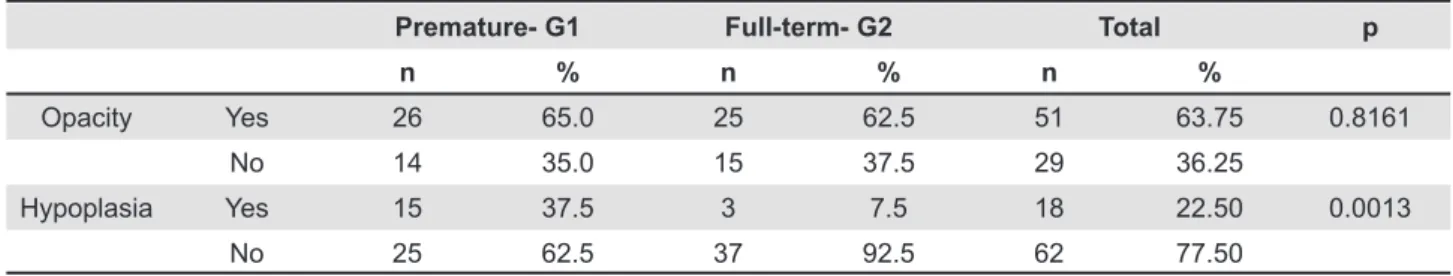
The percentage of children in G1 and G2 who presented opacity and hypoplasia was presented at Table 1. The percentage of primary and permanent teeth with opacity and hypoplasia in G1 and G2 was detailed at Table 2 and 3, respectively.
There was a statistically signiicant difference between the groups for marked opacity (p=0.0174)
Table 1- Percentage of children in G1 and G2 who presented opacity and hypoplasia
Premature- G1 Full-term- G2 Total p
n % n % n %
Opacity Yes 26 65.0 25 62.5 51 63.75 0.8161
No 14 35.0 15 37.5 29 36.25
Hypoplasia Yes 15 37.5 3 7.5 18 22.50 0.0013
No 25 62.5 37 92.5 62 77.50
Table 2- Percentage of primary teeth with opacity and hypoplasia in G1 and G2
Sample Groups of teeth Opacity Hypoplasia
n % n %
Total Incisor 2 0.7 23 8
Canine 17 6.1 10 3.6
Molar 43 7.5 13 2.3
G1 (premature) Incisor 2 1 23 11.8
Canine 12 8.4 9 6.3
Molar 29 9.8 10 3.4
G2 (full-term) Incisor 0 0 0 0
Canine 5 5.5 1 0.75
and hypoplasia (p<0.001), for primary teeth (Figure 1). However, there was no signiicant difference for any type of defect for permanent teeth (Figure 2).
Controlling for the effects of the variables that could be considered a probable risk factor for the occurrence of defects in the enamel such as: mother’s level of education (0.5777), per capita income (0.2442), trauma (p=0.9784), infectious diseases (p=0.4906), and rashes (p=0.9571), the type of birth was not a risk factor for the occurrence
of opacity (p=0.8161) with odds ratio equaling 1.11 (0.45 to 2.77). However, the type of birth was a risk factor for the occurrence of hypoplasia (p=0.0034) with odds ratio equaling 7.4 (1.94 to 28.25).
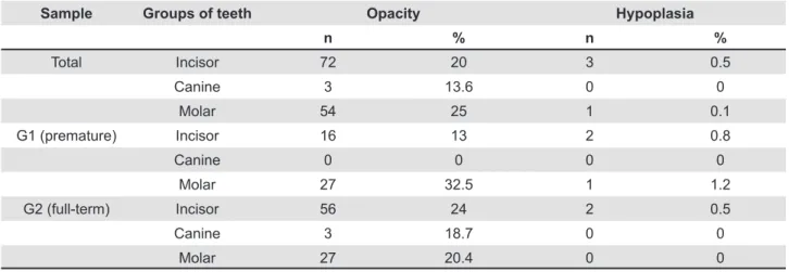
Table 3- Percentage of permanent teeth with opacity and hypoplasia in G1 and G2
Sample Groups of teeth Opacity Hypoplasia
n % n %
Total Incisor 72 20 3 0.5
Canine 3 13.6 0 0
Molar 54 25 1 0.1
G1 (premature) Incisor 16 13 2 0.8
Canine 0 0 0 0
Molar 27 32.5 1 1.2
G2 (full-term) Incisor 56 24 2 0.5
Canine 3 18.7 0 0
Molar 27 20.4 0 0
Figure 1- Percentage of marked opacity, diffuse opacity and hypoplasia in primary teeth
DISCUSSION
In Brazil, developmental defects of enamel are not studied enough although they result in esthetic problems, dental sensibility, and are predisposing factors for dental caries. Prematurity has been described as one of the causes for the appearance of enamel defects6,8. The etiological factors for these problems are multiple, and they range from the conception of the baby through the irst years of life, making it dificult to demonstrate the association between the variables. The scarcity of studies that evaluate permanent dentition relects the dificulty in following premature children until the change of dentition.
Comparisons of the results of the present study, characterized as a cross-sectional study of speciic samples, with other studies available in the literature and discussed below, must be done with caution owing to the differences in sample delineation, environmental inluences, and different methodologies.
This study evaluated information from medical records of the pregnancy through behavioral and social factors of the children and their families which could impact on their oral health. The maternal disorders registered as principal causal factors for prematurity were: hypertension (42%), preeclampsia (14.5%), premature labor (12.5%), anemia (8%), detached placenta (5%), gestational diabetes (5%), and cardiopathy (4%). In addition to these complications, 5 births were twins and 1 birth was a pregnancy with triplets8.
In premature children, the most frequent neonatal complications were respiratory distress, hyaline membrane disease, jaundice, pneumonia, osteopenia of prematurity, anemia, and non-speciic infections. Various drug therapies were necessary, among them antibiotic therapy, ventilatory assistance, parenteral nutrition, and prescription vitamin supplements containing iron and calcium. The need for these procedures was directly related to the seriousness of the general state of health of the child.
The systemic diseases in the first 3 years of the children’s life, which were related by the responsible parties, were pneumonia, tonsillitis, ear infections, chicken pox, rubella, and measles. This period coincides with the time of mineralization of permanent teeth. In this study, there was no signiicant association between infectious diseases (OR=1.45, IC=0.50-4.22) and rashes (OR=0.96, IC=0.34-2.79) with the presence of structural defects in the enamel. However, we must not disregard the possibility of biased information. These results disagree with Chaves, et al.2 (2007), who found association between post-natal infectious episodes and the prevalence of defects in the
enamel (OR=2.89, IC=0.84-10.03). The children in group 1 (premature) and group 2 (full-term) presented mean scores from 5 to 10 permanent teeth, respectively, which should take into account the difference in mean age between the groups. These indings support the study by Seow18 (1996), in which differences between dental age and chronological age were found when children from 6 to 8 years of age, born with low birth weight, were compared with children of normal birth weight (p<0.001). However, when children at 9 years of age were analyzed there was no more difference between the groups.
In the present study, approximately 72.5% of the sample presented at least one tooth with enamel defect. These indings agree with the study by Chaves, et al.2 (2007), who found a prevalence of defects in the enamel in 78.9% of the population studied. Oliveira, et al.14 (2006) found a prevalence of defects in the enamel in approximately 50% of the sample.
Lunardelli and Peres12 (2005) obtained a result of 24.4% of children with opacity and hypoplasia. However, there may have been an underestimation in the diagnoses of these patients, due to the absence of prior prophylaxis and drying of the examined surfaces.
No significant difference was found in the prevalence of enamel defects when children from G1 and G2 were divided by gender. These results coincide with those cited by Chaves, et al.2 (2007) and Oliveira, et al.14 (2006) who also found no difference.
In relation to the number of children from groups G1 and G2 who presented structural enamel defects in both dentitions, the difference was signiicant only for hypoplasia (Table 1). These results are supported by those of previous works1,4,5,8,11, who also found signiicant results for hypoplasia alone. From these indings, prematurity associated with low birth weight can be considered a risk factor for disorders in enamel mineralization.
of these defects4,8,18.
When opacity was the defect analyzed in the primary teeth, an inversion of prevalence in relation to hypoplasia occurred: molars (7.5%), canines (6.1%) and incisors (0.7%) (Table 2). In the irst study, the teeth most affected went in the following order: canines, molars and incisors. The inversion in the percentage of affected canines and molars may have been due to the greater number of teeth present in the current examination, because all the children who were examined had the 8 primary molars exposed. This is in contrast to the previous study8 in which they presented an average of 2.7 molars. The greater prevalence of this defect in the molars and canines may be explained by the mineralization chronology of these teeth, which occurs approximately around the 9th month of pregnancy. In children born prematurely who presented teeth with defects, the process of amelogenesis may have been interrupted or temporarily impaired. As a consequence, an enamel defect which was dependent upon the stage of amelogenesis occurring in the tooth at that moment would appear.
After categorizing the variables into the three types of defects, marked opacity appeared to be the most prevalent defect, occurring in 43% of the total sample. This was followed by hypoplasia, which occurred in 22.5% of the sample. The children in G1 presented 43 primary teeth with marked opacity (7.0%) and 42 with hypoplasia (6.0%). Of those in G2, 19 (3.7%) and 4 (0.8%) presented those defects, respectively (Figure 1). These indings disagree with those of Chaves, et al.2 (2007) and Oliveira, et al.14 (2006), who found diffuse opacity to be the most frequent defect, followed by marked opacity and hypoplasia. Lunardelli and Peres12 (2005), analyzing a sample of 431 public preschool children, found a prevalence of 17.9% of diffuse opacity, 11.1% of hypoplasia and 6.1% of marked opacity. Many factors must be considered together in order to justify the occurrence of these defects, without discarding the possibility of diffuse opacity being due to inadequate intake of luoride thus causing luorosis in the examined teeth. In relation to the permanent teeth, the dental groups most affected by enamel defects went in the following order: molars (25.5%), incisors (21.4%) and canines (13.6%). These indings support the study by Seow18 (1996), in which the most affected teeth were the molars followed by the incisors. As the mineralization of the permanent teeth begins at birth, or some months after the premature delivery, it may be hypothesized that persistent systemic problems lead to malformations in the enamel following birth.
Analyzing the permanent teeth affected by structural defects between the two groups, the
premature children (G1) presented 43 teeth with opacity (20%) and 2 teeth with hypoplasia (0.9%), while in G2 there were 86 teeth with opacity (22.7%) and 2 teeth with hypoplasia (0.5%). The number of permanent teeth with opacity and hypoplasia between the two groups was not statistically different (Figure 2). These results disagreed with those of Seow18 (1996), who found 17% opacity and 3% hypoplasia in the group of very low birth weight, and 8% opacity and 3% hypoplasia in the group of normal birth weight, there being a statistically signiicant difference in the total prevalence of defects between the groups (p<0.02).
The most affected permanent teeth, in a descending order, by the opacity defect in G1 were the molars (32.5%) and incisors (13%); and in G2 were the incisors (24%), molars (20.4%) and canines (18.7%) (Table 3).
Only 3 permanent incisors presented hypoplasia in the total sample, and all had history of trauma in the homologous primary teeth.
It was observed, through multivariate analysis, that the type of birth was a risk factor for the occurrence of hypoplasia (p=0.0034) in primary dentition. Premature children had 7.4 times more chance of having hypoplasia than full-term children, with a conidence interval of 95% ranging from 1.93 to 28.25.
It is very dificult to distinguish the etiological factors for enamel alterations, because pre-, neo-, and post-natal complications may occur together, interacting among themselves. enamel defects result from severe cumulative events associated with external factors such as social, nutritional state, and life-style questions, which must be considered because they make a strong impact on health and have repercussions on the quality of life of the individual3.
CONCLUSIONS
REFERENCES
1- Aine L, Backström MC, Mäki R, Kuusela AL, Koivisto AM, Ikonen RS, et al. enamel defects in primary and permanent teeth of children born prematurely. J Oral Pathol Med. 2000;29(8):403-9. 2- Chaves AMB, Rosenblatt A, Oliveira OF. enamel defects and its relation to life course events in primary dentition of Brazilian children: a longitudinal study. Community Dent Health. 2007;24(1):31-6.
3- Cruvinel VR, Gravina, DB, Azevedo TD, Bezerra AC, Toledo AO. Prevalence of dental caries and caries-related risk factors in premature and term children. Braz Oral Res. 2010;24(3):329-35. 4- Drummond BK, Ryan S, O'Sullivan EA, Congdon P, Curzon Me. enamel defects of the primary dentition and osteopenia of prematurity. Pediatr Dent. 1992;14(2):119-21.
5- Fearne JM, Bryan EM, Elliman AM, Brook AH, Williams DM. enamel defects in the primary dentition of children born weighing less than 2000 g. Br Dent J. 1990;168(11):433-7.
6- Federation Dentaire Internationale. Commission on Oral Health, Research and epidemiology. A review of the developmental defects of enamel index (DDe index). Int Dent J. 1992;42(6):411-26. 7- Franco KMD, Peres SR, Moura-Ribeiro MVL. Prenatal and neonatal variables associated with enamel hypoplasia in deciduous teeth in low birth weight preterm infants. J Appl Oral Sci. 2007;15(6):518-23.
8- Gravina DB, Cruvinel VR, Azevedo TD, Toledo OA, Bezerra AC. Prevalence of dental caries in children born prematurely or at full term. Braz Oral Res. 2006;20(4):353-7.
9- Horowitz HS. Research issues in early childhood caries. Community Dent Oral epidemiol. 1998;26(Suppl 1):67-81. 10- Lai PY, Seow WK, Tudehope DI, Rogers Y. enamel hypoplasia and dental caries in very-low birthweight children: a case-controlled, longitudinal study. Pediatr Dent. 1997;19(1):42-9. 11- Li Y, Navia JM, Bian JY. Prevalence and distribution of developmental enamel defects in primary dentition of Chinese children 3-5 years old. Community Dent Oral epidemiol. 1995;23(2):72-9.
12- Lunardelli Se, Peres MA. Prevalence and distribution of developmental enamel defects in the primary dentition of preschool children. Braz Oral Res. 2005;19(2):144-9.
13- Norén JG, Ranggård L, Klingberg G, Persson C, Nilsson K. Intubation and mineralization in the enamel of primary teeth. Acta Odontol Scand. 1993;51(5):271-5.
14- Oliveira AF, Chaves AM, Rosenblatt A. The inluence of enamel defects on the development of early childhood caries in a population with low socioeconomic status: a longitudinal study. Caries Res. 2006;40(4):296-302.
15- Sawyer DR, Nwoku AL. Malnutrition and the oral health of children in Ogbomosho, Nigeria. ASDC J Dent Child. 1985;52(2):141-5.
16- Schröder U, Lindström LG, Olsson L. Interview or questionnaire? A comparison based on the relationship between caries and dietary habits in preschoolchildren. Community Dent Oral epidemiol. 1981;9(2):79-82.
17- Seow WK. A study of the development of the permanent dentition in very low birthweight children. Pediatr Dent. 1996;18(5):379-84.
18- Seow WK, Humphrys C, Tudehope DI. Increased prevalence of developmental dental defects in low-birth-weight, prematurely born children: a controlled study. Pediatr Dent. 1987;9(3):221-5. 19- Silva-Sousa YTC, Peres LC, Foss MC. Are there structural alterations in the enamel organ of offspring of rats with alloxan-induced diabetes mellitus? Braz Dent J. 2003;14(3):162-7. 20- Slayton RL, Warren JJ, Kanellis MJ, Levy SM, Islam M. Prevalence of enamel hypoplasia and isolated opacities in the primary dentition. Pediatr Dent. 2001;23(1):32-6.
21- World Health Organization. International Statistical Classiication of Diseases and Related Health Problems. 10th