ABSTRACT
Inluence of lingual bracket position on microbial
and periodontal parameters
in vivo
Maria Francesca SFONDRINI1, Maurizia DEBIAGGI2, Francesca ZARA2, Roberto BRERRA2, Mario COMELLI3, Marco
BIANCHI4, Sara Ramella POLLONE5, Andrea SCRIBANTE6
1- MD, DDS, PhD, Professor, Department of Orthodontics, University of Pavia, Pavia, Italy. 2- MD, Professor, Department of Microbiology, University of Pavia, Pavia, Italy.
3- MD, Professor, Department of Statistics, University of Pavia, Pavia, Italy.
4- DDS, Graduate Fellow, Department of Orthodontics, University of Pavia, Pavia, Italy. 5- DDS, Graduate Fellow, Department of Orthodontics, University of Pavia, Pavia, Italy.
6- DDS, PhD, Research Resident, Department of Orthodontics and Department of Surgical Sciences, University of Pavia, Pavia, Italy.
Corresponding address: Dr. Andrea Scribante - Istituto di Discipline Odontostomatologiche “S. Palazzi” - IRCCS Policlinico S. Matteo - P.le Golgi 2 - 27100 Pavia - Italy - Phone/fax: +39-0382-516223 - e-mail: [email protected]
Received: October 18, 2010 - Modiication: August 4, 2011 - Accepted: August 16, 2011
O
bjective: Lingual orthodontics is becoming more popular in dental practice. The purpose of the present investigation was to compare plaque formation on teeth bonded with the same bracket onto buccal or lingual surface, with non-bonded control teeth, via an in vivogrowth experiment over a 30-day period. Material and Methods: A randomized controlled trial with split-mouth design was set up enrolling 20 dental students. Within each subject sites with buccal and lingual brackets and control sites were followed. Clinical periodontal parameters (periodontal pocket depth: PPD; bleeding on probing: BOP) were recorded at baseline and on days 1, 7 and 30. Microbiological samples were taken from the brackets and the teeth on days 1, 7 and 30 to detect colony-forming units (CFU). Total CFU, streptococci
CFU and anaerobe CFU were measured. Results: No signiicant differences (P>0.05) were
found between buccal and lingual brackets in terms of clinical periodontal parameters and
microbiological values. Conclusion: Bracket position does not have signiicant impact on
bacterial load and on periodontal parameters.
Key words: Microbiology. Orthodontics. Dental plaque. Periodontal diseases.
INTRODUCTION
Gingivitis and periodontal disease are frequent concomitant phenomena of orthodontic treatment with ixed appliances29. It seems that the main factor
for an increased accumulation of dental plaque and inlammatory response is the appearance of new retentive places around the components of ixed appliances attached to the teeth1. Several studies
have addressed the impact of ixed, removable, and myofunctional orthodontic/orthopedic appliances or retainers in relation to supragingival plaque accumulation and gingivitis3,11,12.
The quantity, as well as the quality of plaque, is influenced by many factors including surface characteristics23,24, surface roughness and surface free
energy21 and bracket design33, frequency of sucrose
exposition25. The presence of gingival inlammation
will further increase plaque growth22,26.
Because of its outstanding aesthetic preconditions and its growing practicability, lingual orthodontics accounts for an ever-increasing percentage of orthodontic treatments20,28. Oral hygiene is even
more important for therapy with lingual brackets than for therapy with labial brackets because control is more dificult from the lingual face than from the buccal face, and plaque accumulations, gingivitis, and demineralization are not detected by the patient14. Most microbiological investigations have
been performed during orthodontic treatment4,9,15,33
but there are no studies dealing with the difference between buccal and lingual brackets.
hypothesis of the study was that there is no signiicant difference in terms of microbiological environment and clinical periodontal parameters between buccal and lingual brackets.
MATERIAL AND METhODS
Subjects
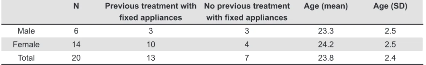
Twenty dental students (14 females and 6 males, Caucasians aged between 20 and 32 years) were involved in the study (Table 1). They were given a written explanation of the background of the study and its objectives. After screening for their suitability and after good comprehension of the protocol, they all gave their written informed consent. During the experiment, the participants could always contact the researcher for questions or remarks. Before the study, all students received oral hygiene instructions in order to ensure a healthy periodontal situation.
The initial placement of the brackets was performed via a randomized protocol by means of concealed envelopes. The students were selected to fulill the following inclusion criteria: no smoking, absence of extensive dental restorations or adhesive-ixed partial dentures, a sulcus bleeding index18 of <0.3 and no
antibiotics during or up to 4 months before the study. The students were also asked whether they had already received an orthodontic treatment with ixed appliances because this might have consequences for smoothness of the buccal enamel10 and as such on the
microbial adhesion in the early formation of a dental plaque ilm23,24. The ethics Committee approved the
design of this study.
Experimental procedure Experimental design
The study had a randomized, examiner-blind, split-mouth design33. In every student, the mouth
was divided into four quadrants, two of which served as controls. One type of bracket (2D, Forestadent, Pforzheim, Germany) was bonded in two different
sites: buccal and lingual. For the split-mouth comparison, 8 sites were deined, namely the canine and the irst premolar of each quadrant. The brackets were placed in contralateral antagonistic quadrants. The irst quadrant used for bracket placement and the order in which the brackets were placed were randomly chosen by means of concealed envelopes, the second one was at the other side of the mouth in the antagonistic jaw. The buccal and lingual bonded teeth were alternated, giving rise to four different experimental settings (Figure 1):
-buccal position in the irst quadrant, lingual position in the third quadrant;
-buccal position in the second quadrant, lingual position in the fourth quadrant;
-buccal position in the third quadrant, lingual position in the irst quadrant;
-buccal position in the fourth quadrant, lingual position in the second quadrant.
The teeth bonded with the different brackets were compared with each other and with the non-bonded control sites.
During the study period, the students visited the clinic three times: T0 (baseline): to record the status of the periodontium (periodontal pocket depth: PPD; bleeding on probing: BOP), to collect samples of dental plaque from the teeth and to place the brackets; T1 (day 7): to record the periodontal status (PPD and BOP) and to collect from the test teeth and from control teeth; T2 (day 30): to record the periodontal status (PPD and BOP), to collect samples of dental plaque from the test teeth and the control teeth, and to remove the brackets.
Bracket placement/removal
The teeth were rinsed, dried with an oil-free air syringe, and etched with 37% phosphoric acid for 30 s. After a thorough washing, they were completely dried with an oil-free air syringe. Then, stainless steel brackets (2D, Forestadent, Pforzheim, Germany) were bonded to the selected teeth with an adhesive system (Transbond XT, 3M, Monrovia, CA, USA), according to the manufacturer’s directions. After applying the primer on the etched enamel, a small amount of composite resin was placed on the mesh pad of the bracket. The bracket was positioned on the buccal or lingual surface of the teeth with suficient pressure to squeeze excess adhesive, which was
B B L L
L L B B
Figure 1- The four different clinical conditions of the split-mouth design
N Previous treatment with
ixed appliances
No previous treatment with ixed appliances
Age (mean) Age (SD)
Male 6 3 3 23.3 2.5
Female 14 10 4 24.2 2.5
Total 20 13 7 23.8 2.4
Table 1- Study population with data on gender distribution, previous orthodontic treatment and age
removed from the margins of the bracket base with an explorer before polymerization. Bracket was then light-cured with a visible light-curing unit (Ortholux XT, 3M Unitek) for 10 s on the mesial side of the bracket and for 10 s on the distal side (total cure time 20 s). All brackets were bonded by the same operator. Verbal and written instructions regarding the appliance care and hygiene protocols were issued to each patient, along with a speciic request to return if a bracket became loose or if any problem arose with the appliance.
Periodontal parameters
PPD and BOP were scored at baseline, day 7 and day 30. To record the PPD, a millimeter probe (HU-friedly Pc puns, Chicago, IL, USA) was inserted in the gingival sulcus. The pocket depths were measured at the buccal, lingual, mesial and distal sides of the tooth and rounded off to the nearest 0.5 mm. BOP was recorded (0: absent; 1: present) 24 s after determination of PPD. The examiner was blinded to previous scores.
Culture techniques
At baseline, on days seven and thirty microbial samples were also taken using a sterile curette (SG 5/6 HU-Friedy) from the test and the control teeth. The supra-gingival dental plaque was taken away with sterile curettes. This was carried out without traumatizing the gingiva and without disturbing the plaque ilm on the remaining sites33. The supragingival
plaque samples were transferred into lip-capped vials containing 250 µL of reduced transport luid (RTF)30.
All samples were transferred to the laboratory and processed within 3 h.
The samples were pooled in 250 µL of RTF and serial ten-fold dilutions were prepared in the same medium. Dilutions of 10-3–10-5 were plated in duplicate
using a spiral plater onto three different media: Mitis-salivarius (MS) agar was used to determine the
total count of streptococci, mitis-salivarius-bacitracin (MSB) agar was used as the selective medium for differentiation and enumeration quantiication of S.
mutans and CDC Anaerobe 5% sheep blood agar
(BD) for enumeration of total recoverable anaerobic bacteria.
After 3 days of aerobic incubation at 37°C for MS and MSB agar plates and 6 days of anaerobic incubation (Gas Pack eZ system, BD) at 37°C for blood agar plates, the number of colony-forming units (CFU) was counted. The total count of microorganism was determined on countable (from 30 to 300 colonies) plates.
Statistical analysis
The P values report concern the interaction between time and position of the bracket, included in mixed linear models itted to the microbiological and clinical outcomes. The models included other possibly relevant ixed effects (e.g. side of the mouth), and the patient identity as a random effect. The R statistical package (R Development Core Team, Wien, Austria) was used for computation. The signiicance level was set at 5%.
RESULTS
Microbiological parameters
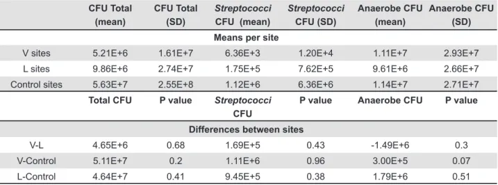
The numbers of streptococci, anaerobic and total CFU in supragingival plaque samples during the experimental period showed no signiicant differences days combined (Table 2). Buccal sites in general allowed equal plaque formation than the lingual sites (P>0.05).
In Table 3, the results are separately depicted
per day and per material. Buccal sites showed no signiicant differences from lingual sites (P>0.05) for either streptococci, anaerobe or total CFU counts.
CFU Total (mean)
CFU Total (SD)
Streptococci
CFU (mean)
Streptococci
CFU (SD)
Anaerobe CFU (mean)
Anaerobe CFU (SD)
Means per site
V sites 5.21E+6 1.61E+7 6.36E+3 1.20E+4 1.11E+7 2.93E+7
L sites 9.86E+6 2.74E+7 1.75E+5 7.62E+5 9.61E+6 2.66E+7
Control sites 5.63E+7 2.55E+8 1.12E+6 6.36E+6 1.14E+7 2.71E+7
Total CFU P value Streptococci
CFU
P value Anaerobe CFU P value
Differences between sites
V-L 4.65E+6 0.68 1.69E+5 0.43 -1.49E+6 0.3
V-Control 5.11E+7 0.2 1.11E+6 0.96 3.00E+5 0.07
L-Control 4.64E+7 0.41 9.45E+5 0.38 1.79E+6 0.51
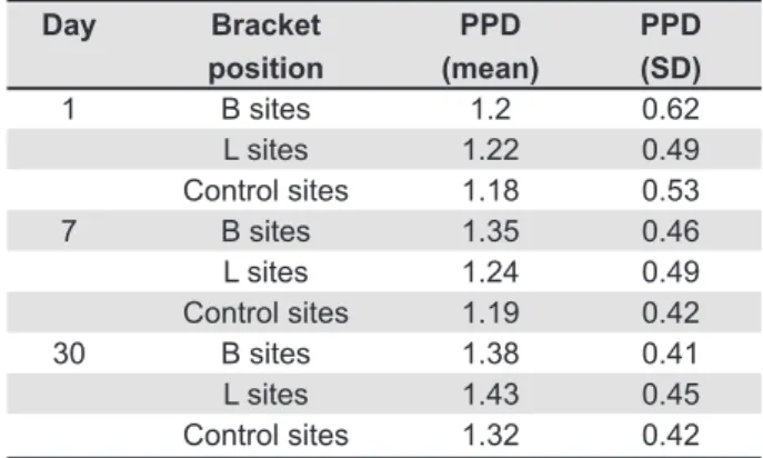
Periodontal parameters PPD
No signiicant inter-material differences in PPD were present among the various groups. No signiicant increase in PPD was recorded (P>0.05) on days 7 and 30 for either buccal or lingual bracket position and for control sites.
BOP
Only few BOP sites were recorded during the experimental period. No significant differences (P>0.05) in BOP sites were detected among the different groups at different times.
No adverse events were reported.
DISCUSSION
The null hypothesis of the study was partially rejected. The present experiment with split-mouth design did not detect any signiicant difference in periodontal and microbiological parameters between the bonded teeth and the non-bonded control teeth or between the groups with brackets bonded onto buccal and lingual sides.
A number of studies have investigated the inluence of orthodontic therapy and appliances on the oral microbial lora. These changes could potentially have a signiicant impact on patient oral health, including gingival inlammation and demineralization of teeth2. Moreover, in literature, orthodontic therapy
was associated with 0.03 millimeters of gingival recession, 0.13 mm of alveolar bone loss and 0.23 mm of increased pocket depth when compared with no treatment6. The effects of orthodontic therapy on
gingivitis and attachment loss are inconsistent across studies6.
Anhoury, et al.2 (2002) evaluated microbial proile
on metallic and ceramic bracket materials and found that composition of dental plaque formed on each bracket type is very similar between the two bracket types and may be of limited clinical signiicance. Furthermore, the differences detected do not favor
one bracket type over another with respect to bacterial accumulation.
Other authors33 evaluated inluence of bracket
design on microbiological and periodontal parameters showing both anaerobe and aerobe colony-forming units signiicantly higher for self-ligating brackets than for conventional brackets. No signiicant differences for bleeding on probing were observed.
Microbial and periodontal parameters have been evaluated also for orthodontic bands7, conventional
stainless steel brackets19, ceramic attachments2,
and self-ligating brackets. In literature there are no studies about lingual brackets. These brackets have been investigated about their laboratory and clinical processes and for particular recommendations on oral hygiene14,20,28. Those authors showed that special
emphasis should be placed on the oral hygiene of patients with lingual brackets and that excellent oral hygiene is possible in patients with lingual devices after instruction and motivation14.
The results of the present investigation showed no signiicant differences in total CFU, streptococci CFU and anaerobe CFU counts among buccal, lingual and control sites. This is in agreement with a previous investigation that evaluated CFU of buccal brackets versus control non-bonded sites and showed no signiicant differences between the two groups32.
There are also authors that found signiicant CFU count decrease29 or increase7,16,19,27 in bracket sites
compared with control sites. This variability is probably due to the different testing conditions and multiple variables correlated with clinical researches.
Moreover, in the present study, PPD and BOP measurements were analyzed and showed no signiicant differences among buccal, lingual and control sites. This is in agreement with previous studies that evaluated these periodontal parameters using conventional brackets: all of them showed no signiicant increase of BOP12,14 and PPD12,19,33 after
brackets placement. There are also studies that detected signiicant increase of PPD27 and BOP19,33
after bracket placement. This variability could be due to different orthodontic bracket materials that have dissimilar clinical manifestations.
The limits of the present study would be that the population of the present investigation was represented by dental students, who are expected to maintain better oral hygiene than the general population. Hygiene regimen of subjects in the trial can be reached also in the general population only if adequately motivated8. In fact, even if in recent
decades, decreasing prevalence in dental caries has been observed worldwide5 correct teaching of hygiene
protocols is crucial, especially during orthodontic treatment. Therefore, even if in the present investigation buccal or lingual bracket position did not have signiicant impact on PPD and BOP periodontal parameters, dental health promotion should be fully
Day Bracket
position
PPD (mean)
PPD (SD)
1 B sites 1.2 0.62
L sites 1.22 0.49 Control sites 1.18 0.53
7 B sites 1.35 0.46
L sites 1.24 0.49 Control sites 1.19 0.42
30 B sites 1.38 0.41
L sites 1.43 0.45 Control sites 1.32 0.42
integrated into broadly based health-promoting strategies. Oral hygiene remain the major factor when evaluating caries prevention and dental bioilm13,17,31.
Another issue is represented by the presence of wires used during the regular orthodontic treatment. The wires and brackets work together to limit hygiene and therefore a further development of the present study could include also this aspect.
Future investigations should be performed to visualize the potentially different periodontal parameters correlated with different orthodontic bracket systems so that brackets can be designed to reduce plaque adhesion.
CONCLUSIONS
This study demonstrated the following: 1. buccal or lingual bracket position does not have a signiicant impact on PPD and BOP periodontal parameters; buccal or lingual bracket position does not have signiicant impact on streptococci, anaerobe and total CFU counts.
ACKNOwLEDgEMENTS
The authors wish to thank 3M and Forestadent for providing the materials tested in this study.
REFERENCES
1- Alexander SA. effects of orthodontic attachments on the gingival health of permanent second molars. Am J Orthod Dentofacial Orthop. 1991;100:337-40.
2- Anhoury P, Nathanson D, Hughes CV, Socransky S, Feres M, Chou LL. Microbial proile on metallic and ceramic bracket materials. Angle Orthod. 2002;72:338-43.
3- Artun J, Urbye KS. The effect of orthodontic treatment on periodontal bone support in patients with advanced loss of marginal periodontium. Am J Orth Dentofacial Orthop. 1998;93:143-8. 4- Atack Ne, Sandy JR, Addy M. Periodontal and microbiological changes associated with the placement of orthodontic appliances. A review. J Periodontol. 1996;67:78-85.
5- Baldini V, Tagliaferro eP, Ambrosano GM, Meneghim MD, Pereira AC. Use of occlusal sealant in a community program and caries incidence in high- and low-risk children. J Appl Oral Sci. 2011;19(4):396-402. 6- Bollen AM, Cunha-Cruz J, Bakko DW, Huang GJ, Hujoel PP. The effects of orthodontic therapy on periodontal health: a systematic review of controlled evidence. J Am Dent Assoc. 2008;139:413-22. 7- Boyd R, Baumrind S. Periodontal consideration in the use of bonds or bands on molar in adolescents and adults. Angle Orthod. 1992;62;117-26.
8- Carvalho VF, Okuda OS, Bernardo CC, Pannuti CM, Georgetti MA, De Micheli G, et al. Compliance improvement in periodontal maintenance. J Appl Oral Sci. 2010;18(3):215-9.
9- Diamanti-Kipioti A, Gusberti FA, Lang NP. Clinical and microbiological effects of fixed orthodontic appliances. J Clin Periodontol. 1987;14:326-33.
10- eliades T, Gioka C, eliades G, Makou M. enamel surface roughness following debonding using two resin grinding methods. eur J Orthod. 2004;26:333-8.
11- Glans R, Larsson e, Øgaard B. Longitudinal changes in gingival condition in crowded and noncrowded dentitions subjected to ixed orthodontic treatment. Am J Orth Dentofacial Orthop. 2003;124:679-82.
12- Gomes SC, Varela CC, Veiga SL, Rösing CK, Oppermann RV. Periodontal conditions in subjects following orthodontic therapy. A preliminary study. eur J Orthod. 2007;29:477-81.
13- Gontijo L, Cruz RA, Brandão PR. Dental enamel around ixed orthodontic appliances after luoride varnish application. Braz Dent J. 2007;18:49-53.
14- Hohoff A, Stamm T, Kühne N, Wiechmann D, Haufe S, Lippold C, et al. effects of a mechanical interdental cleaning device on oral hygiene in patients with lingual brackets. Angle Orthod. 2003;73:579-87.
15- Huser MC, Baehni PC, Lang R. Effects of orthodontic bands on microbiologic and clinical parameters. Am J Orthod Dentofacial Orthop. 1990;97:213-8.
16- Lee SM, Yoo SY, Kim HS, Kim KW, Yoon YJ, Lim SH, et al. Prevalence of putative periodontopathogens in subgingival dental plaques from gingivitis lesions in Korean orthodontic patients. J Microbiol. 2005;43:260-5.
17- Maltz M, Jardim JJ, Alves LS. Health promotion and dental caries. Braz Oral Res. 2010;24(Suppl 1):18-25.
18- Mühlemann HR, Son S. Gingival sulcus bleeding – a leading symptom in initial gingivitis. Helv Odontol Acta. 1971;15,107-13. 19- Naranjo AA, Triviño ML, Jaramillo A, Betancourth M, Botero Je. Changes in the subgingival microbiota and periodontal parameters before and 3 months after bracket placement. Am J Orthod Dentofacial Orthop. 2006;130:275.e17-22.
20- Poon KC, Taverne AA. Lingual orthodontics: a review of its history. Aust Orthod J. 1997;15:101-4.
21- Quirynen M, Bollen CM. The inluence of surface roughness and surface free energy on supra- and subgingival plaque formation in man. A review of the literature. J Clin Periodontol. 1995;22:1-14. 22- Quirynen M, Dekeyser C, Van Steenberghe D. The inluence of gingival inlammation, tooth type, and timing on the rate of plaque formation. J Periodontol. 1991;62:219-22.
23- Quirynen M, Marechal M, Busscher HJ, Weerkamp AH, Arends J, Darius PL, et al. The inluence of surface free-energy on planimetric plaque growth in man. J Dent Res. 1989;68:796-9.
24- Quirynen M, Marechal M, Busscher HJ, Weerkamp AH, Darius PL, Van Steenberghe D. The inluence of surface free energy and surface roughness on early plaque formation. An in vivo study in man. J Clin Periodontol. 1990;17:138-44.
25- Raggio DP, Braga MM, Rodrigues JA, Freitas PM, Imparato JC, Mendes FM. Reliability and discriminatory power of methods for dental plaque quantiication. J Appl Oral Sci. 2010;18:186-93.
26- Ramberg P, Axelsson P, Lindhe J. Plaque formation at healthy and inlamed gingival sites in young individuals. J Clin Periodontol. 1995;22:85-8.
27- Ristic M, Vlahovic Svabic M, Sasic M, Zelic O. Effects of ixed orthodontic appliances on subgingival microlora. Int J Dent Hyg. 2008;6:129-36.
28- Rummel V, Wiechmann D, Sachdeva RCL. Precision inishing in lingual orthodontics. J Clin Orthod. 1999;13:101-13.
29- Speer C, Pelz K, Hopfenmuller W, Holtgrave EA Investigations on the inluencing of the subgingival microlora in chronic periodontitis, a study in adult patients during ixed appliances therapy. J Orofac Orthop. 2004;65,34-47.
30- Syed SA, Loesche WJ. Survival of human dental plaque lora in various transport media. Appl Microbiol. 1972;24:638-44. 31- Teles RP, Teles FR. Antimicrobial agents used in the control of periodontal bioilms: effective adjuncts to mechanical plaque control? Braz Oral Res. 2009;23(Suppl 1):39-48.
32- Thornberg MJ, Riolo CS, Bayirli B, Riolo ML, Van Tubergen eA, Kulbersh R. Periodontal pathogen levels in adolescents before, during, and after ixed orthodontic appliance therapy. Am J Orthod Dentofacial Orthop. 2009;135:95-8.
33- Van Gastel J, Qurynen M, Teughels W, Coucke W, Carels C. Inluence of bracket design on microbial and periodontal parameters