ABSTRACT
Qualitative SeM/eDS analysis of microleakage
and apical gap formation of adhesive root-illing
materials
Soraia de Fátima Carvalho SOUZA1, Carlos FRANCCI2, Antonio C. BOMBANA3, Silvia KENSHIMA4, Lúcia P. BARROSO5, Liz Z. D’AGOSTINO6, Alessandro D. LOGUERCIO7
1- DDS, MSc, PhD, Adjunct Professor, School of Dentistry, Federal University of Maranhão, São Luiz, MA, Brazil. 2- DDS, MSc, PhD, Associate Professor, Department of Dental Materials, University of São Paulo, São Paulo, SP, Brazil. 3- DDS, MSc, PhD, Honorary Professor, Department of Restorative Dentistry, University of São Paulo, São Paulo, SP, Brazil. 4- DDS, MSc, PhD, Department of Dental Materials, University of São Paulo, São Paulo, SP, Brazil.
5- DDS, MSc, PhD, Associate Professor, Department of Mathematics and Statistics, University of São Paulo, São Paulo, SP, Brazil. 6- MSc, Department of Mining and Petroleum Engineering, University of São Paulo, São Paulo, SP, Brazil.
7- DDS, MSc, PhD, Adjunct Professor, State University of Ponta Grossa, Ponta Grossa, PR, Brazil.
Corresponding address: Soraia de Fatima Carvalho Souza - Faculdade de Odontologia - Universidade Federal do Maranhão (UFMA) - Av. dos Portugueses s/n - 65085-580 - Bacanga - São Luis - MA - Brasil - Phone: +55 98 3301 8575 - e-mail: [email protected] - [email protected]
Received: September 18, 2010 - Modiication: August 14, 2011 - Accepted: September 15, 2011
O
bjective: The aim of this study was to compare the correspondence between gapformation and apical microleakage in root canals illed with epoxy resin-based (AH Plus)
combined or not with resinous primer or with a dimethacrylate-based root canal sealer (epiphany). Material and Methods: Thirty-nine lower single-rooted human premolars were
illed by the lateral condensation technique (LC) and immersed in a 50-wt% aqueous silver
nitrate solution at 37oC (24 h). After longitudinal sectioning, epoxy resin replicas were made
from the tooth specimens. Both the replicas and the specimens were prepared for scanning electron microscopy (SeM). The gaps were observed in the replicas. Apical microleakage was detected in the specimens by SeM/energy dispersive spectroscopy (SeM/eDS). The data were analyzed statistically using an Ordinal Logistic Regression model and Analysis
of Correspondence (α=0.05). Results: epiphany presented more regions containing gaps
between dentin and sealer (p<0.05). There was correspondence between the presence of
gaps and microleakage (p<0.05). Microleakage was similar among the root-illing materials (p>0.05). Conclusions: The resinous primer did not improve the sealing ability of AH Plus
sealer and the presence of gaps had an effect on apical microleakage for all materials.
Key words: electron microscopy. Leakage. Resin cements. Root canal obturation. Silver nitrate.
INTRODUCTION
It is generally accepted that microleakage
between the illing materials and root canal walls
might adversely affect the outcome of root canal treatment13. Therefore, it is critical the complete
sealing of the root canal system after cleaning and shaping in order to avoid the bacterial penetration and re-infection of the root and periapical tissues3.
The association of gutta-percha cones and root canal sealer has been traditionally used for this
purpose. However, in the last decade, the dentin
adhesive technology has been incorporated into the
root canal illing techniques to reduce apical and
coronal leakage by bonding to root canal walls28.
etch-and-rinse adhesives have been tested with resin cements10 and the combination of a
dentin-bonding agent and an epoxy resin-based root canal
sealer signiicantly reduced apical leakage12.
In Restorative Dentistry, the self-etch adhesive systems have shown less technique sensitivity, with reliable long-term performance of a two-step mild self-etch adhesive5,30. Following this trend, Pentron
sealer and a polyester-based thermoplastic
root-illing material (Resilon™; Resilon Research LLC,
Madison, CT, USA). According to Shipper, et al.23
(2004) this material has been shown to be more resistant to bacterial leakage than epoxy
resin-based sealers for illing root canals.
In endodontics, the controversy about the performance of adhesive systems inside the root canal remains1,21,26. Despite that, other possibility
is combining Epiphany primer with AH Plus in an
attempt to add the hybridization capacity to the gold standard endodontic sealer. The reason for this is that the removal of the smear layer with ethylenediamine tetraacetic acid (eDTA) does not provide the same etching pattern usually associated with the hybrid layer, which is considered an important factor for dentin bonding15,19.
The majority of studies have evaluated apical or coronal microleakage, and few have focused gap formation at the dentin/sealer interface1,26-28. So
far, no correlation between microleakage and gap formation has been established in the literature. Thus, it is reasonable to believe that the association of both methods would provide a more precise evaluation of the adhesive interface.
The aim of this study was to compare the apical
sealing and gap formation of AH Plus/gutta-percha
with epiphany system. In addition, the opportunity was taken to assess the effect of the association of
Epiphany primer and AH Plus/gutta-percha on apical
sealing and gap formation. The null hypotheses tested were as follows: (1) there is no difference in apical microleakage or (2) apical gap formation among the experimental groups and (3) there is no correspondence between apical microleakage and the presence of gaps.
MATERIAL AND METhODS
Specimen preparation
Thirty-nine lower single-rooted human premolars with straight root canals and fully developed apices (Local ethics Committee approval 177/05) were cleaned and submitted to 18.5 KGy gamacell radiation (Nuclear energy Research Institute, São Paulo, SP, Brazil), and stored in saline solution at 4°C. After endodontic access, the real working length (RWL) was established 1 mm short of the apical foramen. A crown-down technique was
used (up to K-ile #50) under constant irrigation
with 0.5% NaOCl. The smear layer was removed with 17% eDTA (5 mL) and 0.5% NaOCl (5 mL)31
followed by saline solution (15 mL). Root canals were dried with paper points. Afterwards, foramen
diameter was standardized (#30 K-ile). Three coats
of nail polish were applied to external root surfaces except for the apical 2 mm.
The teeth were randomly (http://www.random.
org) divided into 3 experimental groups (n=11). This web-site allowed generating randomized sequences of integers. Six additional teeth were used for control. The endodontic sealers were prepared according to the manufacturer’s instructions and
the cold lateral condensation illing technique (LC)
was used according with the following description:
group Ah Plus
An ISO #50 master gutta-percha cone was
lightly coated with AH Plus sealer (AH Plus®;
Dentsply DeTrey, Konstanz, Germany) and placed
into the canal to RWL. A size B inger spreader
(Dentsply Maillefer, Ballaigues, Switzerland) was then inserted into the canal to a level approximately 1 mm short of RWL. LC with accessory gutta-percha
cones was performed until the entirely illed root
canal. The excess gutta-percha was removed with a heated plugger and then compacted vertically.
group Ah Primer
Adhesive-modified technique was used for
bonding AH Plus to intraradicular dentin. A paper
point soaked with epiphany primer was used to etch dentin (30 s) and the excess was removed with
paper points. The illing technique was the same of the group AH Plus.
group epiphany
Dentin was etched as described for the AH
Primer group. The LC was performed with Resilon cones and epiphany sealer. The coronal surface of
the root illing was light-cured for 40 s (600 mW/
cm2).
Positive and negative controls
The positive controls (n=3) were left unilled and
coated as described earlier. The negative controls
(n=3) were illed and totally coated, including the
apical foramina.
The openings were sealed (Cavit™ W, 3M eSPe, St Paul, MN, USA), and stored in a chamber held at 100% humidity and 37°C for 7 days. Next, all teeth were immersed in a 50-wt% aqueous silver nitrate solution (AgNO3, pH≈7.0) in the darkness that was buffered using NaOH 0.1 N35 for 24 h at 37°C. The
silver-impregnated teeth were rinsed and placed
in photodeveloper (8 h) in luorescence light to
reduce the silver ions into metallic silver4. They were
dispersive spectroscopy (SeM/eDS); or the protocol 2 for gaps analysis using SeM according to the following description:
Protocol 1
Specimens were ixed, dehydrated in ascending grades of ethanol and inal chemical drying in HMDS (Hexamethyldisilazane, Sigma-Aldrich Inc.,
St. Louis, MO, USA) for 10 min20, and covered with
carbon (Sputter Coater SCD 050, BAL-TeC AG, Balzers, Liechtenstein). The apical 5 mm of the root
canal illing were divided into 5 regions of 1 mm to
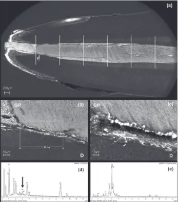
evaluate microleakage by SeM LeO Stereoscan 440 (LeO electron Microscopy Ltd., Cambridge, england) using back scattered electrons (BSe) mode (Figure 1a). The eDS (Inca software, Oxford, UK, england)
was performed in lower magniication within a
pre-determined area (300 mm2) (Figure 1b), and in a
higher magniication, the identiication of silver was
made punctually to determine its exact location (Figure 1c). each 1 mm region was classified according to the following scores: 0 (absence of leakage in both interfaces) and 1 (presence of leakage in at least one of the interfaces). In Figures 1d and 1e typical spectrum showed higher or less Ag inside the interface.
Protocol 2
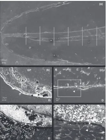
Impressions of polyvinyl siloxane (Aquasil™,
ULV, Dentsply DeTrey) were made of the interfaces and the surfaces replicated with epoxy resin (epon-Thin™, Buhler). The replicas were covered with carbon to investigate the presence of gaps at the dentin/sealer and sealer/cone interfaces, as described previously, using secondary electrons (Se) mode. As the gaps were not continuous, for each 1 mm-region, two interfaces were analyzed (Figure 2a) and classified as: type 0: both interfaces were gap-free, type 1: gap at dentin/ sealer interface (Figure 2b), type 2: gap at sealer/ cone interface (Figure 2c), and type 3: both types of gaps present. Therefore, for each experimental group, 11 replicas were prepared; 55 regions were
evaluated and accordingly classiied.
The data obtained for apical microleakage and types of gaps were statistically analyzed (α=0.05) using the Ordinal Logistic Regression model9 and
Correspondence Analysis7 in the MINITAB Statistical
software program (Minitab Inc. Release 14 for Windows 2003, State College, PA, USA).
The counting of regions analyzed for gaps was also computed and adjusted in a Generalized Linear Model with Poisson distribution and logarithmic link
function. The sealers groups (AH Plus, AH Primer
and epiphany) and the type of gap were considered
as explanatory variables and AH Plus and the type
0 as references.
RESULTS
The Ordinal Logistic Regression Analysis demonstrated that there was no statistically
signiicant difference (p>0.05) among the groups
tested and types of gaps over the logit of microleakage. The model was adjusted to identify the effect of the type of gap on microleakage. The type 3 data were not considered isolated in the analysis (very few regions) but joined with type 2 data (Figure 2c; Table 1). The adjustment tests of the model presented high p-values for Pearson’s test
(p=0.900) and Deviance (p=0.852). The adjusted model demonstrated that the effect of the type of gap on the logit of microleakage is signiicant when
compared to the presence of types 1 and 2 (Figure 2b and 2c, respectively) of gap with the absence of gap (p=0.047). The Correspondence Analysis also showed the association between microleakage and both types of apical gaps (Figure 3).
The Generalized Linear Model with Poisson distribution (Table 1) found no difference between
AH Plus and AH Primer (p=0.497) in distribution
of each type of gap, but they presented more
type 0 regions (p<0.001). There was a signiicant
interaction between epiphany and the type of gap (p<0.003) and for this group, there were more type 1 regions.
Figure 2- Scanning electron microscopy (SEM) micrographs using secondary electrons (SE) and back scattered electrons (BSE) mode of the replica and the section of the same specimen, respectively. (a) - The apical 5 mm of the root canal illing divided into 1 mm-regions. Classiication of the types of gaps: (b) – Type 1: gap between dentin/sealer (pointer); Air voids were present within the sealer (open arrow); (b’) - Silver penetration was evident along the dentin/sealer interface, into dentinal tubules in a reticular form and granular aspect on the dentin surface (arrowhead); (c) – Type 2: gap between sealer/cone (pointer) - Cohesive fracture of the sealer (open arrow); (c’) - the area marked in igure C at higher magniication: Silver deposition into the dentinal tubules in a reticular form and granular aspect within the sealer layer (arrowhead) could be seen; (The sealer thickness was indicated between black/white arrowheads. GP=Gutta-percha; D=Dentin; R=Resilon)
Figure 3- Correspondence analysis of microleakage vs. type of gap: (Inf.0) - absence of leakage; (Inf.1) - leakage up to the 1st millimeter; (Inf.2) – leakage up to the 2nd mm; (Inf.3) – leakage up to the 3rd millimeter; (Inf.5) – leakage up to the 5th millimeter; (F.0) – absence of gaps; (F.1) – gaps type 1; (F.2) - gaps types 1 and 2. The absence of leakage is associated with the absence of gap; leakage up to the 1st millimeter is associated with type 1; and more extensive leakage is associated with types 1 and 2
Groups n Frequency of gaps (%)*
Type 0 Type 1 Type 2
AH Plus 55 42 (76) 11 (20) 2 (4)
AH Primer 55 36 (65) 14 (26) 5 (9)
Epiphany 55 14 (25) 32 (58) 9 (17)
Table 1- Frequency and type of gaps in the apical 5 mm of root canal illings
DISCUSSION
Based on the indings of this study, the irst null
hypothesis could not be rejected. Recently some
studies have shown similar results between AH Plus
and epiphany on apical sealing ability using the
luid iltration method18,22. Consistent with these
studies, our results indicate that the epiphany
system is as effective as AH Plus/gutta-percha in
preventing microleakage based on the chemical tracer penetration analysis by SeM and the use of epiphany primer does not reduce the microleakage
of AH Plus.
Nevertheless, other researches pointed out that the epiphany system provided the greatest
resistance to the movement of luids25,29 or dye
leakage test16, when compared to epoxy resin-based
root canal sealers,but others have just indicated the opposite8,17. These apparent discrepancies can
perhaps be explained by the methodology used and their variables6,16.
These results agree with those of previous studies in which, microleakage markers were used to evaluate apical sealing of the same endodontic sealers28,32. The tracer selected was AgNO
3, which
may penetrate into dentinal tubules due to their physical and chemical characteristics including: concentration, smaller molecular size, its neutral
pH, diffusion coeficient34 and service life up to 168
h post-preparation time of solution4. Another reason
for this choice is that metallic silver deposits may be observed by SeM using back scattered electrons (BSe) mode35.
On the other hand, the results of the current study
disagree with the indings of Shipper, et al.23 (2004)
and Leonardo, et al.11 (2007). An explanation for this
would be that their studies have evaluated coronal and not apical bacterial leakage. Indeed, coronal seal
might be favorably inluenced by photoactivation of
the material which is unlikely to occur in the apical third14. Furthermore, it is speculated that in in vivo
studies (dogs), the high release of calcium hydroxide (41.46 mg/L) that would occur during the process of epiphany sealer solubilization, would make the medium alkaline, resulting in acceleration of the periapical tissue repair process33.
This study has shown that the presence of apical gaps at the sealer/dentin or sealer/cone interfaces had an effect on apical microleakage. The epiphany group presented more type 1 gaps than the other groups (Table 1), rejecting the second null hypothesis. These results corroborate with previous observation of gaps in the apical 4 mm of epiphany
or AH Plus-illed root canals28. Stress generated
during the polymerization shrinkage of epiphany
sealer has probably inluenced the integrity loss at
the sealer/dentin interface24. In addition, the cavity
coniguration factor (C-factor) is highly unfavorable
for adhesion inside root canals26. Moreover, it is known
that analysis of gap formation of vertically sectioned
root illed teeth hides the risk of artifacts during
sectioning. Therefore, in this study, the sections were made at low speed under water cooling. The resin-epoxy replicas at the dentin/sealer and sealer/ cone interfaces were made before the specimens had been prepared for SeM examination in order to differentiate genuine gaps from artifactual gaps created after vacuum desiccation in conventional scanning electron microscopes. Besides, the replica analysis has to be regarded with caution20.
Few studies have focused on apical sealing from two separate perspectives, such as microleakage and apical gap formation. This inter-relationship has not been fully demonstrated in the literature. SeM was used to evaluate these perspectives1,26-28, but it was
restricted to a descriptive approach.
A s y s t e m a t i z a t i o n o f t h e o b s e r va t i o n s , complemented by a statistical analysis, such as the one performed in this study, could provide greater methodological accuracy and impartiality for comparing the experimental groups. The Correspondence of Analysis between the types of gaps and linear extent of AgNO3 leakage (Fig. 3)
conirms the effect of gaps on apical microleakage,
expressed by the silver deposition into gaps that extended deeply inside dentinal tubules (Fig. 2b’ and c’), rejecting the third null hypothesis.
Regarding to statistical analysis, the Ordinal Logistic Regression Analysis Model was used due to microleakage was considered categorized ordinal response variable (ranging from 0 – no leakage
to 5 – leakage up to the ifth millimeter). The
Correspondence Analysis is a method that leads to visualize the association between two categorical variables, in this case microleakage and the type of gap. The Generalized Linear Model with Poisson which is an extension of usual regression model was performed to quantify the distribution of types of gaps. This result could be explained by two hypotheses: (1) the low degree of conversion of the epiphany sealer2 and (2) the formation of hydrogel
at the bond interface resulting from the incomplete evaporation of the epiphany primer solvent3. The
second hypothesis could also explain the ineficacy of
epiphany primer in improving the sealing capacity of
AH Plus. Moreover, there is a chemical incompatibility
between these two materials, since the epoxy resin sealers do not copolymerize with methacrylate resin-based adhesives28. Other studies should be carried
out to clarify the sealing ability of the root-illed
materials studied.
CONCLUSIONS
epiphany primer did not improve the sealing capacity
of AH Plus sealer. The presence of gaps had an effect
on apical microleakage for all materials. Comparing the materials, epiphany system presented more regions containing gaps between the dentin and the
sealer (type 1). In view of these indings, clinically, it can be suggested that AH Plus would provide a
better apical seal.
ACKNOwLEDgEMENTS
This study was partially supported by CAPeS (Coordenação de Aperfeiçoamento de Pessoal de
Nível Superior)/Institutional Qualiication Program
(PQI no: 0090/03-4).
REFERENCES
1- Bergmans L, Moisiadis P, De Munck J, Van Meerbeek B, Lambrechts P. effect of polymerization shrinkage on the sealing capacity of resin
illers for endodontic use. J Adhes Dent. 2005;7:321-9.
2- Beriat NC, ertan A, Cehreli ZC, Gulsahi K. Time-dependent conversion of a methacrylate-based sealer polymerized with different light-curing units. J endod. 2009;35:110-2.
3- Bouillaguet S, Shaw L, Barthelemy J, Krejci I, Wataha JC.
Long-term sealing ability of Pulp Canal Sealer, AH-Plus, GuttaFlow and
epiphany. Int endod J. 2008;41:219-26.
4- Costa JF, Siqueira WL, Loguércio AD, Reis A, Oliveira e, Alves CM, et al. Characterization of aqueous silver nitrate solutions for leakage tests. J Appl Oral Sci. 2011;19:254-9.
5- De Munck J, Van Landuyt K, Peumans M, Poitevin A, Lambrechts P, Braem M, et al. A critical review of the durability of adhesion to tooth tissue: methods and results. J Dent Res. 2005;84:118-32. 6- Dultra F, Barroso JM, Carrasco LD, Capelli A, Guerisoli DM, Pécora JD. evaluation of apical microleakage of teeth sealed with four different root canal sealers. J Appl Oral Sci. 2006;14:341-5. 7- Greenacre M. Correspondence analysis in pratice. London:
Chapman & Hall; 2007.
8- Hirai VH, Silva Neto UX, Westphalen VP, Perin CP, Carneiro E, Fariniuk LF. Comparative analysis of leakage in root canal illings performed with gutta-percha and Resilon cones with AH Plus and
epiphany sealers. Oral Surg Oral Med Oral Pathol Oral Radiol endod. 2010;109:e131-5.
9- Hosmer DW, Lemeshow S. Applied logistic regression. New York:
John Wiley & Sons; 1989.
10- Leonard Je, Gutmann JL, Guo IY. Apical and coronal seal of roots obturated with a dentine bonding agent and resin. Int endod J. 1996;29:76-83.
11- Leonardo MR, Barnett F, Debelian GJ, Pontes Lima RK, Bezerra
da Silva LA. Root canal adhesive illing in dogs' teeth with or
without coronal restoration: a histopathological evaluation. J endod. 2007;33:1299-303.
12- Mannocci F, Ferrari M. Apical seal of roots obturated with laterally condensed gutta-percha, epoxy resin cement, and dentin bonding agent. J endod. 1998;24:41-4.
13- Mannocci F, Innocenti M, Bertelli e, Ferrari M. Dye leakage and
SEM study of roots obturated with Thermaill and dentin bonding
agent. endod Dent Traumatol. 1999;15:60-4.
14- Nagas e, Cehreli ZC, Durmaz V, Vallittu PK, Lassila LV. Regional push-out bond strength and coronal microleakage of Resilon after different light-curing methods. J endod. 2007;33:1464-8.
15- Nunes VH, Silva RG, Alfredo E, Sousa-Neto MD, Silva-Sousa YT. Adhesion of Epiphany and AH Plus sealers to human root dentin
treated with different solutions. Braz Dent J. 2008;19:46-50.
16- Oddoni PG, Mello I, Coil JM, Antoniazzi JH. Coronal and apical
leakage analysis of two different root canal obturation systems. Braz Oral Res. 2008;22:211-5.
17- Onay EO, Ungor M, Unver S, Ari H, Belli S. An in vitro evaluation
of the apical sealing ability of new polymeric endodontic illing
systems. Oral Surg Oral Med Oral Pathol Oral Radiol endod. 2009;108:e49-54.
18- Paqué F, Sirtes G. Apical sealing ability of Resilon/epiphany
versus gutta-percha/AH Plus: immediate and 16-months leakage.
Int endod J. 2007;40:722-9.
19- Pashley DH, Ciucchi B, Sano H, Carvalho RM, Russell CM. Bond
strength versus dentine structure: a modelling approach. Arch Oral Biol. 1995;40:1109-18.
20- Perdigão J, Lambrechts P, Van Meerbeek B, Vanherle G, Lopes
AL. Field emission SEM comparison of four postixation drying
techniques for human dentin. J Biomed Mater Res. 1995;29:1111-20.
21- Perdigão J, Lopes MM, Gomes G. Interfacial adaptation of adhesive materials to root canal dentin. J endod. 2007;33:259-63.
22- Raina R, Loushine RJ, Weller RN, Tay FR, Pashley DH. Evaluation
of the quality of the apical seal in Resilon/epiphany and
Gutta-Percha/AH Plus-illed root canals by using a luid iltration approach.
J endod. 2007;33:944-7.
23- Shipper G, Ørstavik D, Teixeira FB, Trope M. An evaluation
of microbial leakage in roots illed with a thermoplastic synthetic polymer-based root canal illing material (Resilon). J Endod.
2004;30:342-7.
24- Souza SFC, Bombana AC, Francci C, Goncalves F, Castellan C,
Braga RR. Polymerization stress, low and dentine bond strength of
two resin-based root canal sealers. Int endod J. 2009;42:867-73.
25- Stratton RK, Apicella MJ, Mines P. A luid iltration comparison of
gutta-percha versus Resilon, a new soft resin endodontic obturation system. J endod. 2006;32:642-5.
26- Tay FR, Loushine RJ, Lambrechts P, Weller RN, Pashley DH.
Geometric factors affecting dentin bonding in root canals: a theoretical modeling approach. J endod. 2005;31:584-9.
27- Tay FR, Loushine RJ, Monticelli F, Weller RN, Breschi L, Ferrari M, et al. effectiveness of resin-coated gutta-percha cones and a dual-cured, hydrophilic methacrylate resin-based sealer in obturating root canals. J endod. 2005;31:659-64.
28- Tay FR, Loushine RJ, Weller RN, Kimbrough WF, Pashley DH,
Mak YF, et al. Ultrastructural evaluation of the apical seal in roots
illed with a polycaprolactone-based root canal illing material. J
endod. 2005;31:514-9.
29- Tunga U, Bodrumlu e. Assessment of the sealing ability of a new root canal obturation material. J endod. 2006;32:876-8.
30- Van Meerbeek B, De Munck J, Yoshida Y, Inoue S, Vargas M, Vijay P, et al. Buonocore memorial lecture. Adhesion to enamel and dentin: current status and future challenges. Oper Dent. 2003;28:215-35.
31- Vasconcelos BC, Luna-Cruz SM, De-Deus G, Moraes IG, Maniglia-Ferreira C, Gurgel-Filho eD. Cleaning ability of chlorhexidine gel and sodium hypochlorite associated or not with eDTA as root canal irrigants: a scanning electron microscopy study. J Appl Oral Sci. 2007;15:387-91.
32- Verissimo DM, Vale MS, Monteiro AJ. Comparison of apical
leakage between canals illed with gutta-percha/AH-Plus and the Resilon/Epiphany System, when submitted to two illing techniques.
J endod. 2007;33:291-4.
33- Versiani MA, Carvalho-Junior JR, Padilha MI, Lacey S, Pascon eA, Sousa-Neto MD. A comparative study of physicochemical
properties of AH Plus and Epiphany root canal sealants. Int Endod
J. 2006;39:464-71.
34- Wu MK, Wesselink PR. endodontic leakage studies reconsidered. Part I. Methodology, application and relevance. Int endod J. 1993;26:37-43.