Letters to the Editor
Radiol Bras. 2015 Mar/Abr;48(2):126–130
130
Dear Editor,
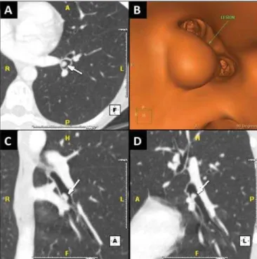
A 45 year-old female patient presented with a previous his-tory of abdominal pain whose onset had occurred one year earlier. Pelvic ultrasonography demonstrated a suspiciously malignant left adnexal complex cyst, confirmed by pelvic magnetic resonance imaging. After surgical resection, the histological diagnosis was ovarian endometrioid adenocarcinoma. As lymph nodes were nega-tive and there was no clinical or radiological evidence of distant metastasis, the disease stage was IA and the surgery was consid-ered curative. Chest multidetector computed tomography (CT) performed at six-month follow-up demonstrated an unusual find-ing of a small endobronchial lesion of 6.7 mm in the left lower lobe causing significant bronchial narrowing (Figure 1). The pa-tient had neither complaints nor symptoms and follow-up positron emission tomography/CT did not reveal any abnormal fluoro-2-deoxy-glucose uptake, including by the endobronchial lesion. Transbronchial biopsy of the lesion confirmed a mucin-produc-ing adenocarcinoma positive for CA-125, compatible with me-tastasis from the ovarian adenocarcinoma. The patient underwent left lower lobectomy followed by chemotherapy. Five years after treatment completion follow-up exams showed no evidence of disease activity.
Endobronchial metastases from ovarian carcinoma are rare, and few cases are described in the literature. Clinical and radio-logical findings are quite similar to those of endobronchial pri-mary lung cancer, so a histopathological diagnosis is critical in such cases(1). Endobronchial metastasis is defined as primary in-volvement of the bronchial epithelium, originating from extra-pulmonary malignant tumors(2). Salud et al. showed that breast, colorectal and renal carcinomas were the most common primary sites of endobronchial metastasis(3). None of these studies dem-onstrated ovarian tumor as the primary site as occurred in the present case.
Endobronchial metastases may be asymptomatic without any other signs of dissemination from the primary tumor, as dem-onstrated in the present case(4). Symptoms may be associated with airway obstruction (dyspnea, wheezing), mucosal irritation and ulceration (cough, hemoptysis), or direct invasion and in-volvement of adjacent structures (recurrent laryngeal nerve palsy, dysphagia).
Currently, multidetector CT is the best imaging modality for the diagnosis and characterization of endobronchial metastases. Multiplanar two-dimensional and three-dimensional image recon-struction techniques, including virtual bronchoscopy, can be eas-ily generated to complement conventional axial CT imaging. In addition, virtual bronchoscopy provides a noninvasive method to evaluate airway lesions and plays an essential role as a comple-ment to conventional bronchoscopy, facilitating planning and guidance of bronchoscopic interventions(5). CT is relatively accu-rate in the evaluation of bronchial abnormalities, and in patients with endobronchial metastases may be used as a complement to bronchoscopy to evaluate lesion extent(6).
Rafael Marques Franco1, Marcos Duarte Guimaraes2, Bruno Lima Moreira2, Almir Galvão Vieira Bitencourt2, Bruno Hoch-hegger3, Edson Marchiori4
1. Hospital Heliópolis, São Paulo, SP, Brazil. 2. A.C.Camargo Cancer Cen-ter, São Paulo, SP, Brazil. 3. Universidade Federal de Ciências da Saúde de Porto Alegre (UFCSPA), Porto Alegre, RS, Brazil. 4. Universidade Fede-ral do Rio de Janeiro (UFRJ), Rio de Janeiro, RJ, Brazil. Mailing Address: Dr. Rafael Marques Franco. Rua Cônego Xavier, 276, Cidade Nova Heliópo-lis. São Paulo, SP, Brazil, 04231-030. E-mail: rafaelmarquesfranco@ gmail.com.
http://dx.doi.org/10.1590/0100-3984.2013.0020
Figure 1.A: CT image shows an endobronchial nodule (arrows) in the lateral basal bronchus of the left lower lobe. B: This nodule measures 6.7 mm. C: CT coronal reconstruction demonstrating > 50% decrease of the endobronchial lu-men (arrow). D: Virtual bronchoscopy confirms the endobronchial narrowing.
Finally, endobronchial metastases are rare and have clinical and radiological findings similar to those of primary endobronchial tumors. Although uncommon, oncologists and radiologists should be alert to the occurrence of endobronchial metastases in early follow-up of patients previously treated for ovarian carcinoma, in order to avoid delay in the diagnosis and to allow for proper thera-peutic planning.
REFERENCES
1. Merrill CR, Hopkirk JA. Late endobronchial metastasis from ovarian tu-mour. Br J Dis Chest. 1982;76:253–4.
2. Ikezoe J, Johkoh T, Takeuchi N, et al. CT findings of endobronchial metastasis. Acta Radiol. 1991;32:455–60.
3. Salud A, Porcel JM, Rovirosa A, et al. Endobronchial metastatic disease: analysis of 32 cases. J Surg Oncol. 1996;62:249–52.
4. Sørensen JB. Endobronchial metastases from extrapulmonary solid tu-mors. Acta Oncol. 2004;43:73–9.
5. Lee KS, Boiselle PM. Update on multidetector computed tomography imaging of the airways. J Thorac Imaging. 2010;25:112–24. 6. Ko JH, Jung GS, Kim SM, et al. Endobronchial metastasis: CT findings
and its usefulness in bronchoscopic correlation. J Korean Radiol Soc. 2000;43:179–84.
Enhancing survival with early surgical resection of endobronchial metastasis in a follow-up of ovarian carcinoma