J Bras Pneumol. 2013;39(1):113-115
To the Editor:
We report the case of a 33-year-old white female patient. Her personal history was unremarkable. She presented with a three-month history of progressive dysphagia, which was accompanied by vomiting after eating, weight loss (8 kg), and asthenia. Physical examination was normal. Laboratory test results showed microcytic hypochromic anemia (hemoglobin, 10.2 g/dL; mean corpuscular volume, 69 fL; and mean corpuscular hemoglobin, 22 pg). No other changes were found.
Upper gastrointestinal endoscopy revealed punctal stenosis 23 cm from the maxillary dental arch, at the transition between the upper third and the middle third of the esophagus, the esophageal mucosa showing mild nodosity and stiffness upstream of the stenosis but no typical signs of neoplastic infiltration into the area of stenosis. The stenosis hindered the progression of the endoscope. Esophageal mucosal biopsies showed edematous esophagitis.
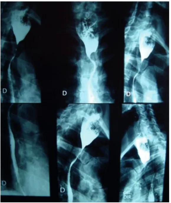
An esophagogram showed stenosis of the esophagus at the cervicothoracic junction and esophageal dilation upstream of the stenosis, food residue being probably present (Figure 1).
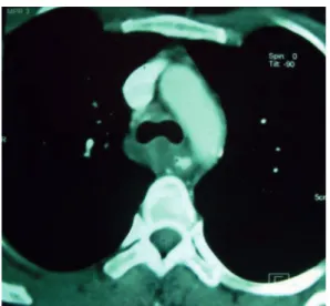
A CT scan of the chest showed solid tissue in the posterosuperior mediastinum, as well as circumferential involvement of the trachea and esophagus, together with significant narrowing of the esophageal lumen and esophageal dilation upstream (Figure 2). Diagnostic hypotheses included lymphoproliferative disease and gastrointestinal stromal tumor.
Fiberoptic bronchoscopy showed no endotracheal lesion. Thoracoscopy showed a tracheoesophageal lesion. The lesion had ill-defined margins and was approximately 5 cm in diameter, as well as being white, hard, and fibrous. Biopsies of the lesion showed no malignancy.
Given the diagnostic uncertainty and the persistence of the symptoms, a thoracotomy was performed, the entire lesion being resected and the thoracic esophagus being freed. Frozen
section analysis was suggestive of gastrointestinal stromal tumor. The histopathological findings were consistent with a nonspecific chronic inflammatory process. Immunohistochemistry was positive for CD20, CD3 (SP7), and smooth muscle actin, and negative for β-catenin, CD30, CD15, and CD245, findings that were consistent with inflammatory pseudotumor.
After surgery, the patient progressed favorably, the symptoms having disappeared. At this writing, 1 year had passed since oncologic control had been achieved.
Inflammatory pseudotumor was first described by Brunn in 1932 and was so designated by Umiker et al. in 1954 because it can mimic malignancy clinically and radiologically.(1) Since
then, inflammatory pseudotumor has been reported in the literature under different names, such as histiocytoma, plasma cell granuloma, xanthoma, and inflammatory myofibroblastic tumor.(2,3)
Inflammatory pseudotumor is a rare tumor of unknown origin that predominantly affects the lung and the orbit. However, inflammatory pseudotumor has been reported to occur in a variety of anatomical sites, including the gastrointestinal tract and the central nervous system.(3,4) Mediastinal involvement is rare.(5)
The etiopathogenesis of inflammatory pseudotumor is unknown, and it has been suggested that inflammatory pseudotumor is a lesion of infectious or autoimmune origin.(6,7)
Although inflammatory pseudotumor most commonly affects the lungs, it can affect any organ.(3,8) Inflammatory pseudotumor affects both
genders and all races equally, and the age of onset ranges from 1 year to 73 years.(2,9) In most
cases, inflammatory pseudotumor is a benign tumor; it rarely invades neighboring structures or shows multifocal development.(8)
The clinical manifestations of inflammatory pseudotumor vary according to the site of origin.(8)
Radiologically, inflammatory pseudotumor usually appears as a well-demarcated lesion.(4,10) The
diagnosis of inflammatory pseudotumor is based
Inflammatory pseudotumor of the posterior mediastinum
Pseudotumor inflamatório do mediastino posterior
Izabella Nobre Queiroz, Renata Mendonça Moreira Penna,
Emanuelly Botelho Rocha Mota, Rafael Turano Mota, Vinícius Turano Mota
114 Queiroz IN, Penna RMM, Mota EBR, Mota RT, Mota VT
J Bras Pneumol. 2013;39(1):113-115
unresectable cases. Steroids are the drugs of first choice for the treatment of orbital inflammatory pseudotumor; however, the therapeutic response is unpredictable.(4,5)
In conclusion, inflammatory pseudotumor of the posterior mediastinum is rare and therefore difficult to diagnose. Clinical and radiological findings suggest other neoplasms, and preoperative test results are usually inconclusive, complete resection of the lesion being required for appropriate treatment and definitive diagnosis. on histopathology, histological findings including
acute and chronic inflammatory cells, with varying degrees of fibrosis.(4,11)
Complete surgical resection is the treatment of choice for all inflammatory pseudotumors except orbital inflammatory pseudotumors.(3,4,8,12,13)
Surgical resection remains the method of choice in cases of recurrence.(3,4)
There have been reports of spontaneous regression and distant metastases.(4,8) Radiation
therapy and chemotherapy have been used in
Inflammatory pseudotumor of the posterior mediastinum
J Bras Pneumol. 2013;39(1):113-115
115
References
1. Umiker WO, Iverson L. Postinflammatory tumors of the lung; report of four cases simulating xanthoma, fibroma, or plasma cell tumor. J Thorac Surg. 1954;28(1):55-63. PMid:13175281.
2. Gorospe L, Fernández-Gil MA, Torres I, Tovar J, García-Miguel P, Tejerina E. Misleading lead: inflammatory pseudotumor of the mediastinum with digital clubbing. Med Pediatr Oncol. 2000;35(5):484-7. http://dx.doi. org/10.1002/1096-911X(20001101)35:5<484::AID-MPO7>3.0.CO;2-T
3. Kim JH, Cho JH, Park MS, Chung JH, Lee JG, Kim YS, et al. Pulmonary inflammatory pseudotumor--a report of 28 cases. Korean J Intern Med. 2002;17(4):252-8. PMid:12647641.
4. Narla LD, Newman B, Spottswood SS, Narla S, Kolli R. Inflammatory pseudotumor. Radiographics. 2003;23(3):719-29. Erratum in: Radiographics. 2003;23(6):1702. PMid:12740472. http:// dx.doi.org/10.1148/rg.233025073
5. Alongi F, Bolognesi A, Gajate AM, Motta M, Landoni C, Berardi G, et al. Inflammatory pseudotumor of mediastinum treated with tomotherapy and monitored with FDG-PET/CT: case report and literature review. Tumori. 2010;96(2):322-6. PMid:20572593.
6. Arber DA, Weiss LM, Chang KL. Detection of Epstein-Barr virus in inflammatory pseudotumor. Semin Diagn Pathol. 1998;15(2):155-60. PMid:9606806.
7. Fernández Villar A, Mosteiro Añón M, Corbacho Abelaira D, Acuña Fernández A, Piñeiro Amigo L. Pulmonary inflammatory pseudotumor: report of 2 cases and review of the literature [Article in Spanish]. An Med Interna. 1997;14(9):469-72.
8. Medina-Achirica C, Gutiérrez de la Peña C, Gómez Menchero J, Gutiérrez Cafranga E, López Hurtado M, Gil Quiros J, et al. Multicentric inflammatory pseudotumor [Article in Spanish]. Cir Esp. 2007;81(3):150-2. http:// dx.doi.org/10.1016/S0009-739X(07)71287-3 9. Topçu S, Taştepe I, Alper A, Ozdülger A, Albayrak M,
Bozkurt D, et al. Inflammatory pseudotumors of the lung: a clinical study of eleven patients. J Thorac Cardiovasc Surg. 2000;119(1):180-2. http://dx.doi.org/10.1016/ S0022-5223(00)70240-6
10. Agrons GA, Rosado-de-Christenson ML, Kirejczyk WM, Conran RM, Stocker JT. Pulmonary inflammatory pseudotumor: radiologic features. Radiology. 1998;206(2):511-8. PMid:9457206. 11. Batsakis JG, el-Naggar AK, Luna MA, Goepfert H.
“Inflammatory pseudotumor”: what is it? How does it behave? Ann Otol Rhinol Laryngol. 1995;104(4 Pt 1):329. 12. Jenkins PC, Dickison AE, Flanagan MF. Cardiac
inflammatory pseudotumor: rapid appearance in an infant with congenital heart disease. Pediatr Cardiol. 1996;17(6):399-401. PMid:8781093. http:// dx.doi.org/10.1007/s002469900088
13. Rose AG, McCormick S, Cooper K, Titus JL. Inflammatory pseudotumor (plasma cell granuloma) of the heart. Report of two cases and literature review. Arch Pathol Lab Med. 1996;120(6):549-54.
Izabella Nobre Queiroz Medical Student,
Universidade Estadual de
Montes Claros
– Unimontes, State University at Montes Claros –Montes Claros, Brazil
Renata Mendonça Moreira Penna Medical Student,
Universidade Estadual
de Montes Claros
– Unimontes, State University at Montes Claros –Montes Claros, Brazil
Emanuelly Botelho Rocha Mota Resident in Clinical Medicine, Clemente de Faria University Hospital,
Montes Claros, Brazil
Rafael Turano Mota Thoracic Surgeon,
Santa Casa de Montes Claros
, Montes Claros, BrazilVinícius Turano Mota Thoracic Surgeon,
Santa Casa de Montes Claros
, Montes Claros, BrazilFigure 2 - Chest CT scan showing solid tissue in the posterosuperior mediastinum, together with circumferential involvement of the trachea and esophagus.