1N e u rology and2Radiology Division of the Internal Medicine Department, Hospital de Clínicas, Universidade Federal do Paraná,
Curitiba PR, Brazil.
Received 30 June 2005, received in final form 26 June 2006. Accepted 31 July 2006.
D r. Pedro André Kowacs - Serviço de Neurologia, Departamento de Clínica Médica - Hospital de Clínicas da Universidade Federal do Paraná - Rua General Carneiro 181 / 12º andar / sala 1236 - 80060-900 Curitiba PR - Brasil. E-mail: [email protected]
THE ROLE OF THE IRRITATIVE ZONE AND OF THE
NUMBER AND DISTRIBUTION OF CALCIFICATIONS
IN THE SEVERITY OF EPILEPSY ASSOCIATED WITH
INTRACRANIAL CALCIFICATIONS
Pedro André Kowacs
1, Enio Rogacheski
2, Juliano Muzzio
1, Lineu César Werneck
1ABSTRACT -Objective:To determine the influence of the location of the irritative zone, and the number and the distribution of the intracranial calcifications in the severity of epilepsy associated with intracra-nial calcifications. Method:We studied 47 patients with epilepsy and intracranial calcifications, 24 with normal (Group A) and 23 with abnormal interictal EEGs (Group B), a control group (n=21) with abnormal interictal EEGs and normal CT-scans (Group C). Clinical, electroencephalographic and neuro r a d i o l o g i c a l f e a t u reswere compared among gro u p s . Results:Temporal lobe interictal EEG abnormalities were found in 23/24 Group B patients, and in all Group C patients. Most Group B and Group C patients presented tem-poral lobe seizure symptomatology, whereas in most Group A patients symptomatology was ro l a n d i c (p=0.0001). Epilepsy was more severe in Group B and Group C patients than in Group A patients (p=0.0001 and p=0.0054). No relationship was found between the number of calcifications and epilepsy severity.
Conclusion:An irritative zone at the temporal lobe is more relevant in determining the severity, sympto-matology and frequency of seizures than the number and location of calcifications.
KEY WORDS: epilepsy-localized, epilepsy-temporal lobe, neuro c y s t i c e rcosis, cysticercosis, calcifications-intracranial.
Papel da zona irritativa e do número e distribuição das calcificações na gravidade da epilepsia associada com calcificações intracerebrais
RESUMO - Objetivo:Testar a relevância do lobo temporal na epilepsia associada a neurocisticercose inati-v a . Método:Foram selecionados 47 pacientes com epilepsia e calcificações intracerebrais, 22 com EEGs i n t e rcríticos normais (Grupo A) e anormais em 23 (Grupo B) e um grupo controle (n=21) com EEGs inter-críticos anormais sem calcificações intracerebrais (Grupo C). Características clínicas, eletrencefalográficas e n e u roradiológicas foram comparadas entre os gru p o s . Resultados:Encontramos anormalidades eletre n c e-falográficas temporais em 23/24 dos pacientes do Grupo B e em todos do Grupo C. Na maioria dos pacientes do Grupo B e Grupo C a sintomatologia ictal foi interpretada como temporal, porém no Grupo A como rolândica (p=0,0001) A epilepsia foi mais grave nos grupos Grupo B e Grupo C que no Grupo A (p=0,0001 e 0,0054). Não houve relação direta entre o número de calcificações e a gravidade da epilepsia. C o n c l u s ã o :
Uma zona irritativa sobre o lobo temporal é mais relevante na gravidade, sintomas e freqüência das crises que o número e a localização das calcificações.
PA L AV R A S - C H AVE: epilepsia localizada, epilepsia do lobo temporal, neuro c i s t i c e rcose, cisticercose, calcifi-cações intracerebrais.
As in other Latin American countries, neuro c y s-t i c e rcosis has been idens-tified in Brazil as s-the single main abnormality found on ancillary investigation of adults with epilepsy in primary health care serv i c-es, university hospitals, and radiological clinic
sur-v e y s1 - 4. Although some studies have been carried out
with the aim of determining the prognostic factors
of epilepsy related to neuro c y s t i c e rc o s i s5 - 7, it has
recently been shown that, in many cases, parenchy-mal neuro c y s t i c e rcosis leaves no clue for later
diag-n o s i s8. However, in these countries, calcified lesions
a re the most common clinical presentation of
neuro-c y s t i neuro-c e rneuro-cosis assoneuro-ciated with epileptineuro-c seizure s1 , 3 , 9 - 1 1,
occurring in 70-90% of patients with neurocysticer-cosis and epilepsy.
Carpio et al.1 2pointed to the lack of studies with
pa-tients with acute symptomatic seizures, chronic re c u r-rent seizures and newly diagnosed re c u rr-rent seizures. We have carried out this study in an attempt to clarify the factors related to the severity of epilepsy associated with intracranial calcifications.
METHOD
Files for two sets of patients were selected from the patients’ database at the HC-UFPR Hospital, in Curitiba, s o u t h e rn Brazil. One of the files i ncluded all the patients with intracranial calcifications and epilepsy that had attend-ed the general neurology out-patient clinic during 1993. Inclusion criteria were age between 15 and 65 years, pre s-ence of calcifications on computerized tomography (CT-scan) images of the brain and follow-up of one year. Exclu-sion criteria were presence of idiopathic or symptomatic generalized epilepsy1 3, presence of active systemic or neu-rological disease, seizures related to specific conditions and
presence of severe mental retardation. All of the patients selected were interviewed by one of the investigators (PA K ) , and submitted to an inventory, according to the protocol. Of the 51 patients selected serially, four dropped out for the following reasons: mental retardation (n=2); intracra-nial neoplasm (n=1); and lack of will to participate (n=1). Patients with intracerebral calcifications and a normal ro u-tine EEG were submitted to another tracing after 24 hours of sleep deprivation and subdivided into two groups - the G roup A(absence of an interictal EEG focus), andG roup B ( p resence of an interictal EEG focus). A control gro u p (Group C)of patients with normal CT-scan images but ab-normal EEGs was selected from the same out-patient clin-ic (n=15) and from the epilepsy out-patient clinclin-ic (n=6) (Ta-ble 1).
Although the pro p o rtion of females was higher in both g roups with abnormal EEGs compared with that of the G roup A, the diff e rences were not statistically significant ( Tables 2, 3 and 4). Only one patient from the Group B had been treated for neuro c y s t i c e rcosis with albendazole.
Sei-Table 1. Main criteria for the Groups A, B and C.
Group A Group B Group C
CT-scan calcifications calcifications normal Inter-ictal EEGs* normal focal spiking focal spiking * for patients with a normal interictal EEG a second EEG tracing after sleep deprivation was done.
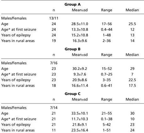
Table 2. Demographics of Group A, Group B and C patients.
Group A
n Mean±sd Range Median
Males/Females 13/11
Age 24 28.5±11.0 17–56 25.5
Age* at first seizure 24 13.3±10.8 0.4–44 12
Years of epilepsy 24 15.2±10.8 1–48 13
Years in rural areas 19 16.3±9.6 2–36 14
Group B
n Mean±sd Range Median
Males/Females 7/16
Age 23 30.2±9.2 15–52 29
Age* at first seizure 23 9.3±7.6 0.7–25 7
Years of epilepsy 23 20.9±8.6 3–35 22.5
Years in rural areas 18 16.6±11.4 0.6–41 17.5
Group C
n Mean±sd Range Median
Males/Females 7/14
Age 21 33.5±10.1 21–55 30
Age* at first seizure 21 11.7±10.3 0.1–38 10
Years of epilepsy 21 21.8±9.1 5–42 23
Years in rural areas 11 23.5±16.4 1–51 24
z u res were classified according to the 1981 ILAE re v i s e d p ro p o s a l1 4. Seizure frequency was classified according to the last year of follow-up as no seizures, few seizures (less than one seizure a month), frequent seizures (at least one s e i z u re a month) and very frequent seizures (one seizure or more a week). Severity of epilepsy was classified by com-bining average seizure frequency with seizure type (m i n o r =p a rtial;major =secondarily generalized) and classi fied as controlled, for no seizures in the last year, or as mild, moderate and severe, according to modified Pazzaglia cri-teria15.
C T-scans of the head were perf o rmed with a 10º angle. Ten millimeter and 6 mm intervals were used for imaging above and below the tentorium, re s p e c t i v e l y. Images were transposed to sketches of the tomograms by a board - c e r-tified neuroradiologist. Quantity of calcifications was clas-sified as isolated (n=1), sparse (1<n5), and diffuse (n>5), in accordance w ith the nomenclature used by t he associ-ated radiologist (ER). For Group B patients, location of cal-cifications was compared to location of surface EEG foci. Calcifications of the patients in each group were superim-posed in a single sketch, and the Group A sketch was com-pared with the Group B sketch.
Table 4. Group B and Group C interictal EEG abnormalities.
Group B Group C Total (n=23) (n=21) (n=44) Location / Abnormalities
Temporal lobe foci*
Unilateral 23 21 44
sharp waves and spikes 17 16 33
slow waves 6 5 11
Bilateral 7 5 12
sharp waves and spikes 4 4 8
slow waves 3 1 4
Extratemporal foci
sharp waves and spikes 1 1 2
slow waves 1 1
Lateralized abnormalities
sharp waves and spikes 1 1 2
slow waves 1 1
n, number; *temporal lobe foci with or without associated extratem-poral foci; note: some patients presented more than a single EEG abnor-mality.
Table 3. Initial ictal symptomatology.
Groups
Initial ictal Group A Group B Group C Group B + Group C
symptoms n=24 n=21* n=19* n=40*
Rolandic 11# 1 1
Temporal lobe 6 14# 15# 29
Mixed 5 7 2 9
Others 2 1 1
* Two Group B patients did not re p o rt initial ictal symptoms, as was also observed for the Group C; Rolandic: simple motor or somatosensory symptoms; Temporal lobe: complex partial seizures associat-ed to automatisms and/or autonomic, olfatory, psychic, or vertiginous symptoms; Mixassociat-ed: association between rolandic and temporal lobe symptoms, or of either type with other symptoms; n: number;
#: p=0.0001.
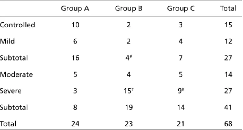
Table 5. Severity of epilepsy in Group A, Group B and Group C patients.
Group A Group B Group C Total
Controlled 10 2 3 15
Mild 6 2 4 12
Subtotal 16 4# 7 27
Moderate 5 4 5 14
Severe 3 15‡ 9# 27
Subtotal 8 19 14 41
Total 24 23 21 68
The possible relationship between the number of cal-cifications and severity of epilepsy was studied. The level of significance was determined by the Qui-square test cor-rected by Yates and by Fisher’s exact test.
The publication of the study was approved by the local regulatory institution.
RESULTS
In the Group A, patients without seizures in the last year prior to the study or with infrequent (low f requency) secondarily generalized seizures were m o re common than patients with frequent or very f requent (high frequency) secondarily generalized s e i z u res (p<0.005). No diff e rence, however, was found between high frequency or low frequency simple and complex partial epileptic seizures in the same group.
In the Group B, patients with frequent or very fre-quent simple and complex partial seizures pre d o m-inated over those without seizures or with infre q u e n t s e i z u res (p=0.001) in the last year prior to the study. In the Group C, no diff e rence was found in seizure f requency between partial and secondarily general-ized seizures.
F requent or very frequent partial seizures were m o re common among Group B patients than Gro u p A patients (p<0.05). Group B patients and Group C patients re p o rted frequent or very frequent second-arily generalized seizures significantly more often than Group A patients (p<0.05).
When only the partial symptoms of seizures were analyzed, those symptoms suggestive of temporal
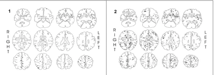
lobe seizures were re p o rted more frequently by the G roup B patients and Group C patients, while ro l a n d i c s e i z u re symptomatology was more frequently des-cribed by Group A patients (p=0.0001, Table 3). No d i ff e rence was found when the location of EEG foci w e re compared between Group B and Group C. Most of the electroencephalographic abnormalities occurr-ed over the temporal lobe in both groups (Table 4). Average number of calcifications was higher for the Group A (mean=12.1) than for the Group B (mean =9.8). Figures 1 and 2 respectively show all the in-tracranial calcifications for the Group A and Gro u p B patients transposed from their original CT-scan ima-ges to sketches of the brain, with one set of sketches for each group. There was no diff e rence between the two groups in terms of the number of cases in which isolated, sparse or diffuse calcifications were found. With re g a rd to severity of epilepsy, when con-t rolled and mild epilepsy were compared againscon-t mo-derate and severe epilepsy within the three gro u p s , G roup B patients presented a significantly more se-v e re epilepsy than Group A patients (p<0.005). When c o n t rolled, mild and moderate epilepsy were com-p a red to severe ecom-pilecom-psy, the diff e rence between the G roup A and the Group B increased, and in the Gro u p C, epilepsy was found to be significantly more severe than in the Group A (p=0.0001 and 0.005, re s p e c t i v e-ly). No diff e rence was found between severity of epi-lepsy in Group B and Group C patients.
The data re g a rding severity of epilepsy can be appreciated in Table 5. When the number of calcifi-cations was plotted against severity of epilepsy tak-ing Group A and Group B together, there was an in-verse relationship between the number of calcifica-tions and severity of epilepsy. Figures 3 and 4 show examples of disparities between the number of cal-cifications and severity of epilepsy.
DISCUSSION
N e u ro c y s t i c e rcosis is the main cause of adult-onset
s e i z u res in the developing world1 6 - 2 3. Calcified
cys-t i c e rci have been re p o rcys-ted as cys-the moscys-t common find-ing in tomograms of epileptic patients in Brazil, pro b-ably because Brazil includes a number of underd
e-veloped areas17-20.
Nonetheless, an association between the number of lesions that became calcified and the persistence
of seizures has been described5 - 7. In these studies,
however, follow-ups were carried out after therapy for cysticercosis, and the use of steroids during cysti-cidal therapy might have changed the natural histo-ry of the disorder.
F rom anecdotal re p o rts, collected series, and pre-vious observations in patients with seizures, perile-sional edema related to calcifications appears to be relatively frequent, ranging from 23% to about 35% in patients with calcified neuro c y s t i c e rcosis and a
his-t o ry of seizure s2 4. When present, these areas of
per-ilesional seizure - related transient edema are thought to re p resent the epileptogenic zone. Indeed, seizure s may be a late manifestation of neuro c y s t i c e rcosis e v e n after adequate treatment, because cicatricial tissue
remains as a permanent sequela25,26. Two
retrospec-tive studies published in 1992 found striking diff e-rences in the evolution of seizures in favor of
anti-parasitic treatment6,27, but in 1995 a controlled trial
showed no diff e rences among patients treated with
albendazole, praziquantel or cort i c o s t e roids alone2 8.
In Brazil, however, the majority of patients diagnosed as having round intracranial calcifications on CT- s c a n images and epilepsy had never been submitted to
therapy with praziquantel or albendazol2 , 9 , 2 9. In these
patients,c y s t i c e rciusually degenerated
spontaneous-l y. We couspontaneous-ld not find a direct respontaneous-lationship between the number of calcifications and the severity of epi-lepsy in our patients, but the inverse relationship bet-ween the number of calcifications and more severe e p i l e p s y, although significant, might have been bias-ed by Group B patients, who also had active interic-tal foci. Studies suggest a lack of a re l a t i o n s h i p between the burden of the neuro c y s t i c e rcotic lesions and the severity of the epilepsy. Some patients with s e v e rere f r a c t o ryepilepsy have only one calcified le-sion; by contrast, other patients have many cysts or calcifications but no epilepsy. Local inflammation and immunity studies in the brain point to significant dif-f e rences in both disorders in the didif-fdif-f e rent areas odif-f the brain, thus adding other sources of local
varia-tion in the host-parasite re l a t i o n s h i p2 6. Because of
the high prevalence of epilepsy and neuro c y s t i c e rc o-sis in less developed countries, there could be a causal as well as fortuitous relationship between the two
disorders30,31.
The presence of interictal epileptiform activity on EEG tracings was associated with a more severe epi-l e p s y, since there was no diff e rence between Gro u p B and Group C in terms of severity of epilepsy, but in both groups epilepsy was more severe than in the G roup A. In some studies, active interictal epilepti-f o rm activity has been epilepti-found to be associated with
a worse pro g n o s i s3 2, and in others this was true for
a b n o rmalities re c o rded over the frontal and
tempo-ral lobes3 3. Previous studies of epilepsy and
interictal EEG abnormalities and severity or pro g n o-sis of epilepsy associated with neuro c y s t i c e rcoo-sis, but most of them included both patients with acute symptomatic seizures and those with unpro v o k e d s e i z u res due to remote symptomatic epilepsy. Al-though our study was not designed to evaluate pro g-nosis, its results suggest that careful EEG investiga-tion should be perf o rmed in these patients, since in-terictal abnormalities over the temporal lobes may have prognostic significance.
I n t e r p reting the pathophysiology of epilepsy asso-ciated with intracranial calcifications as secondary to i n c reased firing of the neurons around the lesion is an oversimplification. This becomes evident in cases presenting a mismatch between the location of the calcifications and the EEG foci or the seizure symp-toms. Indeed, Benabis and Lüders stated that the
syn-d romic approach to epilepsy hasyn-d been unsyn-deru s e syn-d3 4.
Another study noted that 18% of patients studied p resented complex partial seizures, and re p o rted that EEG abnormalities were associated with simple and
complex partial seizure s3 5. In most of the studies,
h o w e v e r, little attention was paid to a syndro m i c a p p roach. Bittencourt et al analyzed 24 patients with epilepsy and neuro c y s t i c e rcosis and compared them with 17 patients with temporal lobe epilepsy alone. They concluded that there was no significant diff e r-ence in the rates of co-localization of the pre s u m p-tive epileptogenic foci between the patients with temporal lobe epilepsy and those with neuro c y s t i c e r-cosis36.
The coexistence of hippocampal atrophy and ex-trahippocampal pathologic abnormalities is re f e rre d to in the literature as “dual pathology” and has been re p o rted in 5 to 30% of patients with medically
re-f r a c t o rypartial seizure s3 7 , 3 8. More re c e n t l y, the
asso-ciation between neuro c y s t i c e rcosis and mesial tempo-ral lobe epilepsy has been re p o rted in anecdotal form
in the literature3 9 , 4 0, but in a larger series the
neuro-pathologic findings indicated that patients with hip-pocampal sclerosis and calcified cysticercosis have a p a t t e rn of hippocampal cell loss and an amount of fascia dentate mossy fiber re o rganization
indistin-guishable from classic cases of hippocampal sclero s i s4 1.
Some new perspectives on the subject have al-ready been investigated in Brazil. In a brief commu-nication, Martinez et al. re p o rted 11 cases of epilep-sy surg e ry on patients with intracranial calcifications, with mesial temporal sclerosis being described in
s o m e4 2. More re c e n t l y, Jorge et al. compared the
clin-ical presentation and surgclin-ical outcome of patients
with mesial temporal sclerosis with and without asso-ciated intracranial calcifications, and found no signi-ficant diff e rences between the two groups. These finding were confirmed by Leite et al. who also dis-c o v e red that the pathologidis-c findings at the hippo-campus were similar for the two groups, and conclud-ed that the presence of calcificonclud-ed lesions did not influ-ence the profile of patients with hippocampal atro-p h y, and that these atro-patients resatro-ponded to tematro-poral lobectomy as well as the patients without
intracra-nial calcifications4 3. The aforementioned works
sup-p o rt the view that we should not be bound to the old concepts of calcification=epileptogenic lesion and/or focus, but rather search for other markers of severity in re f r a c t o rycases of epilepsy and intracrnial calcifications, such as the syndromic pre s e n t a-tion, the interictal EEG findings and MRI imaging. Fi-n a l l y, we would like to emphasize a view that is Fi-now acceptable, namely that the presence of intracranial calcifications on CT-scan images of the brain should not be a criterion for excluding patients from epilep-sy surgery protocols.
Acknowledgements – The authors express their thanks to Dr. Duilton de Paola and Dr. Mara Balliana for EEG interpretation, and to Dr. Carlos Silvado for his sugges-tions.
REFERENCES
1. Medina MT, Rosas E, Rubio-Donnadieu F, Sotelo J. Neuro c y s t i c e rc o s i s as the main cause of late-onset epilepsy in Mexico. A rch Intern Med 1990;150:325-327.
2. A r ruda WO. Etiology of epilepsy: a prospective study of 210 cases. A rq Neuropsiquiatr 1991;49:251-254.
3. Teive HAG, Tsubouchi MH, Ferreira MVC, Minguetti M. Análise de 1000 tomografias computadorizadas do crânio em pacientes com crises epilépticas. Arq Neuropsiquiatr 1992;50:70.
4. Gracia AK. Avaliação preliminar de casos de neuro c i s t i c e rcose diag-nosticada em um serviço de tomografia em Curitiba-PR nos anos de 1992 e 1993. In: Anais do I Encontro Cone Sul e Seminário Latino-Americano sobre Teníase e Cisticercose, Curitiba, SESA: UFPR: OPA S : MS/FNS, 1994:180-181.
5. Medina MT, Cordova S, Genton P, Sotelo J, Dravet C, Montoya MC. P rognosis in neuro c y s t i c e rcosis: the effect of anticysticercal tre a t m e n t . Epilepsia 1991;32(Suppl 1):S110-S111.
6. Vasquez v, Sotelo j. The course of seizures after treatment for cerebral cysticercosis. N Engl J Med 1992;327:696-701.
7. Del Brutto, OH. Prognostic factors for seizure re c u r rence after with-drawal of antiepileptic drugs in patients with neuro c y s t i c e rcosis. Neu-rology 1994;44:1706-1709.
8. G a rcia HH, Gilman RH, Catacora M, Verastegui M, Gonzalez AE, Ts a n g VCW, and the Cysticercosis Working Group in Peru. Serologic evolu-tion of neuro c y s t i c e rcosis patients after antiparasitic therapy. J Infect Dis 1997;175:486-489.
9. Takayanagui OM. Neuro c i s t i c e rcose: evolução clínico-laboratorial de 151 casos. Arq Neuropsiquiatr 1990;48:1-10.
10. Palacio LG, Jiménez I, Garcia HH, et al. Neuro c y s t i c e rcosis in persons with epilepsy in Medellín, Colombia. Epilepsia 1998;39:1334-1339. 11. Carpio A. Hauser WA. Neuro c y s t i c e rcosis and epilepsy. In: Singh G,
Prabhakar S (eds).Taenia soliumc y s t i c e rcosis. Wa l l i n g f o rd, UK: CABI
Publishing, 2002:211-220.
13. Commission on Classification and Terminology of the International League Against Epilepsy. Proposal for revised classification of epilep-sies and epileptic syndromes. Epilepsia 1989;30:389-399.
1 4 . Commission on Classification and Terminology of the International Legue Against Epilepsy. Proposal for revised clinical and electro e n c e p h a-lographic classification of epileptic seizures. Epilepsia 1981;22:489-501. 15. Pazzaglia P, D’Allessandro R, Lozito A, Lugaresi E. Classification of partial epilepsies according to the symptomatology of seizures: prac-tical value and prognostic implications. Epilepsia 1982;23:343-350. 16. G a rcia HH, Pretell J, Gilman RH, et al. A trial of antiparasitic tre a t m e n t
to reduce the rate of seizures due to cerebral cysticercosis. N Engl J Med 2004;350:249-258.
17. Narata A P, A r ruda WO, Uemura E, et al. Neuro c i s t i c e rcose: diagnós-tico tomográfico em pacientes neurológicos. A rq Neuro p s i q u i a t r 1998;56:245-249.
18. A r ruda WO, Camargo NJ, Coelho RC. Neuro c y s t i c e rcosis: an epidemi-ological survey in two small rural communities. A rq Neuro p s i q u i a t r 1996;48:419-424.
19. Gonçalves Coelho TD, Coelho MDG. Cerebral Cysticercosis in Campina Grande, Paraíba - northern Brazil. Arq Neuropsiquiatr 1996;54:94-97. 20. Teive HAG, Minguetti G, Sasaki MGM, Lopes CER, Carvalho MTM,
Szpeiter N. Neurocisticercose. Rev Bras Neurol 1997;33:147-153. 21. Riley T, White AC Jr. Management of neuro c y s t i c e rcosis. CNS Dru g s
2003;17:577-591.
22. Sotelo J. Neuro c y s t i c e rcosis. InA m i n o ff MJ, Daro ff RB (eds). Ency-clopedia of the neurological sciences. San Diego, Calif.: Academic Pre s s , 2003:474-475.
23. Proaño JV, Madrazo I, Avelar F, López-Felix B, Díaz G, Grijalva I. Me-dical treatment for neuro c y s t i c e rcosis characterized by giant subarach-noid cysts. N Engl J Med 2001;345:879-885.
24. Fleury A, Dessein A, Preux PM, et al. Symptomatic human neurocys-t i c e rcosis: age, sex and exposure facneurocys-tors relaneurocys-ting wineurocys-th disease heneurocys-tero- hetero-geneity. J Neurol 2004. 251:830-837
2 5 . Pradhan S, Kathuria MK, Gupta RK. Perilesional gliosis and seizure out-come: a study based on magnetization transfer magnetic resonance imag-ing is patients with neuro c y s t i c e rcosis. Ann Neurol 2000:48:181-187. 26. Nash TE, Del Brutto OH, Butman JA, et al. Calcific neurocysticercosis
and epileptogenesis. Neurology 2004;62:1934-1938.
27. Del Brutto OH, Santibanez R, Noboa CA, A g u i r re R, Diaz E, A l a rc o n TA. Epilepsy due to neuro c y s t i c e rcosis: analysis of 203 patients. Neurology 1992;42:389-392.
28. Carpio A, Santillan F, Leon P, Flores C, Hauser WA. Is the course of n e u ro c y s t i c e rcosis modified by treatment with antihelminthic agents? Arch Intern Med 1995;155:1982-1988.
29. Quagliato EMAB. Forma epiléptica da cisticercose encefálica, análise de 96 casos. Tese. Campinas, 1987.
30. B romfield EB, Vonsattel J-P. Weekly Clinicopathological Exercises: Case 24-2000: A 23-year-old man with seizures and a lesion in the left tem-poral lobe. N Engl J Med 2000; 343:420-427.
31. Sakamoto AC, Bustamante VCT, Garzón E, et al. Cysticercosis and e p i l e p s y. In Kotagal P, Luders HO (eds). The epilepsies: etiologies and prevention. San Diego: Academic Press, 1999:275-282.
32. Rodin EA. Various prognostic aspects in patients with epilepsy. Folia Psychiatr Neurol Jpn 1978;32:407-418.
33. The Group for the Study of Prognosis of Epilepsy in Japan. Natural history and prognosis of epilepsy: report of a multi-institutional study in Japan. Epilepsia 1981;22:35-53.
34. Benbadis SR, Lüders HO. Epileptic syndromes: an underutilized con-cept. Epilepsia 1996;37:1029-1034.
35. M o n t e i ro L, Nunes B, Mendonça D, Lopes J. Spectrum of epilepsy in n e u ro c y s t i c e rcosis: a long-term follow-up of 143 patients. Acta Neuro l Scand 1995;92:33-40.
36. Goldsmith P, Sandmann MC, Souza DS, Mazer S, Antoniuk A, Bittecourt PRM. The relationship between parasite location and electro c l i n-ical abnormality in neuro c y s t i c e rcosis. Neurol Infect Epidemiol 1996; 1:127-133.
37. Cendes F, Cook MJ, Watson C, et al. Frequency and characteristics of dual pathology in patients with lesional epilepsy. Neurology 1995; 45:2058-2064.
38. Raymond AA, Fish DR Stevens JM, Cook MJ, Sisodiya SM, Shorvon SD. Association of hippocampal sclerosis with cortical dysgenesis in patients with epilepsy. Neurology 1994;44:1841-1845.
39. Chung CK, Lee SK, Chi JG. Temporal lobe epilepsy caused by intrahip-pocampal calcified cysticercus: a case report. J Korean Med Sci 1998; 13:445-448.
40. G a rg RK, Karak B, Mohan Kar A. Neuroimaging abnormalities in Indian patients with uncontrolled partial seizures. Seizure 1998;7:497-500. 41. Leite JP, Terra-Bustamante VC, Fernandez RMF, Santos AC, Chimelli
L, Sakamoto AC, Assirati JA, Takayanagui OM. Calcified neuro c y s-t i c e rcos-tic lesions and poss-tsurgery seizure cons-trol in s-temporal lobe epilepsy. Neurology 2000;55:1485-1491.
42. Martinez JAG, Cukiert A, Marino R Jr, Yacubian EM. Ciru rgia de epilep-sia em pacientes com calcificações neuro c i s t i c e rcóticas. Braz J Epilep Clin Neurophysiol 1995;1:131.