Detection of serum amyloid-A concentration in the calf clinically diagnosed with pneumonia, enteritis and pneumoenteritis. 293
Detection of serum amyloid-A concentration in the calf clinically
diagnosed with pneumonia, enteritis and pneumoenteritis
A detecção de amilóide-A concentração sérica em bezerros com diagnóstico clínico de pneumonia, enterite e pneumoenterites
Mustafa KabuI* Bulent ElitokI Ismail KucukkurtII ISSN 0103-8478
ABSTRACT
The aim of this study is to determine serum amyloid-A (SAA) concentration in the cases of pneumonia, pneumoenteritis, and enteritis which are frequently encountered in calves in veterinary medicine. Although a great deal of experimental studies
has been conducted in this field, studies on naturally infected
calves are quite few. Eighty calves at the age of 0-6 months were used in the study and the calves were divided into four groups. Due to the clinical examination, the calves diagnosed with pneumonia (Group P; n=20), with pneumoenteritis (Group PE; n=20) and with enteritis (Group E; n=20) formed the disease group as the healthy ones formed the control (Group C; n=20) group. After the body temperatures of all calves were taken, blood samples were obtained from Jugular vein for haematological and biochemical measurements. As haematological, white blood cell (WBC), red blood cell (RBC), hemoglobin (Hb) and hematocrit (Hct) measurements were performed in Veterinary Hematology Analyzer.
Serum amyloid-A (SAA), interleukin 1 1β), interleukin 6 (IL-6), tumor necrosis factor-α (TNF-α) concentration measurements
were carried out with ELISA reader by using commercial kits. Aspartate aminotransferase (AST), alanine aminotransferase (ALT), albumin (ALB), total bilirubin (T. Bil), total protein (TP),
gamma glutamyltransferase (GGT), blood urea nitrogen (BUN)
concentration measurements were conducted in autoanalyzer by using commercial kits. In all disease groups (P, PE, and E) body temperature, haematologic parameters (WBC, RBC, Hb and Hct), serum biochemical parameters (AST, ALT, ALB, T. Bil, TP,
GGT and BUN), SAA concentration and serum concentrations of cytokines (IL-1β, IL-6 and TNF-α) were determined to be higher
in comparison to the control group (P<0.005). According to these
findings, routine measurement of serum SAA concentration in veterinary medicine is considered to be beneficial in determining
the severity of the disease, in selecting the proper treatment, in monitoring the applied treatment, and detecting subclinical
diseases. In the light of these findings we acknowledge that
routine measurements of serum SAA concentration from the
moment the calves are diagnosed with pneumonia, enteritis and pneumoenteritis in veterinary medicine until the actual cause is determined (bacteria, virus, parasites, etc.) would avail the clinician to, identify the severity of the disease, select the appropriate treatment and monitor the effectiveness of the treatment.
Key words: serum amyloid-A, calves, pneumonia, enteritis, pneumoenteritis.
RESUMO
O objetivo deste estudo é determinar amilóide A (SAA) da concentração sérica nos casos de pneumonia, pneumoenterites e enterite, que são frequentemente encontrados em bezerros em medicina veterinária. Apesar de uma grande quantidade de estudos experimentais terem sido realizados neste campo, os estudos sobre animais com infecção natural são muito poucos. 80 vitelos com a idade de 0-6 meses de idade foram utilizados no estudo e os animais foram divididos em quatro grupos. Devido ao exame clínico, os bezerros diagnosticados com pneumonia (Grupo P; n=20), com pneumoenterites (Grupo PE; n=20) e com enterite (Grupo E; n=20) formaram o grupo de doença, como as saudáveis, formando o grupo controle (Grupo C; n=20). Após as temperaturas corporais de todos os bezerros, foram tomadas amostras de sangue que foram obtidas de veia jugular para medições hematológicas e bioquímicas. Como hematológica, glóbulos brancos (WBC), glóbulos vermelhos (RBC), hemoglobina (Hb) e hematócrito (HCT) foram realizados em Hematologia Veterinária Analyzer. O soro
amilóide-A (SAA), a interleucina 1 (IL-1β), interleucina 6 (IL-6), fator de necrose tumoral (TNF-α) medidas de concentração foram
efectuadas com um leitor de ELISA, utilizando kits comerciais. Aspartato aminotransferase (AST), alanina aminotransferase (ALT), albumina (ALB), bilirrubina total (T. Bil), proteína total
(TP), gama glutamiltransferase (GGT), ureia (BUN) às medições
das concentrações foram realizadas em auto-analisador por utilizandos kits comerciais. Em todos os grupos de doenças (P, PE,
IDepartment of Internal Medicine, Faculty of Veterinary Medicine, Afyon Kocatepe University, ANS Campus, 03200, Afyonkarahisar,
Turkey. E-mail: [email protected]. *Corresponding author.
IIDepartment of Biochemistry, Faculty of Veterinary, Afyon Kocatepe University, Afyonkarahisar, Turkey.
e E), a temperatura corporal, parâmetros hematológicos (WBC, RBC, Hb e Hct), parâmetros bioquímicos de soro (AST, ALT, ALB,
T. Bil, TP, GGT e BUN), a concentração SAA e as concentrações séricas de citocinas (IL-1β, IL-6 e TNF-α) foram determinadas a
ser mais elevadas em comparação com o grupo controle (P<0,005). De acordo com estas descobertas, a medição rotineira da concentração de soro SAA em medicina veterinária é considerada
benéfica para determinar a gravidade da doença, na selecção de
um tratamento adequado, no seguimento do tratamento aplicado e para a detecção de doenças subclínicas. À luz desses resultados, reconhecemos que as medições de rotina da concentração SAA soro do momento em que os bezerros são diagnosticadas com pneumonia, enterite e pneumoenterites em medicina veterinária, até que a causa real seja determinada (bactérias, vírus, parasitas,
etc.), seria aproveitar o clínico, a fim de identificar a gravidade da
doença, além de seleccionar o tratamento adequado e monitorar a
eficácia do tratamento.
Palavras-chave: soro-amiloide A, bezerros, pneumonia, enterite, enterite pneumo.
INTRODUCTION
In veterinary medicine, pneumonia, enteritis and pneumoenteritis cases comprise a large part of the diseases in calves. The large frequency in the occurrence of these calfhood diseases causes substantial impacts on several commercial dairy activities. As well as the costly treatment procedures
of sick calves, the financial losses may include
increased mortality, reduced growth, and increased
age and difficulty at first calving (STANTON et al.,
2012; WINDEYER et al., 2014). Some previously conducted studies indicated over all calfhood
morbidity at the rates of 35% (WALTNER-TOEWS et
al., 1986) with precise risks of neonatal calf diarrhea
(NCD) and bovine respiratory disease (BRD) of 29 and 39%, respectively (VAN DONKERSGOED
et al., 1993; DONOVAN et al., 1998). The risk of
mortality during the very first year of life ranged
from 2.1 to 14% on grounds of the year, population
and age of calves (WALTNER-TOEWS et al., 1986;
GULLIKSEN et al., 2009). Diarrhea and BRD were considered to be involved in reduced weight gain
(VIRTALA et al., 1996).
It has been reported that even though
serum amyloid-A (SAA) level is quite low in
healthy calves, the concentration can go up to about
ten times in the event of inflammation; and therefore
it can be regarded as an acute phase protein which
is highly sensible to inflammation (PETERSEN
et al., 2004). Several studies reported that SAA increased in the events of bacterial infections
(HORADAGODA et al., 1993) and viral infections (ECKERSALL & BELL, 2010; CECILIANI et al.,
2012). Some studies by some researchers indicated
that the diversity in SAA concentrations was a
significant marker in detecting and monitoring acute, subacute and chronic infections (GRUYS et al., 1994; PETERSEN et al., 2004).
Proinflammatory cytokines such as interleukin-6 (IL-6), tumor necrosis factor (TNF-α) and IL-1β are basic mediators of AFPs synthesized by the liver. While IL-6 is more efficient in the hepatic acute phase response, IL-1β and TNF-α are efficient in extra hepatic cases. These cytokines are
mainly produced by macrophages; yet, in case of internal or external stimuli they might be produced
by the other cells. IL-1β is produced by activated
monocytes and macrophages. TNF is a polypeptide produced by macrophages which are stimulated by
the lipopolysaccaride (MURATA et al., 2004).
New markers are needed in veterinary medicine for clinical diagnosis of the diseases and
monitoring of prognosis. Previous studies on SAA are mostly experimental. However, the present
study is about the clinical use of SAA in naturally infected calves. For this purpose, pneumonia, enteritis and pneumoenteritis cases which are frequently observed in neonatal calves were used. With the occurrence of clinical symptoms, SAA concentration was determined.
MATERIAL AND METHODS
In the study 80 calves at the ages of 0-6 months were used and they were divided into four groups. A thorough clinical examination was conducted in which respiratory rate were determined, rectal temperature was measured and a description of respiratory sounds and appearance and amount of nasal discharge were recorded. Coughing was noted during the clinical examination, and calves were examined for diarrhea; and haematological
examinations were done. Healthy calves whose clinical examinations proved no pathological findings
and haematological data were normal, therefore
formed the Control group (Group C; n=20). On the
basis of clinical examination, the calves diagnosed with abnormal sound in lung auscultation, dyspnea, cough, nasal discharge, anorexia, depression, hyper salivation, lacrimation and accessible mucosal surface
lesions were selected for pneumonia group (Group P, n=20); the ones diagnosed with anorexia, depression,
accessible mucosal surface lesions and diarrhea
formed enteritis group (Group E, n=20) as the
diarrhea were chosen for the Pneumoenteritis group (Group PE, n=20). From all calves in all groups,
blood samples from jugular vein were collected in dry biochemistry tube for serum, and in tubes containing EDTA for plasma and haematological measurements.
With blood samples collected in EDTA containing tubes, measurements for leukocytes
(WBC), red blood cell (RBC), hemoglobin (Hb) and hematocrit (Hct) concentrations were performed in 9/5 Veterinary Hematology Analyzer (Modern Laboratory Services, Inc). Blood samples
obtained for biochemical parameters and without anticoagulant were centrifuged at 5000rpm and room temperature. Serum was stored at -20°C until the time of measurement. On the serum obtained from the blood collected, aspartate aminotransferase
(AST), alanine aminotransferase (ALT), albumin (ALB), total bilirubin (T.bil), totalprotein (TP), gamma glutamyltransferase (GGT), blood urea nitrogen (BUN) concentration measurements were performed in autoanalyser (Roche Cobas C111
Germany) by using the Roche Diagnostics Germany commercial kit. On the serum obtained from blood
sample; SAA (Tridelta Development LTD, Ireland) (ALSEMGEEST et al.1994), interleukin 1(IL-1β), interleukin 6 (IL-6) and tumor necrosis factor-α (TNF-α) concentration measurements were measured using the Bovine Interleukin 1, 6 and TNF-α ELISA Kits (Cusabio Biotech CO.LTD, China) as
recommended by the manufacturer.
Statistical analysis was performed by
using the SPSS (Windows) program via the computer.
Shapiro-Wilk test was applied on the data and a heterogeneous distribution was seen. Minding the
number of the subjects in each group (n=20) and the
heterogeneous distribution; Kruskal-Wallis test was
applied to determine whether there was a significant diversity among the C, P, PE and E groups in terms
of measured parameters. Mann-Whitney U test was used for pairwise comparison of the groups which
showed difference. Significance level was set at P<0.05. To avoid Type 1 alpha error, a
Bonferroni-corrected Mann-Whitney U test was used for pairwise comparisons. Values on the table were given as mean ± standard error.
RESULT
The average body temperatures (0C) of the
calves in the presented study groups, in a statistically
significant manner (P<0.0001), were detected as
38.12±0.12oC in the Control group (C), 40.21±0.13oC
in Pneumonia Group (P), 40.27±0.18oC in enteritis
group (E), and 40.14±0.14oC in pneumoenteritis
group (PE) (Table 1). As being statistically significant (P<0.0001), haematological findings for WBC (µL), RBC (x106 µL-1), Hemoglobin (g/dl) and Hematocrit
(HCT/%) concentrations were determined to be higher in P, PE and E groups than in the group C. Highest concentrations were noted for WBC (µL) in group E (16187±81,04) , for RBC (106 µL-1) in group PE
(9,720±0,29), for HB (g dl-1) in group P (14,81±1,43),
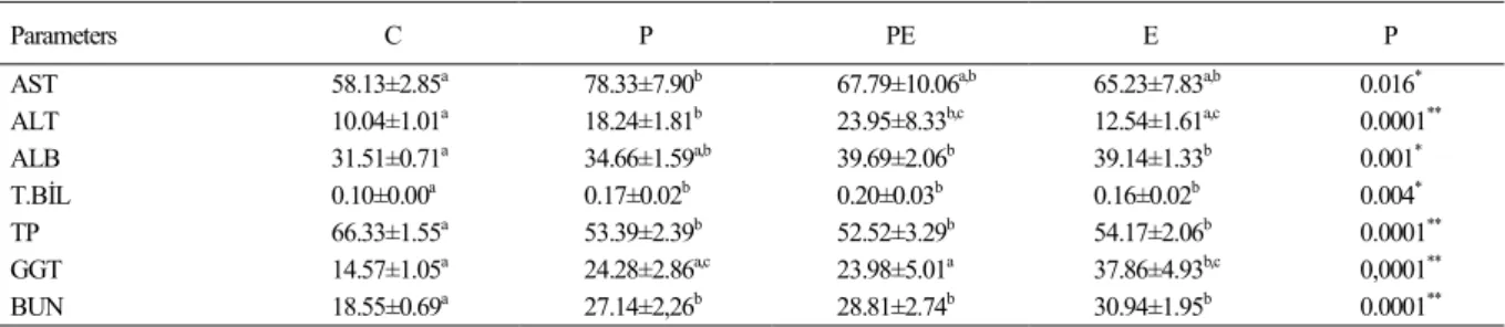
and for HCT (%) in group E (46,93±0,51) (Table 1). Regarding the biochemical findings; serum aspartate aminotransferase (AST,U/L),
alanine aminotransferase (ALT, U L-1), albumin
(ALB g dl-1), total bilirubin (T. Bil, mg dl-1), gamma
glutamyltransferase (GGT, U L-1), blood urea
nitrogen (BUN, mg dl-1) concentrations in P, PE and
E groups were determined to be higher than Group
C (P<0.05) while Total Protein (TP) concentration
was higher in the group C than in the other groups. AST concentration was determined to be the highest
in group P (78.33±7.90); ALT, ALB and T.BİL
concentrations were seen to be the highest in group
PE (23.95±8.33; 39.69±2.06; 0.20±0.03) while the
highest GGT and BUN concentrations were detected
in group E (37.86±4.93; 30.94±1.95) (Table 2). In our study, statistically significant (P<0.0001) variations were identified in Serum Amyloid-A (SAA, mg/L) concentration between
the groups. Whereas the lowest SAA concentration
was ascertained in group C (17.64±2.50), the highest level was in group E (205.65±22.70) (Table 3). As being statistically significant (P<0.0001), the lowest serum tumor necrosis factor-α (TNF-α) concentration was measured in group C (0.10±0.01) while the highest was in group P (0.31±0.03) (Table 3). Statistically (P<0.0001), Serum Interleukin-1β (IL-1β) concentration was determined to be the lowest in group C (14.17±1.04) and the highest in group PE (57.26±4.40) (Table 3). Serum Interleukin-6 (IL-6) concentration (p<0.0001) was identified as the lowest in group C (5.44±0.82) and as the highest in group E (22.14±2.82) (Table 3).
DISCUSSION
In the presented study, mean body
temperatures (oC) were determined, as statistically
significant (P<0.0001), the lowest in the control group (C) and the highest in enteritis group (E).
As a conducted study reported that rectal body temperatures were about 39.0-39.23 in healthy
with diarrhea were elevated in relation to the severity
of the infection and inflammation (RISALDE et
al., 2011). Body temperature was reported to be elevated during bovine respiratory diseases in
calves (GÜNEŞ et al., 2013). HANZLICEK et al. (2010) ascertained a rise in fever in the experimental
pneumonia they created in calves. In the conducted study, body temperature was determined to be
statistically (P≤0.05) higher in groups P, PE and E
than in the control group.
In our study, on haematological
examination WBC, RBC, Hemoglobin and Hematocrit concentrations were determined, as statistically significant (P<0.0001), to be higher in groups P, PE and E in comparison to group C. WBC
concentration was detected at reference ranges in
the control (7275±27.90) group. In the calves with experimental pneumonia, high WBC, RBC, Hct and Hb values were detected; those rises were reported to be related to infection (HANZLICEK et al.,
2010). As those values were detected to be higher
in group P than in the control group in our study
as well, we suggest that this rise may be infection related. We detected highest WBC concentration in
group E (16187±81.04). Depending on the fact that
RBC, Hb and Hct values were higher in groups E and PE than in the control group in our study, it was
concluded that this situation could be associated to
excessive fluid loss.
In groups P, PE and E; the concentrations
of serum AST, ALT, ALB, T.Bil, GGT and BUN
were statistically (P<0.05) higher than in group C. Concentration of Serum TP was higher in group C
than in the other groups. The highest concentrations
detected were AST in group P (78,33±7,90), ALT, ALB and T.BİL in group PE (23.95±8.33;
39.69±2.06; 0.20±0.03) , GGT and BUN in group
E (37.86±4.93; 30.94±1.95). In our study, although
concentrations of serum AST, ALT, ALB, T. Bil,
BUN were higher in P, PE and E groups than in group C, they remained at reference ranges (BRUN-HANSEN et al., 2006; MERCK MANUAL, 2013).
The concentration of GGT in group E presented above the reference ranges. We suggest that this resulted is related tothe negative impact of diarrhea on the gastrointestinal system and the liver.
Statistically significant (P<0.0001)
variations between the groups were determined for
the concentration of Serum Amyloid-A (SAA, mg
L-1). The concentration of SAA was detected to be Table 1 - In Control (C), Pneumonia (P), Pneumoenteritis (PE) and Enteritis (E) groups; Leukocyte (WBC µL-1), Erythrocyte (RBC/x106 µL-1), Hemoglobin
(HB g dl-1), Hematocrit (HCT/%) concentrations and Mean Body Temperatures (oC) (Mean ±StdDev).
Parameters C P PE E P
WBC 7275±27.90a 16045±87.16b,c 13337±60.38b 16187±81.04c 0.0001**
RBC 7.48±0.25a,c 8.75±0.23b,d 9.720±0.29b 8.33±0.30c,d 0.0001**
HB 11.27±0.36a 14.81±1.43b 13.66±0.58b 14.59±0.37b 0.0001**
HCT 28.46±1.03a 37.60±1.86b 45.68±1.49c 46.93±0.51c 0.0001** 0C 38.12±0.12a 40.21±0.13b 40.14±0.14b 40.27±0.18b 0,0001** *P<0.05, **P<0.0001. The variations between the groups were indicated with letters (a,b,c,d).
Table 2 - In Control (C), Pneumonia (P), Pneumoenteritis (PE) and Enteritis (E) groups; AST (U L-1), ALT (U L-1), ALB (g dl-1), T.BİL (mg dl-1), TP (g dl-1), GGT (U L-1) and BUN (mg dl-1) concentrations (Mean ±StdDev).
Parameters C P PE E P
AST 58.13±2.85a 78.33±7.90b 67.79±10.06a,b 65.23±7.83a,b 0.016*
ALT 10.04±1.01a 18.24±1.81b 23.95±8.33b,c 12.54±1.61a,c 0.0001**
ALB 31.51±0.71a 34.66±1.59a,b 39.69±2.06b 39.14±1.33b 0.001*
T.BİL 0.10±0.00a 0.17±0.02b 0.20±0.03b 0.16±0.02b 0.004*
TP 66.33±1.55a 53.39±2.39b 52.52±3.29b 54.17±2.06b 0.0001**
GGT 14.57±1.05a 24.28±2.86a,c 23.98±5.01a 37.86±4.93b,c 0,0001**
the lowest in group C (17.64±2.50) as it was the highest in group E (205.65±22.70). It was reported
that in calves which experimentally pneumonia was created via bovin respiratory syncytial virus (BRSV),
the concentration of SAA increased between the day 4 and 8 together with pathological sounds detected
in lung auscultation (HEEGAARD et al., 2000).
On the other hand, some studies noted that the concentrations of SAA and haptoglobin remained at normal ranges during experimentally created
bacterial and aseptic infections (ALSEMGEEST et
al., 1994; NAKAGAWA et al., 1997).
In the study of NIKUNEN et al. (2007) on 84 calves with bovine respiratory disease (BRD),
trachea bronchial lavage samples were examined for bacteria and the serum samples were tested for antibodies against the virus. In the same study, the calves were clinically diagnosed with diarrhea, high fever and an increase in respiratory rate; WBC value presented no variations in calves with viral and bacterial pneumoenteritis whereas the concentration of SAA proved to be high in pneumonia and pneumoenteritis cases based on Pasteurella
(NIKUNEN et al., 2007). Several researchers have reported that SAA is the first acute phase protein
increase in calves with respiratory system diseases
(ANGEN et al., 2009; ORRO et al., 2011). In our
study, the concentration of SAA was determined to be higher in the calves clinically diagnosed with pneumonia, pneumoenteritis and enteritis than in the control group. We consider that the reason for this
is inflammation is the same case that in the studies
mentioned above.
BURCIAGA-ROBLES et al. (2010)
performed experimental BVDV, mannheimia
hemaloytica (mh) and BVDV+mh applications on
calves, thus created pneumonia, pneumoenteritis and enteritis in calves. Then, in all groups experimental infection was created, the concentrations of serum
TNF- α, IL-1β and IL-6 were found to be higher than in the control group. In our study, the lowest (P<0.0001)
concentrations of serum TNF-α, IL-1β and IL-6 were
determined in group C, as well. The highest Serum
TNF- α concentration was found in group P, the highest serum IL-1β concentration was seen in group PE, and
serum IL-6 concentration was the highest in group E. Some researchers indicated that on the 4th day the
concentration of serum IL-6 increased in the calves which had experimental bovine respiratory syncytial
virus (BRSV) was performed while the concentration
of serum IL-6 remained the same in these animals during the reinfection occurring on day 98th (GRELL
et al., 2005). In another study on calves, experimental pneumonia and pneuomoenteritis was created and monitored for 14 days resulting in no change in
the concentration of serum IL-1β (RISALDE et al., 2011). In our study, proinflammatory cytokines (TNFα, IL-1β ve IL-6) were detected to be higher in
the calves with clinical pneumonia, pneumoenteritis and enteritis than in the control group. We suggest
that the existing inflammation in the studied group is
the cause for this.
Regarding the results obtained from
the presented study, it has been identified that
haematological and biochemical parameters accompanied clinical symptoms of the elevation in serum SAA concentration in calves with pneumonia, pneumoenteritis and enteritis. SAA was detected at very low concentrations in the healthy animals
in the control group. In the light of these findings
we acknowledge that routine measurements of serum SAA concentration from the moment the calves are diagnosed with pneumonia, enteritis and pneumoenteritis in veterinary medicine until the
actual cause is determined (bacteria, virus, parasites,
etc.), we also believe that more studies on the clinical use of SAA are needed.
ACKNOWLEDGEMENT
The research was supported in part by a grant (project
number;11.VF.04) from Afyon Kocatepe University.
Table 3 - In Control (C), Pneumonia (P), Pneumoenteritis (PE) and Enteritis (E) groups; SAA (mg L-1), TNF-α (ng ml-1), IL-1 (pg ml-1) and IL-6 (pg ml-1) concentrations (Mean ±StdDev).
Parameters C P PE E P
SAA 17.64±2.50a 112.19±10.55b 136.89±16.35b,c 205.65±22.70c 0.0001**
TNF-α 0.10±0.01a 0.31±0.03b 0.28±0.02b 0.29±0.05b 0.0001** IL-1 β 14.17±1.04a 40.83±3.75b 57.26±4.40b 55.60±7.91b 0.0001**
BIOETHICS AND BIOSSECURITY COMMITTEE APPROVAL
We authors of the article entitled “The detection of serum amyloid-A concentration in the calf clinically diagnosed with pneumonia, enteritis and pneumoenteritis” declared, for all due purposes, that the project that gave rise to the present data has not been submitted for evaluation of the Ethics Committee of
the “Afyon Kocatepe University”, but we are aware of Brazilian
content resolutions of the National Council for Control of Animal
Experimentation - CONCEA <http://www.mct.gov.br/index.php/
content/view/310553.html> if it involves animals.
Thus, the authors assume full responsibility for the presented data and are available for possible questions, if they would be required by the competent authorities.
REFERENCES
ALSEMGEEST, S.P.M. et al. Concentrations of SAA (SAA) and haptoglobin (Hp) as parameters of inflammatory diseases in cattle.
Veterinary Quarterly, v.16, p.21-23, 1994. Available from: <http:// www.ncbi.nlm.nih.gov/pubmed/8009814>. Accessed: nov. 20, 2012. ANGEN, Ø. et al. Respiratory disease in calves: Microbiological
investigations on trans-tracheally aspirated bronchoalveolar fluid
and acute phase protein response. Veterinary Microbiology, v.137,
p.165-171, 2009. Available from: <http://www.sciencedirect.com/ science/article/pii/S037811350800610X>. Accessed: May 28, 2009. doi: 10.1016/j.vetmic.2008.12.024.
BRUN-HANSEN, H.C. et al. Hematologic values in
calves during the first 6 months of life. Veterinary Clinical Pathology, v.35, n.2, p.182-187, 2006. Available
from:
<http://onlinelibrary.wiley.com/doi/10.1111/j.1939-165X.2006.tb00111.x/abstract>. Accessed: Mar. 5, 2008. doi: 10.1111/j.1939-165X.2006.tb00111.x.
BURCIAGA-ROBLES, L.O. et al. Effects of exposure to calves persistently infected with bovine viral diarrhea virus type 1b and subsequent infection with mannheima haemolytica on clinical signs and immune variables: model for bovine respiratory disease via viral and bacterial interaction. Journal Animal Science,
v.88, n.6, p.2166-2178, 2010. Available from: <https://www.
animalsciencepublications.org/publications/jas/articles/88/6/2166>. Accessed: December 4, 2014. doi: 10.2527/jas.2009-2005. CECILIANI, F. et al. Acute phase proteins in ruminants. Journal of Proteomics, v.75, p.4207-4231, 2012. Available from: <http://
www.sciencedirect.com/science/article/pii/S1874391912002102>. Accessed: Jul. 19, 2012. doi: 10.1016/j.jprot.2012.04.004. DONOVAN, G.A. et al. Calf and disease factors affecting growth
in female Holstein calves in Florida, USA. Preventive Veterinary Medicine, v.33, p.1-10, 1998.
ECKERSALL, P.D.; BELL, R. Acute phase proteins: Biomarkers of infection and inflammation in veterinary medicine. Veterinary Journal, v.185, p.23-27, 2010. Available from: <http://www.
sciencedirect.com/science/article/pii/S1090023310001176>. Accessed: July 2010. doi: 10.1016/j.tvjl.2010.04.009.
GRELL, S.N. et al. Marked induction of IL-6, haptoglobin and IFN gamma following experimental BRSV infection in young calves. Veteterinary Immunology and Immunopathology, v.103, n.3-4, p.235-245, 2005.
GRUYS, E. et al. Diagnostic significance of the major acute phase
proteins in veterinary clinical chemistry. A Revue Veterinary Bull, v.64, p.1009-1018, 1994. Available from: <http://www.ncbi.
nlm.nih.gov/pmc/articles/PMC1390650/>. Accessed: Oct. 28, 2005. doi: 10.1631/jzus.2005.B1045.
GULLIKSEN, S. et al. Calf mortality in Norwe-gian dairy herds. Journal of Dairy Science, v.92, p.2782-2795, 2009.
Available from: <http://www.sciencedirect.com/science/article/ pii/S0022030209705958>. Accessed: June 2009. doi: 10.3168/ jds.2008-1807.
GÜNEŞ, V. et al. Neonatal buzağilarin solunum sistemi hastalikları.Türkiye klinikleri. Journal of Veterinary Science, v.4, n.1, p.86-94, 2013.
HANZLICEK, G.A. et al. Serial evaluation of physiologic,
pathological, and behavioral changes related to disease progression of experimentally induced Mannheimia haemolytica pneumonia in postweaned calves. American Journal of Veterinary Research, v.71, p.359-369, 2010.
HEEGAARD, P.M.H. et al. The acute phase response of haptoglobin and serum amyloid A (SAA) in cattle undergoing
experimental infection with bovine respiratory syncytial virus.
Veterinary Immunology and Immunopathoogy, v.77,
p.151-159, 2000. Available from: <http: www.sciencedirect.com/science/
article/pii/S0165242700002269>. Accessed: Nov. 23, 2000. doi:
10.1016/S0165-2427(00)00226-9.
HORADAGODA, A. et al. Purification and quantitative
measurement of bovine SAA. Research in Veterinary Science, v.55, p.317-325, 1993.
MERCK MANUAL. Serum biochemical reference ranges, 2013.
Available from: <http://www.merckmanuals.com/vet/appendixes/
reference_guides/serum_biochemical_reference_ranges.html>. Online. Accessed: Nov. 2013.
MURATA, H. et al. Current research on acute phase proteins in
veterinary diagnosis: an overview. Veterinary Journal, v.168,
p.28-40, 2004. Available from: <http://www.sciencedirect.com/
science/article/pii/S1090023303001199>. Accessed: July 2004.
doi: 10.1016/S1090-0233(03)00119-9.
NAKAGAWA, H. et al. Detection of serum haptoglobin by enzyme-linked immunosorbent assay in cows with fatty liver.
Research in Veterinary Science, v.62, p.137-141, 1997.
Available from: <http://www.sciencedirect.com/science/article/
pii/S0034528897901351>. Accessed: March–April 1997. doi:
10.1016/S0034-5288(97)90135-1.
NIKUNEN, S. et al. Association of bovine respiratory disease with clinical status and acute phase proteins in calves. Comperative Immunology Microbiology Infectious Diseases, v.30, n.3,
p.143-151, 2007. Available from: <http://www.sciencedirect.com/ science/article/pii/S0147957106001044?np=y>. Accessed: May
2007. doi: 10.1016/j.cimid.2006.11.004.
ORRO, T. et al. Acute phase protein changes in calves during an outbreak of respiratory disease caused by bovine respiratory syncytial virus. Comperative Immunology Microbiology Infectious Diseases, v.34, p.23-29, 2011.
Available from: <http://www.sciencedirect.com/science/
PETERSEN, H.H. et al. Application of acute phase
proteinmeasurements in veterinary clinical chemistry. Veterinary Research, v.35, p.163-187, 2004. Available from: <http://www.
vetres.org/articles/vetres/pdf/2004/02/V4202.pdf>. Accessed: Nov. 18, 2003 doi: 10.1051/vetres:2004002.
PICCIONE, G. et al. Monitoring of physiological and blood
parameters during perinatal and neonatal period in calves.
Arquivo Brasileiro de Medicina Veterinaria e Zootecnia,
v.62, n.1, p.1-12, 2010. Available from: <http://www.scielo.br/ pdf/abmvz/v62n1/v62n1a01.pdf>. Accessed: Nov. 6, 2009 doi:
10.1590/S0102-09352010000100001.
RISALDE, M.A. et al. Response of proinflammatory and anti-inflammatory cytokines in calves with subclinical bovine viral
diarrhea challenged with bovine herpesvirus-1. Veterinary Immunology Immunopathology, v.144, p.135-143, 2011.
Available from: <http://www.sciencedirect.com/science/article/pii/
S0165242711002935>. Accessed: Nov. 15, 2011 doi: 10.1016/j. vetimm.2011.07.022.
STANTON, A.L. et al. The effect of respiratory disease and a
preventative antibiotic treatmenton growth, survival, age at first
calving, and milk production of dairyheifers. Journal of Dairy Science, v.95, p.4950-4960, 2012. Available from: <http://www.
sciencedirect.com/science/article/pii/S0022030212005097>. Accessed: Sept. 2012 doi: 10.3168/jds.2011-5067
VAN DONKERSGOED, J. et al. Epidemiological study of
enzootic pneumonia in dairy calves in Saskatchewan. Canaian Journal of Veterinary Research, v.57, p.247-254, 1993. VIRTALA, A.M. et al. The effect of calf-hood diseases on growth
of female dairy calves during the first 3months of life in New York
State. Journal of Dairy Science, v.79, p.1040-1049, 1996. WALTNER-TOEWS, D. et al. Dairy calf manage-ment morbidity
and mortality in Ontario Holstein herds. I: the data. Preventive Veterinary Medicine, v.4, p.103-124,1986.
WINDEYER, M.C. et al. Factors associated with morbidity, mortality, and growth of dairy heifer calves up to 3 months of age. Preventive Veterinary Medicine, v.113, p.231-240, 2014.
Available from: <http://www.sciencedirect.com/science/article/
Título fonte 8. 1
ISSN 1678-4596 http://dx.doi.org/10.1590/0103-8478crerr20150571
ERRATUM
Artigo “Detection of serum amyloid-A concentration in the calf clinically diagnosed with pneumonia, enteritis and pneumoenteritis” publicado no fascículo v46n2 de fevereiro da Ciência Rural páginas 293-299, onde se lia:
“A detecção de amilóide-A concentração sérica na panturrilha com diagnóstico clínico de pneumonia, enterite e pneumoenterites”
leia-se:
“A detecção de amilóide-A concentração sérica em bezerros com diagnóstico clínico de pneumonia, enterite e pneumoenterites”
Para a versão correta, acesse: