The aim of this study was to quantitatively evaluate the amount of apically extruded debris by single-file reciprocating instruments with different working lengths and apical preparation sizes. Eighty human single-rooted mandibular incisors were used and conventional access cavities were prepared. Then, the specimens were divided into four groups (n=20), according to root canal instrumentation: Reciproc size 25, .08 taper and Reciproc size 40, .06 taper instruments were used at the foramen; Reciproc size 25, .08 taper and Reciproc size 40, .06 taper instruments were used 1 mm short of the foramen. Distilled water was used as an irrigant and the apically extruded debris were collected in pre-weighted glass vials and dried afterwards. The mean weight of debris was weighed with a microbalance and statistically analyzed using one-way analysis of variance and the post hoc Tukey multiple comparison test (p<0.05). The results showed that all experimental groups were associated with debris extrusion. No significant difference was found in the amount of apically extruded debris among all experimental groups (p>0.05). The present study demonstrated that the working length and the apical preparation size did not have a significant effect on debris extrusion when performing single-file reciprocating instrumentation.
Influence of Apical Preparation Size and
Working Length on Debris Extrusion
Emmanuel João Nogueira Leal Silva1,João M. Teixeira2, Nancy Kudsi2, Luciana M. Sassone2, Renato L. Krebs2, Tauby S. Coutinho-Filho2
1Department of Endodontics, Dental
School, UNIGRANRIO - Universidade Grande Rio, Rio de Janeiro, RJ, Brazil 2Department of Endodontics,
Dental School, UERJ – Universidade Estadual do Rio de Janeiro, Rio de Janeiro, RJ, Brazil
Correspondence: Dr. Emmanuel João Nogueira Leal da Silva, Rua Erotides de Oliveira, 61/902, Icaraí, 24230-230 Niterói, RJ, Brasil. Tel: +55-21-98357-5757. e-mail: [email protected]
Key Words: debris, extrusion, foraminal instrumentation, apical preparation, Reciproc.
Introduction
During root canal treatment, aggressive materials and its products, such as dentin chips, microorganisms and their by-products, remaining pulp tissue and irrigants usually extrude into the periradicular tissues. The apical extrusion can be considered an undesirable side effect of the shaping procedures since it may induce inflammation, postoperative pain and delay of periapical healing (1). All currently used root canal preparation techniques and instruments are, at least to some degree, associated with extrusion of debris (2-5); however, it is worthwhile observing that less dentinal debris extrusion has been associated with the use of motor-driven rotary instruments (6).
The recent introduction on the market of single-file reciprocating systems has raised new perspectives for root canal preparation and, to some extent, there are reports showing that they have outperformed conventional continuous rotary nickel-titanium (NiTi) preparation in some aspects. The reciprocating motion relieves the stress on the instrument by special counter-clockwise (cutting action) and clockwise (release of the instrument) movements and, therefore, increases its resistance to fatigue in comparison to the continuous rotation motion (7,8).
In cases of apical periodontitis, recognizing the presence of microorganisms in the apical portion of the canal has contributed to the concept of larger apical preparations and even debridement of the apical foramen (9-11). In fact, these procedures may overcome the potential limitation of irrigation procedures in the apical area, optimizing root canal disinfection (12). However, one main concern while
performing larger apical preparation and/or foraminal debridement is the possibility of more debris, bacteria and irrigants extrusion. Up to now, there are no studies on whether foraminal instrumentation with single-file reciprocating technique provides different results in terms of debris extrusion. Therefore, the purpose of the present study was to evaluate the influence of instrumentation with different WL and different apical preparation sizes on apically extruded debris. The null hypotheses tested were (i) larger apical preparation sizes do not influence debris extrusion and (ii) the foraminal instrumentation does not extrude more debris than instrumentation 1 mm short of the foramen.
Material and Methods
Sample SelectionThis study was revised and approved by the local Ethics Committee. A sample of 80 human mandibular incisors with a single canal and similar root length were collected. Soft tissue remnants and calculus on the external root surface were mechanically removed and disinfected with 0.5% chloramine T, stored in distilled water at 4 °C and used within 6 months after extraction. The selected teeth presented complete root formation and root canal curvature <10°, which was determined based on the angle of curvature starting at the coronal aspect of the apical third of the root using Schneider’s method (13). After acquisition of digital buccolingual and mesiodistal radiographs, the angle of curvature of each specimen was measured using an image-analysis program (AxioVision 4.5; Carl Zeiss Vision,
ISSN 0103-6440
Brazilian Dental Journal (2016) 27(1): 28-31
Braz Dent J 27(1) 2016
29
Preparation size, WL and debris extrusion
Hallbergmoos, Germany). Specimens with initial apical size equivalent to a size 15 K-file (DentsplyMaillefer, Baillaigues, Switzerland) with buccolingual diameter four or more times larger than the mesiodistal diameter (flat oval canals) were selected for the study (n=80). The 80 teeth were matched according to their shapes and dimensions, based on visual examination of the radiographs.
Endodontic access cavities were cut in the lingual surfaces with a diamond bur and a high-speed dental turbine with water-cooling. The working length was determined using a size 15 K-file (DentsplyMaillefer) introduced in all teeth until it was visible at the apical foramen under an operating microscope at a 20× magnification (DFVasconcelos Ltda, Valença, RJ, Brazil). This measurement at foramen determined the root canal length of all groups, in the foraminal instrumentation groups (R25/0 and R40/0), the working length applied was the same of total root canal length. In the other experimental groups (R25/-1 and R40/-1), the root canal length was determined and reduced 1 mm from this measure, in order to instrument 1 mm shorter than the apical foramen.
Root Canal Preparation
The groups were randomly distributed using a computer algorithm (http://www.random.org) to one of the four experimental groups (n=20) as follows:
Group R25/0: Reciproc size 25, .08 taper (VDW, Munich, Germany) instrument used at the foramen - A R25 Reciproc instrument (VDW) was gradually advanced in the root canal until reaching 2/3 of the WL (established at the foramen). The R25 instrument was moved in a slow and gentle in-and-out pecking motion with a 3 mm amplitude. After each set of three complete pecking movements, the instrument was removed from the canal, and its flutes were cleaned. Canal patency was checked with a size10 K-file (DentsplyMaillefer) before using the R25 instrument and after each three complete pecking movements. Root canal instrumentation was complete when the R25 instrument reached the WL.
Group 25/-1: Reciproc size 25, .08 taper (VDW) instrument used 1 mm short of the foramen - The R25 instruments (VDW) were used as in group R25/0, except that WL was established 1 mm short of the foramen.
Group R40/0: Reciproc size 40, .06 taper (VDW) instrument used at the foramen - The R40 instruments (VDW) were used as in group R25/0.
Group 40/-1: Reciproc size 40, .06 taper (VDW) instrument used 1 mm short of the foramen - The R40 instruments (VDW) were used as in group R25/-1.
For all groups, a stainless steel K-file (VDW) was inserted into the canal up to WL. Thus, a glide path with a size 10 K-file (DentsplyMaillefer) was created to assure smooth preliminary preparation, rendering the canal predictably
negotiable. Instruments were used with a VDW Silver motor according to the manufacturer´s instructions. A total volume of 12 mL distilled water was used for each root canal as an endodontic irrigant. The irrigants were delivered by 5-mL disposable plastic syringes (Ultradent Products Inc., South Jordan, UT, USA) with a 31-gauge stainless steel needle Endo-Eze (Ultradent) inserted into the canal as far as possible and retracted 2 mm before the application of irrigation. Aspiration was performed using SurgiTips tips (Ultradent) attached to a high-speed suction pump. Irrigation and aspiration were performed in exactly the same manner for all specimens. Between sets of pecking movements and patency checking, the canals were irrigated with 3 mL of distilled water. At the end of instrumentation, each tooth was flushed with more 3 mL of irrigant. The smear layer was then removed with 3 mL 17% EDTA for 3 min. A total of 3 mL distilled water was then used for 3 min as a final rinse.
Debris Collection
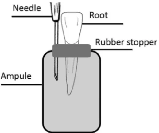
The method used for collection of apically extruded debris was adapted from previous studies (4,5). A 10-mL ampule with a rubber stopper was adjusted for this experiment. Using a heated instrument, a hole was punched through the center of every rubber stopper in which the root was adapted. A 30-G needle was inserted into the rubber stopper to balance internal and external pressures, allowing debris extrusion. Each collection assembly was then individually pre-weighed three times with a 10-5-g precision analytic microbalance (Model 1101; ElbaTech, Isola d’Elba, Italy). The mean weight of each assembly was used as baseline value. All plastic assay tubes were covered with black tape to blind the operator regarding the production of debris during root canal instrumentation. Teeth were instrumented into the collection assembly (Fig. 1).
After instrumentation, the collection assembly was placed in a dry heat oven at 140 °C for 5 h, allowing
Braz Dent J 27(1) 2016
30
E.J.N.L. Silva et al.
evaporation of the irrigant. Three consecutive weight measurements were then taken for each collection assembly, with the mean value recorded. The weight of the extruded debris was determined by subtracting the weight of the pre-weighed collection assembly from the final weight of the collection assembly.
Statistical Analysis
Since the preliminary analysis of the raw pooled data revealed a bell-shaped distribution (D’Agostino-Pearson omnibus normality test), statistical analysis was performed using parametric methods (one-way ANOVA). Post hoc pair-wise comparisons were performed using Tukey’s multiple-comparison test. The alpha-type error was set at 0.05.
Results
The median, minimum, and maximum values and the standard deviation data of each experimental group are shown in Figure 2. No significant difference was found in the debris extrusion among all experimental groups (p>0.05).
Discussion
It is known that apical extrusion of debris may impact treatment outcome and postoperative symptoms. The first results of this study revealed no significant difference in the amount of apically extruded debris comparing different apical preparation sizes (p>0.05). Therefore, the first null hypothesis was accepted. Previous studies, using debris extrusion methodology, also demonstrated no correlation between extrusion and apical diameter (2,14,15). Moreover, a recent study using a bacteria-specific extrusion methodology also demonstrated no significant difference in the amount of apically extruded bacteria in the comparison of different apical preparation sizes performed with Reciproc system (16); however, to the best of the authors’ knowledge, there are no data on apical
debris extrusion using reciprocating single-file systems and different apical preparation sizes. The rationale behind the comparison of different apical preparations sizes is the current trend to promote larger apical preparations with the purpose of optimizing root canal disinfection (17-19) and thus rendering better conditions for tissue repair (9,11). The second results of this study demonstrated no significant differences in debris extrusion between foraminal instrumentation and instrumentation 1 mm short of the foramen (p>0.05). Therefore, the second null hypothesis was also accepted. Some previous studies reported different results from the present study, demonstrating that when instrumentation was performed at the apical foramen, significantly more debris were forced apically than when instrumentation was 1 mm short (6,20). The differences presented herein may be explained by the differences in instrument design, the number of used files, movement kinematics and irrigation protocol. The recent bacteria-specific extrusion study used the similar instrumentation technique and irrigation protocol and also showed absence of differences between foraminal instrumentation and instrumentation 1 mm short of the foramen (16). Foraminal debridement is an important procedure, especially in cases of periapical lesion, as the apical portion of root canal is a niche for bacteria colonization. Hence, preparation up to the foramen has been suggested as the most efficient means of cleaning and disinfecting canals (10,21). It also allows better removal of infected dentin and reduces significantly the bacterial load in the canal system (17,18).
The amount of extruded debris was collected following the Myers and Montgomery method (20), but the collection apparatus was slightly modified to make it more simple, practical and affordable, as previously suggested (4,5). Moreover, this method eliminated the possibility of fingertip contamination throughout the entire experiment procedure because there was no direct contact between the operator’s fingertips and the assembly. It is worth mentioning that the amount of extruded material is extremely low, so the contact of moist or greasy fingertips may significantly alter the weight of extruded debris (4).
It is important to emphasize that the use of extracted teeth has its limitations. The results obtained from this study may be different if applied in a clinical situation, because the in vitro setup had the apex suspended in the
air without any physical back pressure, whereas in vivo,
the apex is surrounded by granulomatous or periradicular tissues that may limit the apical extrusion (22). The use of floral foam to simulate the resistance of periapical tissues has already been suggested (23); however, this approach may absorb irrigant solution and debris. Therefore, no attempt was made to simulate periapical resistance in the present study. The implications of a vital or necrotic pulp
Braz Dent J 27(1) 2016
31
Preparation size, WL and debris extrusion
and the presence of a lesion of endodontic origin in the apical extrusion is also not clear.
The use of single and flat-oval rooted mandibular incisors was intended to produce a reliable and comparable anatomical baseline. Extrusion of debris in mandibular incisors has not been largely studied. Despite the high anatomical variability regarding shape, size and dimensions in the natural morphology of teeth, several attempts were made to ensure better comparison of the 4 experimental groups. Special care was taken to obtain groups that were as balanced as possible in terms of anatomical features, such as root length, angle and radius of root curvature. The standardization of the size of apical foramen is another important issue. Only teeth with compatible foramen to size 15-K files under magnification were selected. Moreover, as previously suggested, matching of teeth was applied when the groups were formed, equalizing levels of challenge and boosting the statistical power of the study (4).
Apical debris extrusion may be an important issue in the outcome of endodontic treatment. Besides bacterial major role in flare-ups (1), mechanical and chemical factors could also be responsible for undesired consequences, such as induction of inflammation and postoperative pain (24). The extrusion of contaminated debris, endodontic materials, or chemically altered tissue proteins, may disrupt the balance of periapical periodontitis, with potential to initiate an acute periapical reaction (1).
It may be concluded that the WL and the apical preparation size did not have significant effect on the debris extrusion when using reciprocating instruments.
Resumo
Este estudo avaliou quantitativamente a quantidade de material extruído apicalmente pela instrumentação com lima única reciprocante, aplicando diferentes comprimentos de trabalho e tamanhos de preparo apical. Para a análise de extrusão foram utilizados oitenta incisivos inferiores unirradiculares humanos. Cavidades de acesso convencionais foram preparadas e os espécimes foram divididos em quatro grupos (n=20), de acordo com o tipo de instrumentação do canal: Reciproc 25, 0.08 e Reciproc 40, 0.06 foram utilizadas na instrumentação até o forame; Reciproc tamanho 25, 0.08 e Reciproc 40, 0,06 foram utilizadas na instrumentação até 1 mm aquém do forame. Água destilada foi empregada como irrigante e o material extruído apicalmente foi coletado em frascos de vidro já pesados e posteriormente submetidos a secagem. O peso médio de detritos foi avaliado com uma microbalança de precisão e os dados submetidos a ANOVA e teste Tukey (p<0,05). Todos os grupos experimentais foram associados à extrusão de debris. Nenhuma diferença significativa foi encontrada na quantidade de material extruído apicalmente entre os grupos (p>0,05). Este estudo demonstrou que o comprimento de trabalho e o tamanho do preparo apical não geraram efeito significativo sobre a extrusão de debris durante a instrumentação reciprocante.
References
1. Seltzer S, Naidorf IJ. Flare-ups in endodontics: I. Etiological factors. J Endod 1985;11:472-478.
2. Al-Omari MA, Dummer PM. Canal blockage and debris extrusion with eight preparation techniques. J Endod 1995;21:154-158.
3. De-Deus G, Barino B, Marins J, Magalhaes K, Thuanne E, Kfir A. Self-adjusting file cleaning-shaping-irrigation system optimizes the filling of oval-shaped canals with thermoplasticized gutta-percha. J Endod 2012; 34:71-75.
4. De-Deus GA, Silva EJNL, Moreira EJ, Neves AA, Belladonna FG, Tameirão M. Assessment of apically extruded debris produced by the Self-Adjusting File system. J Endod 2014;40:526–529.
5. Silva EJ, Sá L, Belladonna FG, Neves AA, Accorsi-Mendonça, Vieira VT, et al.. Reciprocating versus rotary systems for root filling removal: assessment of the apically extruded material. J Endod 2014;40:2077-2080. 6. Beeson TJ, Hartwell GR, Thornton JD, Gunsolley JC. Comparison of debris
extruded apically in straight canals: conventional filing versus profile .04 Taper series 29. J Endod 1998;24:18–22.
7. Yared G. Canal preparation using only one Ni-Ti rotatory instrument: preliminary observations. Int Endod J 2008;41:339-344.
8. Varela-Patiño P, Ibañez-Párraga A, Rivas-Mundiña B, et al. Alternating versus continuous rotation: a comparative study of the effect on instrument life. J Endod 2010;36:157-159.
9. Souza-Filho FJ, Benatti O, Almeida OP. Influence of the enlargement of the apical foramen in periapical repair of contaminated teeth of dog. Oral Surg Oral Med Oral Pathol 1987;64:480-484.
10. Fornari VJ, Silva-Sousa YT, Vanni JR, Pecora JD, Versiani MA, Sousa-Neto MD. Histological evaluation of the effectiveness of increased apical enlargement for cleaning the apical third of curved canals. Int Endod J 2010;43:988-994.
11. Aminoshariae A, Kulild JC. Master apical file size - smaller or larger: a systematic review of healing outcomes. Int Endod J 2015;48:639047. 12. Card SJ, Sigurdsson A, Ostavik D, Trope M. The effectiveness of increased
apical enlargement in reducing intracanal bacteria. J Endod 2002;28:779-783.
13. Schneider SW. A comparison of canal preparations in straight and curved root canals. Oral Surg Oral Med Oral Path Oral Radiol Endod 1971;2:271-275.
14. Hinrichs RE, Walker WA III, Schindler WG. A comparison of amounts of apically extruded debris using handpiece-driven nickel-titanium instrument systems. J Endod 1998;24:102–106.
15. Lambrianidis T, Tosounidou E, Tzoanopoulou M. The effect of maintaining apical patency on periapical extrusion. J Endod 2001;27:696–698. 16. Teixeira JM, Cunha FM, Jesus RO, Silva EJ, Fidel SR, Sassone LM. Influence
of working length and apical preparation size on apical bacterial extrusion during reciprocating instrumentation. Int Endod J 2015;48:648-653.
17. Coldero LG, McHugh S, MacKenzie D, Saunders WP. Reduction in intracanal bacteria during root canal preparation with and without apical enlargement. Int Endod J 2002;35:437–446.
18. Rollison S, Barnett F, Stevens RH. Efficacy of bacterial removal from instrumented root canals in vitro related to instrumentation technique and size. Oral Surg Oral Med Oral Pathol Oral Radiol Endod 2002;94:366– 371.
19. Aminoshariae A, Kulild JC. Master apical file size – smaller or larger: a systematic review of microbial reduction. Int Endod J 2015 [Epub ahead of print].
20. Myers GL, Montgomery S. A comparison of weights of debris extruded apically by conventional filling and Canal Master techniques. J Endod 1991;17:275–279.
21. Albrecht LJ, Baumgartner JC, Marshal JG. Evaluation of apical debris removal using various sizes and tapers of ProFile GT files. J Endod 2004;30:425–428.
22. Salzgeber RM, Brilliant JD. An in vivo evaluation of the penetration of an irrigating solution in root canals. J Endod 1977;3:394-398.
23. Altundasar E, Nagas E, Uyanik O, Serper A. Debris and irrigant extrusion potential of 2 rotary systems and irrigation needles. Oral Surg Oral Med Oral Pathol Oral Radiol Endod 2011;112:e31-5.
24. Sipavičiūtė E, Manelienė R. Pain and flare-up after endodontic treatment procedures. Stomatologija 2014;16:25-30.