Mestre em Genética Molecular e Biomedicina
Identification and characterization of CteG,
a novel Chlamydia trachomatis
type III secretion effector protein
Dissertação para obtenção do Grau de Doutor em
Biociências Moleculares
Orientador: Luís Jaime Mota, Professor Auxiliar, Faculdade de
Ciências e Tecnologia, Universidade NOVA de Lisboa
Júri:
Presidente: Doutora Maria João de Reis Madeira Crispim Romão Arguentes: Doutora Sandra Manuela Rodrigues Sousa Cabanes
Doutora Isaura Isabel Gonçalves Simões
Vogais: Doutor Luís Manuel Valla Teixeira
Doutora Isabel Maria Godinho de Sá Nogueira Doutor Luís Jaime Gomes Ferreira da Silva Mota
Identification and characterization of CteG,
a novel Chlamydia trachomatis
type III secretion effector protein
Dissertação para obtenção do Grau de Doutor em
Biociências Moleculares
Copyright© by Sara Raquel Vilela Pais, FCT/UNL and UNL.
A Faculdade de Ciências e Tecnologia e a Universidade Nova de Lisboa têm o direito, perpétuo e sem limites geográficos, de arquivar e publicar esta dissertação através de exemplares impressos reproduzidos em papel ou de forma digital, ou por qualquer outro meio conhecido ou que venha a ser inventado, e de a divulgar através de repositórios científicos e de admitir a sua cópia e distribuição com objetivos educacionais ou de investigação, não comerciais, desde que seja dado crédito ao autor e editor.
First, I would like to thank my supervisor Luís Jaime Mota for mentoring me since I first arrived in the laboratory to do my Master’s thesis with almost no laboratory experience. His constant guidance, support and availability and all the fruitful discussions and critics helped me become the researcher that I am today. I will always be grateful.
Also, I would like to acknowledge Isabel Sá Nogueira and Adriano Henriques for taking part on my thesis committee, and our collaborators Vítor Borges and João Paulo Gomes from Instituto Nacional de Saúde Pública Dr. Ricardo Jorge and Charlotte Key and Derek Fisher from Southern Illinois University. I thank the MolBioS PhD Program for accepting me and to Fundação para a Ciência e Tecnologia for financial support.
To all my colleagues of the Infection Biology Lab, Joana Bugalhão, Irina Franco, Filipe Almeida, Nuno Charro, Maria Cunha, Lia Domingues, Catarina Milho, Inês Pereira, Beatriz Costa and Maria Luís, a big thank you. Your companionship and all the helpful advices and discussions made this PhD thesis a more enriching experience. Besides, all the laughs shared inside and out of the laboratory made these PhD years more pleasant and enjoyable. Special thanks to Joana Bugalhão, who was a constant presence. Thanks for all the enthusiastic discussions and for the friendship. Also, thanks to Catarina Milho, Beatriz Costa, Ana Luzia and Inês Pereira for contributing to the work presented here in this thesis. Further, I thank my colleagues with whom I share the “Aquário”, Raquel Portela, Inês Grilo, Bárbara Gonçalves and Cynthia Barroco for the all the relaxing breaks and chats.
I also would like to thank Carolina Cassona and Diana Espadinha, my resilient companions from the MolBioS PhD Program, for all the shared experiences and support.
Finally, I thank my parents and my brother for their constant love and patience and to Ana, Inês and Patrícia for all the support and for all the adventures.
This work was supported by Fundação para a Ciência e a Tecnologia (FCT) through grants PTDC/IMI-MIC/1300/2014 and PTDC/BIA-MIC/28503/2017, and by Unidade de Ciências Biomoleculares Aplicadas (UCIBIO), which is financed by national funds from FCT (UID/Multi/04378/2013) and co-financed by the European Regional Development Fund (ERDF) under the PT2020 Partnership Agreement (POCI-01-0145-FEDER-007728). SVP was supported by PhD fellowship PD/BD/52210/2013 within the scope of the PhD program Molecular Biosciences (PD/00133/2012) funded by FCT.
Chlamydia trachomatis é uma bactéria patogénica, intracelular obrigatória, que em células
eucarióticas reside num vacúolo membranar. Durante o seu ciclo de desenvolvimento, esta bactéria usa um sistema de secreção do tipo III, que permite o transporte de proteínas efetoras para o citoplasma da célula hospedeira. Estas proteínas efetoras modificam vários processos e organelos das células hospedeiras, permitindo que as bactérias sobrevivam no ambiente intracelular.
Para identificar novas proteínas efetoras de Chlamydia, foram infetadas células de mamífero com estirpes de C. trachomatis que expressavam proteínas candidatas com um duplo péptido de hemaglutinina na extremidade C-terminal. Análises por microscopia de imunofluorescência revelaram que a proteína CT105/CteG era transportada para a célula hospedeira infetada. Observou-se que CT105/CteG associava-se inicialmente ao complexo de Golgi e que com a progressão da infeção, acumulava-se na membrana plasmática. Mostrou-se que esta mudança da localização de CT105/CteG é independente da integridade de filamentos de actina e microtúbulos. Além disso, quando diferentes regiões de CT105/CteG (656 resíduos de aminoácidos) foram expressas ectopicamente em células de mamífero, os primeiros 100 resíduos de CT105/CteG foram identificados como contendo uma região que direcionava a proteína para o Golgi. Apesar de uma estirpe de C. trachomatis com ct105/cteG inativado não ter apresentado um defeito no crescimento intracelular da bactéria, a expressão de CT105/CteG em Saccharomyces cerevisiae causou um defeito no transporte de proteínas para o vacúolo. Isto sugere que a função de CT105/CteG seja alterar o transporte vesicular em células eucarióticas. Por fim, a análise de várias estirpes de C. trachomatis sugeriu que o ct105/cteG é apenas transcrito em estirpes causadoras de linfogranuloma venéreo, indicando uma função de CT105/CteG neste contexto.
Em conclusão, neste trabalho foi identificada e caracterizada a primeira proteína efetora de C.
trachomatis que se associa com o complexo de Golgi, ajudando a uma melhor compreensão das
interações bactéria-hospedeiro.
Termos chave: Interações bactéria-hospedeiro; Chlamydia trachomatis; Sistema de secreção do tipo
Chlamydia trachomatis is an obligate intracellular bacterial pathogen that resides in eukaryotic
host cells within a membrane-bounded vacuole, termed inclusion. Throughout its characteristic biphasic developmental cycle, the bacterium uses a type III secretion system, which allows the direct delivery of effector proteins into the host cell cytoplasm. These effector proteins are thought to modify a variety of host cell processes enabling the bacteria to survive and multiply within the intracellular environment.
To identify novel type III secretion effectors of Chlamydia, mammalian host cells were infected by
C. trachomatis strains independently expressing previously identified candidate effectors with a double
hemagglutinin tag at their C-termini. Immunofluorescence microscopy analysis revealed that CT105/CteG (Chlamydia trachomatis effector associated with the Golgi) was delivered into infected host cells, where initially (~ 16 to 20 h post-infection) associated with the Golgi complex and as the developmental cycle progressed (~30 to 40 h post-infection) accumulated at the plasma membrane. The change of subcellular localization of CT105/CteG in the host cell during infection was shown to be independent of intact actin filaments and microtubules. Furthermore, when ectopically expressing different regions of CT105/CteG (656 amino acid residues) in mammalian cells, the first 100 residues of the protein were identified as containing a Golgi-targeting region. Although a C. trachomatis ct105/cteG mutant did not display a defect in intracellular growth, CT105/CteG induced a vacuolar protein sorting defect when expressed in Saccharomyces cerevisiae. This suggested that CteG could function in modifying eukaryotic vesicular transport. Lastly, ct105/cteG was mostly transcribed in C. trachomatis strains causing lymphogranuloma venereum, indicating that CT105/CteG might contribute to the characteristic tropism of these strains.
In conclusion, we have identified and characterized CT105/CteG, the first chlamydial effector found to associate with the Golgi complex, thus expanding the portfolio of known chlamydial effectors and increasing the understanding of host-pathogen interactions.
Keywords: Host-pathogen interactions, Chlamydia trachomatis, Type III secretion, Golgi, membrane
Page Acknowledgements ...…..………... I Funding ………...…… III Resumo ………... V Abstract …...……… VII Table of contents ………...……… IX Figure index ………..………. XIII Table index ..………..………. XV List of abbreviations ……...………...……… XVII
1 INTRODUCTION ……..………... 1
1.1 Discovery of the Chlamydia pathogen ……… 3
1.2 Taxonomy of Chlamydia …...………... 3
1.2.1 The Chlamydiae ………..………...………. 3
1.2.2 The Chlamydiaceae …….……… 4
1.3 The human pathogen Chlamydia trachomatis ……….……..………... 6
1.3.1 C. trachomatis strains ...……….……….…… 6
1.3.2 Medical importance and public health ………...……… 6
1.3.3 Diagnostic, treatment and vaccine ..………..…… 7
1.4 C. trachomatis genetics ………..……….……… 8
1.4.1 C. trachomatis genome ...…………..………...………… 8
1.4.2 The virulence plasmid ………..……… 8
1.4.3 Genetic manipulation of Chlamydia ……….……….. 9
1.4.3.1 Transformation of Chlamydia and development of stable shuttle vectors …... 10
1.4.3.2 Mutagenesis ...………...……….. 11
1.4.3.2.1 Random mutagenesis ...………..………..……… 11
1.4.3.2.2 Site-directed mutagenesis ...…..……….……….… 11
1.5 Chlamydia trachomatis pathogenesis ..………...………..… 12
1.5.1 The type III secretion system of Chlamydia trachomatis ………..…… 14
1.5.1.1 Chlamydial T3S effectors ...………...………… 16
1.5.1.1.1 Inc (inclusion membrane) proteins .……….……… 17
1.5.1.1.2 Non-Inc T3S effector proteins ……….……….……… 17
1.5.1.2 Chlamydial T3S chaperones …..……….……….. 19
1.5.1.3 Chlamydial T3S-independent effectors ……… 20
1.5.2 Modulation of host cells by C. trachomatis ….……….………. 21
1.5.2.3 Subversion of host cell trafficking pathways and nutrient acquisition ……….. 21
1.5.2.4 Golgi fragmentation ……….………... 23
1.5.2.5 Host cell exit ………... 24
1.6 General aims ………... 25
2 MATERIALS AND METHODS ………..……….. 27
2.1 Plasmids, oligonucleotides, and DNA manipulation …..……….. 29
2.2 Cells lines and transient transfection ……….………….……….….. 29
2.3 E. coli and Y. enterocolitica growth conditions …………..……….... 30
2.4 Maintenance and manipulation of C. trachomatis strains ………..…….. 30
2.4.1 Infection of HeLa 229 cells with C. trachomatis ……….………….. 30
2.4.2 Transformation of C. trachomatis ……….………. 31
2.4.3 Clonal isolation of C. trachomatis by plaque assay purification ………. 32
2.4.4 Construction of a C. trachomatis ct105::aadA mutant strain ……….…. 32
2.4.5 Quantification of infectious progeny ………..……… 33
2.5 Y. enterocolitica T3S assays ……….………….. 33
2.6 Yeast strains, invertase assays and toxicity analysis ………..…………. 34
2.7 Antibodies and fluorescent dyes ……….……… 34
2.8 Drug treatments ……….………... 35
2.9 Immunoblotting ………. 35
2.10 Fluorescence microscopy ……….. 36
2.11 Real-time quantitative PCR (RT-qPCR) ……….. 36
3 RESULTS………... 37
3.1 Identification of candidate type iii secretion effectors of Chlamydia trachomatis ………. 39
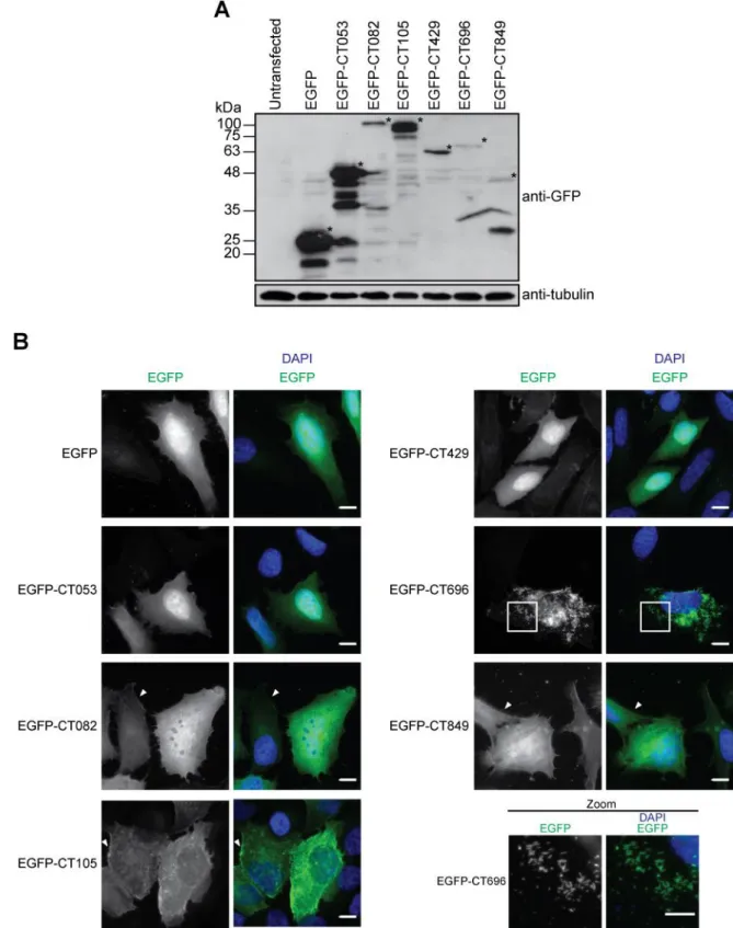
3.1.1 Ectopic expression of candidate T3S effectors of C. trachomatis in mammalian cells ……….. 39
3.1.2 Expression of epitope-tagged candidate chlamydial T3S effectors in C. trachomatis ………...……….. 41
3.1.2.1 Construction of C. trachomatis transformation vectors ………..……… 41
3.1.2.2 Construction of strains with plasmid-encoded candidate C. trachomatis T3S effectors ………. 44
3.1.3 Assessment of the delivery of candidate chlamydial T3S effectors by C. trachomatis into the host cell cytoplasm ………...……….. 45
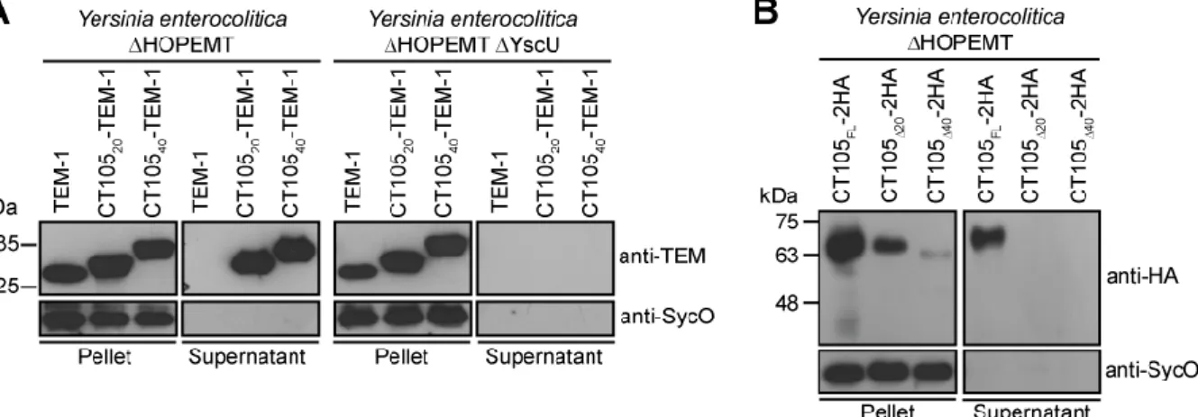
3.1.4 The first 20 amino acids of CT105 contain a T3S signal ……….……… 47 3.1.5 Full-length orthologues of CT105 are only present in C. muridarum and C. suis
venereum (LGV) strains ………..………... 51
3.2 The C. trachomatis effector CT105 associates with the Golgi complex and plasma membrane of infected host cells ………...………. 53
3.2.1 Characterization of the C. trachomatis strain producing plasmid-encoded CT105-2HA ………..……….. 53
3.2.2 Analysis of the delivery of CT105-2HA into host cells during C. trachomatis developmental cycle ……….. 56
3.2.3 CT105-2HA localizes at the Golgi complex in C. trachomatis-infected cells …….. 59
3.2.4 CT105-2HA localizes at the host cell plasma membrane in C. trachomatis- infected cells ………... 61
3.2.5 Bacterially-delivered CT105-2HA changes its main localization in the host cell cytoplasm as the C. trachomatis developmental cycle progresses .……… 62
3.2.6 The change in the main localization of CT105-2HA during infection from the Golgi to the plasma membrane is independent of intact host microfilaments and microtubules ………..…………. 64
3.2.7 The localization of bacterially-delivered CT105-2HA at the host cell plasma membrane is independent of the timing of the protein expression during the C. trachomatis developmental cycle ………... 66
3.2.8 The first 100 amino acid residues of CT105 are sufficient to target the protein to the Golgi in uninfected mammalian cells ……….………... 66
3.2.9 CT105 appears in filamentous structures within host cells ………..…. 73
3.3 The Chlamydia trachomatis effector CT105 can interfere with vesicular trafficking in eukaryotic cells ………...…. 75
3.3.1 CT105 is not essential for intracellular replication of C. trachomatis in infected tissue culture cells ………. 75
3.3.2 CT105 induces a vacuolar protein sorting defect when ectopically expressed in S. cerevisiae ………... 78
4 DISCUSSION AND CONCLUSIONS ………... 81
4.1 Mechanism of delivery of CT105 ………... 83
4.2 CT105 is modified within C. trachomatis ……….….. 84
4.3 CT105 has a dual localization within host cells during C. trachomatis infection ………... 85
4.4 CT105 disrupts vesicular trafficking in eukaryotic cells ………..……. 86
4.5 CT105 is likely relevant for the tropism of LGV strains ……….………... 87
4.6 Outlook on other candidate T3S effectors of C. trachomatis ……….…………. 88
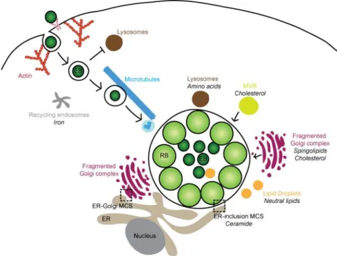
4.7 Hypothetic mode of action for CT105 during C. trachomatis infection ………...… 88
4.8 Future work ……….….. 89
REFERENCES ………..……… 93 ANNEXES ………..……… 111
Page Figure 1.1 Phylogenetic 16S rRNA tree showing relationships among members of the
phylum Chlamydiae ……….……….. 4
Figure 1.2 Phylogenetic reconstruction of the Chlamydia genus ………...…… 5
Figure 1.3 Transmission electron micrograph of different C. trachomatis morphologic forms ………...………. 13
Figure 1.4 Schematic representation of the C. trachomatis developmental cycle ... 14
Figure 1.5 The T3SS of C. trachomatis ………...………... 15
Figure 1.6 Chlamydia-host interactions.……….. …….. 24
Figure 3.1 Localization of candidate T3S effectors of C. trachomatis when ectopically expressed in mammalian cells ...……….…. 40
Figure 3.2 C. trachomatis transformation vectors ……….……... 42
Figure 3.3 Construction of C. trachomatis L2/434-derived strains expressing candidate chlamydial T3S effectors ...……….. …. 45
Figure 3.4 Analysis of the delivery of the candidate T3S effectors by C. trachomatis L2/434 into the host cell ...……….. …….….………….……… 46
Figure 3.5 The first 20 amino acids of CT105 are sufficient and necessary to drive T3S by Y. enterocolitica ………..………...… 48
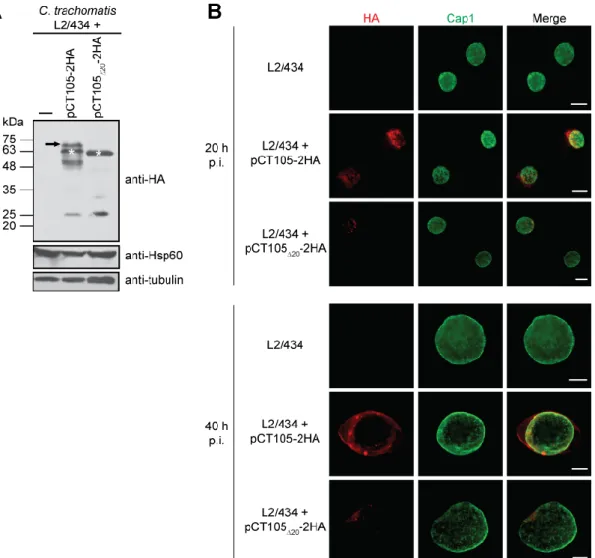
Figure 3.6 The first 20 amino acids of CT105 are necessary for the delivery of the protein into the host cell by C. trachomatis L2/434 ………..……….…. 49
Figure 3.7 mRNA levels of ct105 in different C. trachomatis strains …... 51
Figure 3.8 Putative promoter region of ctl0360/ct105 in different C. trachomatis strains 52
Figure 3.9 Characterization of C. trachomatis L2/434 strain harboring pCT105-2HA …. 54 Figure 3.10 Kinetics of expression of CT105-2HA during infection by C. trachomatis L2/434 55 Figure 3.11 CT105-2HA is modified in C. trachomatis L2/434 ……….. 56
Figure 3.12 C. trachomatis L2/434 produces CT105-2HA early during infection ………... 57
Figure 3.13 The subcellular localization of bacterially-delivered CT105-2HA changes in infected cells as the C. trachomatis developmental cycle progresses ……… 58
Figure 3.14 CT105-2HA expressed under the control of the tetracycline promoter is delivered into the host cell by C. trachomatis L2/434 …………..……….. 59
Figure 3.15 CT105-2HA associates with the Golgi complex in C. trachomatis-infected cells ………..… 61 Figure 3.16 CT105-2HA localizes at the host cell plasma membrane in C.
trachomatis-infected cells ……… 62
infection ……… 63 Figure 3.18 Subcellular localization of CT105-2HA expressed under the control of the
tetracycline promoter in C. trachomatis-infected cells ………... 63 Figure 3.19 CT105-2HA changes in its subcellular localization during C. trachomatis
infection is independent of intact host microfilaments and microtubules …… 65 Figure 3.20 Bacterially-translocated localizes at the host cell plasma membrane
regarding the timing of its expression by C. trachomatis ………...…… 66
Figure 3.21 Ectopic expression of CT105 in HeLa cells ………..……….. 67
Figure 3.22 The first 100 amino acids of CT105 are target to the Golgi complex ………… 69
Figure 3.23 CT105 fusions to the N-terminus of mEGFP …..……… 70
Figure 3.24 The first 100 amino acids of CT105 associate with the trans-Golgi …...…….. 71 Figure 3.25 C. trachomatis L2/434 strains harboring plasmids encoding truncated
versions of CT105 were not detected in the host cell cytoplasm ……...……... 72 Figure 3.26 CT105 localizes in filamentous structures in non-infected and
Chlamydia-infected cells ……… 74 Figure 3.27 Verification of intron insertion in the C. trachomatis ct105::aadA mutant
strain ………. 76 Figure 3.28 A C. trachomatis ct105::aadA insertional mutant is not defective for
intracellular growth in tissue culture cells ………..……….. 77 Figure 3.29 The CT105 Chlamydia trachomatis effector protein does not affect host cell
Golgi morphology during infection ……..……….……… 78 Figure 3.30 CT105 induces a vacuolar protein sorting defect in S. cerevisiae ……… 79 Figure 3.31 Phenotypic characterization of S. cerevisiae strains expressing CT105 ...…. 80 Figure 4.1 Schematic model for CT105 mode of action during C. trachomatis infection
TABLE INDEX
Page Table 3.1 Predicted promoter regions of candidate T3S effectors of C. trachomatis …. 43 Table 3.2 Potential orthologues of C. trachomatis CT105 (CTL0360) in other
Chlamydiae ………...……….. 50
Table A.1 Plasmids used in this work ………...………. 113
Table A.2 Oligonucleotides used in this work ……….………..……… 119
LIST OF ABBREVIATIONS
2HA Double hemagglutinin
AB Aberrant body
ATCC American Type Culture Collection
BFA Brefeldin A
BHI Brain heart infusion
bla Amplicilin resistance gene
cAMP Cyclic adenosine monophosphate
Cas9 CRISPR associated protein 9
CEL I Mismatch specific endonuclease
COPII Coatomer protein II complex
CPAF Chlamydia protease-like activity factor
CPY-Inv Carboxypeptidase Y – invertase
CRISPRi Clustered regularly interspaced short palindromic repeats interference
CyaA Bordetella pertussis adenylate cyclase
DAPI 4',6-diamidino-2-phenylindole
DFA Direct immunofluorescence assays
DMEM Dulbecco’s modified eagle medium
DMSO Dimethyl sulfoxide
DNA Deoxyribonucleic acid
EB Elementary body
ECACC European Collection of Authenticated Cell Cultures
EGFP Enhanced fluorescent protein
EHEC Enterohaemorrhagic Escherichia coli
ELISA Enzyme-linked immunosorbent assays
EMS Ethyl methanesulfonate
ENU N-ethy-N-nitrosurea
EPEC Enteropathogenic Escherichia coli
ER Endoplasmic reticulum
ESCRT Endosomal sorting complexes required for transport
FBS Foetal bovine serum
FITC Fluorescein isothiocyanate
FRAEM Fluorescence-reported allelic exchange mutagenesis
FRU Fructose
GAL Galactose
GCIP Grap2 cyclin D-interacting protein
GFP Green fluorescent protein
GSK Glycogen synthase kinase
HBSS Hank’s balanced salt solution
HRP Horseradish peroxidase
Hsp60 Heat shock protein 60
IFN-γ Interferon gamma
IFU Inclusion forming units
Inc Inclusion membrane
INSA Portuguese National Institute of Health Dr. Ricardo Jorge
Inv Invertase
LGV Lymphogranuloma venereum
MCS Multiple cloning site
mEGFP Monomeric enhanced green fluorescent protein
MHC Major histocompatibility complex
MOMP Major outer membrane protein
mRNA Messenger RNA
rRNA Ribosomal RNA
MVB Multivesicular body
NAAT Nucleic acid amplification tests
NCBI National Center of Biotechnology Information
NEM N-ethylmaleimide
OD600 Optical density at 600 nm
ORF Open reading frame
ori Origin of replication
p.i. Post-infection
PBS Phosphate-buffered saline
PCR Polymerase chain reaction
PDZ Postsynaptic Density95/Disc Large/Zonula Occludens-1
PFA Paraformaldehyde
PGK1 Phosphoglycerate kinase 1
Pmps Polymorphic outer membrane proteins
Ptet Tetracycline promoter
RB Reticulate body
RNA Ribonucleic acid
rRNA Ribosomal RNA
RT-qPCR Real-time quantitative PCR
SET Su(var)3-9, Enhancer-of-zeste and Trithorax
SDS Sodium dodecyl sulphate
SDS-PAGE SDS-polyacrylamide gel electrophoresis
SNX Sorting nexins
SPG Sucrose-phosphate-glutamate buffer
SPI-1 Salmonella pathogenesis island-1
sRNA Small RNA
STI Sexually transmitted infections
T2SS Type II secretion system
T3S Type III secretion
T3SS Type III secretion system
TCA Trichloroacetic acid
TILLING Targeting-induced local lesions in genomes
TincD incD terminator
TSS Transcription start site
WHO World Health Organization
1.1 DISCOVERY OF THE CHLAMYDIA PATHOGEN
In 1907, Ludwig Halberstadter and Stanislaus von Prowazek were examining Giemsa-stained smears of conjunctival scrapings from subjects with trachoma when they identified intracytoplasmatic inclusions arranged around the nucleus like a cloak. This was the first description of the etiological agent of trachoma which was named Chlamydozoa, derived from the Greek word “khlamús”, meaning cloak (Halberstaedter and von Prowazek, 1907). Later, similar inclusions were identified in scrapings from adults with urethritis and cervicitis, from newborns with nongonococcal conjunctivitis (Lindner, 1910), and in samples obtained from patients with lymphogranuloma venereum (LGV) (Durand et al., 1913). In 1935, the agent was classified as a virus because it was able to pass through bacterial filters and unable to grow on agar or liquid media (Miyagawa et al., 1935). This was accepted until 1966, when alongside with the emergence of electron microscopy, Chlamydia was reclassified as bacterium, due to the presence of DNA, RNA, a cell wall similar to Gram-negative bacteria, and the ability to undergo binary fission (Moulder, 1966).
1.2 TAXONOMY OF CHLAMYDIA
1.2.1 The Chlamydiae
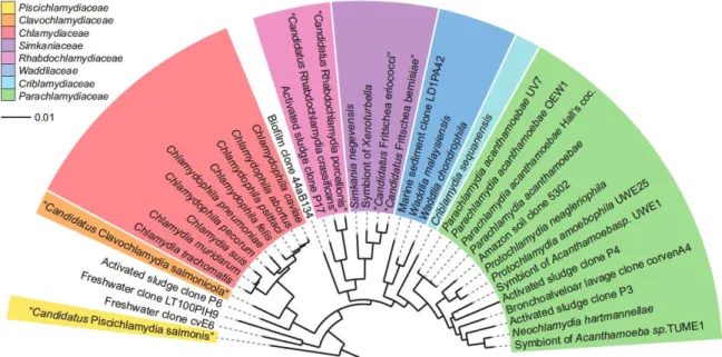
Until the 1990’s, the phylum Chlamydiae was thought to have only one genus, the Chlamydia. However, the improvement and the increase accessibility to DNA sequencing set the grounds to the discovery of additional Chlamydiae. Over the last 25 years, the diversity of the Chlamydiae increased from one to nine families: Chlamydiaceae, Parachlamydia, Waddliaceae, Simkaminiaceae,
Rhabdochlamydiaceae, Criblamydiaceae, Piscichlamydiaceae, Clavichlamydiaceae and
Parilichlamydiaceae (Figure 1.1) (Horn, 2008). Despite covering a great host range and an ubiquitous
distribution in the environment, all Chlamydiae share the obligate intracellular lifestyle within eukaryotic cells, a unique biphasic developmental cycle and more than 80% 16S ribosomal RNA (rRNA) and/or 23S rRNA sequence identity (Everett, 2000). The non-Chlamydiaceae are also referred as Chlamydia-like bacteria or environmental Chlamydia and exist as symbionts of free-living amoeba and of other eukaryotic hosts (Horn, 2008).
Figure 1.1 Phylogenetic 16S rRNA tree showing relationships among members of the phylum
Chlamydiae. Representatives of all recognized families. Bar, 10% estimated evolutionary distance. Reprinted
from Horn 2008.
1.2.2 The Chlamydiaceae
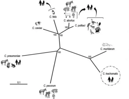
Based on the analysis of rRNA gene sequences, it was proposed the division of Chlamydiacea into two genera, Chlamydia and Chlamydiophyla (Everett et al., 1999). However, this division raised controversy, as the thresholds used were not consistent with those from other bacteria taxa, all genomes were highly similar and there were no distinct phenotypic features between the recently created genera. Therefore, most of the researchers in the field continued to use the one genera nomenclature and a reunification for the genera was proposed (Sachse et al., 2015). Although there are still doubts on the nomenclature, a single Chlamydia genus comprising all the known Chlamydiaceae species is the more accepted classification. The Chlamydiaceae family comprises 11 different species that infect a variety of hosts (Figure 1.2):
Chlamydia trachomatis is an exclusive human pathogen causing ocular and genital infections. It is the most studied and characterized Chlamydia species (see Section 1.3);
Chlamydia pneumoniae is another human pathogen and the major cause of community acquired pneumonia (Brown, 2012). It has been linked to several chronic diseases in humans such as atherosclerosis, osteoporosis, multiple sclerosis, and Alzheimer disease, although scarce data supports these associations. Besides infecting humans, C. pneumoniae infects amphibians, reptiles and other mammals (Roulis et al., 2013);
Chlamydia psittacci is the cause of avian respiratory disease and has a high impact on poultry farming and bird breeding. Transmission from birds to humans has been reported, when humans are in contact with infected birds. C. psitacci has been also isolated from other mammalian hosts, such as dogs, cats, pigs, cattle, sheep and horses (Longbottom and Coulter, 2003);
Chlamydia avium and Chlamydia gallinaceae infect pigeons and poultry, respectively. These were previously included in the C. psitacci species. (Sachse et al., 2014).
Chlamydia pecorum is a pathogen of livestock (cattle, sheep, goats and pigs) (Walker et al., 2015) and wild animals such as the koalas. In fact, C. pecorum is a major cause of the decline of the koalas in Australia (Polkinghorne et al., 2013);
Chlamydia abortus causes abortion in sheep and goat due to infection of the placenta and therefore has a significant economic impact, particularly in Europe. Although rare, transmission to humans has been reported and pregnant women exposed to infected animals are at risk of abortion (Longbottom and Coulter, 2003);
Chlamydia felis is a frequent cause of infection in cats causing conjunctivitis and pneumonitis (Cai
et al., 2002). Infection of humans is rare and does not cause severe illness however transmission
can occur to humans in close contact with cats (Hartley et al., 2001);
Chlamydia caviae is a pathogen of guinea pigs causing conjunctivitis and genital infections. These have been used as a model of sexual transmission of Chlamydia (Rank et al., 2003) and to the study of genital tract infection and disease pathology (Rank and Sanders, 1992);
Chlamydia muridarum infects mice causing pneumonitis and is widely used as a model to study genital chlamydial infections (Barron et al., 1981).
Chlamydia suis is a common pathogen of pigs. It is the only chlamydial species known to have naturally acquired genes that confer antibiotic resistance, as it contains tetracycline resistant genes (tetC islands) (Dugan et al., 2004).
Figure 1.2 Phylogenetic reconstruction of the Chlamydia genus. Chlamydiaceae species and the
respective natural hosts are shown in the figure and arrows depicted the cases of zoonotic transmission. C. suis,
C. avium and C. gallinacea are not represented due to lack of sequence data. Tree topology is based on the
accumulation of single-nucleotide polymorphisms (SNPs) on 600 orthologous genes shared among the species. Reprinted from Nunes et a.l, 2014.
1.3 THE HUMAN PATHOGEN CHLAMYDIA TRACHOMATIS
1.3.1 C. trachomatis strains
C. trachomatis is an exclusive obligate intracellular human pathogen and the most studied Chlamydia
species. C. trachomatis are conventionally divided into 15 major serovars (A to L), depending on the immunoreactivity of the C. trachomatis major outer membrane protein (MOMP), or the genotyping and sequencing of ompA (the gene encoding MOMP). Serovars A to C mainly infect ocular epithelial cells, serovars D to K mainly infect urogenital epithelial cells, and lymphogranuloma venereum (LGV) strains (serovars L1 to L3) are also sexually transmitted but are more invasive and can infect macrophages and monocytes and spread into lymph nodes (Schachter, 1999).
All sequenced genomes of C. trachomatis are similar in size (1.04-1.05 Mb), exhibit more than 99% nucleotide sequence identity and have a high degree of synteny (Stephens et al., 2009; Harris et al., 2012; Seth-Smith et al., 2013). Nevertheless, a small region in the genome (10 to 50 kb corresponding to 45-49 genes) named plasticity zone (PZ) has a high degree of genetic divergence between the different serovars and is likely involved in the different tissue tropisms (Read et al., 2000). For example, the tryptophan operon (trpRBA) and the gene encoding cytotoxin CT166 are localized in this region. The tryptophan operon is present in all C. trachomatis strains, but only in the genital strains the tryptophan synthase (encoded by trpBA) is active and able to synthesize tryptophan from indole, a compound found in the genital tract and thought to be produced by the resident microbiota. During infection, the host immune system produces interferon gamma (IFN-γ), which limits the availability of tryptophan. However, genital strains are able to produce their own tryptophan and can survive in the genital tract environment (A Shaw et al., 2000; Fehlner-Gardiner et al., 2002; Caldwell et al., 2003). Cytotoxin CT166 is a protein involved in the disassembly of actin filaments during bacterial internalization, which has been called the “cytopathic effect” (Thalmann et al., 2010). Although present (complete or truncated) in ocular and genital strains, the gene encoding CT166 is deleted in LGV strains (Carlson et al., 2004). Besides the PZ region, other genes across the genome might contribute to the variability among different C.
trachomatis serovars, such as the highly variable polymorphic outer membrane proteins (Pmps) (Gomes et al., 2006) and different secreted virulence proteins (Lutter et al., 2010; Almeida et al., 2012).
1.3.2 Medical importance and public health
C. trachomatis infections are a public health concern worldwide. Ocular strains are the major
causative agent of preventable infectious blindness (trachoma), whereas urogenital strains are the most common cause of bacterial sexually transmitted infections.
Ocular infections caused by C. trachomatis serovars A to C produce a conjunctival inflammation that can lead to the scarring of the eyelid. Repeated infections can result in severe eye scarring and ultimately cause irreversible blindness. Transmission occurs through the contact with eye or nose discharges of infected people (Taylor et al., 2014). C. trachomatis ocular infections are endemic in the poorest and rural areas of Africa, Central and South America, Asia, Australia and Middle East, mainly
due the lack of hygiene standards. C. trachomatis is responsible for visual impairment or blindness of about 1.9 million people, causing approximately 1.4% of all blindness (Taylor et al., 2014; Trachoma Fact Sheet, World Health Organization (WHO), 2018)
C. trachomatis serovars D to K infect urogenital epithelial cells leading to cervicitis in women and
urethritis in men. Infections are asymptomatic in approximately 70% of women and 50% of men. (Harryman et al., 2014). If the infections are left untreated, bacteria ascend to the upper genital tract in 15 – 40% of the cases and leads to serious sequelae among woman including pelvic inflammatory disease (PID), tubal infertility, ectopic pregnancy, and chronic pelvic pain (O’Connell and Ferone, 2016). The World Health Organization (WHO) estimated that, in 2012, 131 million new cases of C. trachomatis infections occurred among adolescents and adults (15 - 49 years old), which corresponds to a 4.2% prevalence of C. trachomatis infections worldwide (Sexually transmitted infections (STIs) Fact Sheet, World Health Organization (WHO), 2016). In Portugal, the data on C. trachomatis infections is still limit as the report of C. trachomatis infections became mandatory only in 2014. The Portuguese National Institute of Health (INSA) inquiry revealed a prevalence of 2.7% of C. trachomatis infections among the 4,866 people inquired (INSA Inquiry 2015-2016, 2016).
C. trachomatis serovars L1 to L3 cause lymphogranuloma venereum (LGV), an inguinal syndrome
normally characterized by genital ulceration and painful inguinal lymphadenopathy (Mabey and Peeling, 2002). LGV strains are endemic in Africa, Southeast Asia, South America and the Caribbean; however, in recent years, LGV has emerged in Europe and North America, mainly in men who have sex with men, as a leading cause of ulcerative proctitis (Mabey and Peeling, 2002; Martin-Iguacel et al., 2010).
1.3.3 Diagnostic, treatment and vaccine
Antibiotic therapy has been shown to be effective in the treatment of primary ocular and urogenital
C. trachomatis infections. The current guidelines of the WHO recommend the use of azithromycin and
doxycycline (Reyburn, 2010) . Nevertheless, the major concern in C. trachomatis infections is the fact that these are commonly asymptomatic and can go undiagnosed and untreated (van de Laar and Morré, 2007). Repeated infections are also common and associated with a high risk of developing serious sequelae, such as blindness due to the ocular infections and infertility due to the urogenital infections (Darville and Hiltke, 2010). Therefore, tools to diagnose C. trachomatis are of major importance. Infections can be diagnosed either by culturing, direct immunofluorescence assays (DFAs), or enzyme-linked immunosorbent assays (ELISAs). However, the method of choice have been the nucleic acid amplification tests (NAATs), as they are highly sensitive and specific in detection of C. trachomatis in medical samples (Meyer, 2016) .
Currently, there are no licensed vaccines for C. trachomatis and the development of a vaccine still has low priority on the public health agenda, as these bacterial infections cause significant morbidity but no mortality. Several evidences from animal models and human studies suggested that the vaccine is feasible and a MOMP-based vaccine is already in phase I clinical trials (Gottlieb et al., 2016; Poston et
1.4 C. TRACHOMATIS GENETICS
1.4.1 C. trachomatis genome
The first complete chlamydial genome (C. trachomatis serovar D) was published in 1998 (Stephens, 1998). However, with the rapid development of the DNA sequencing technologies, the number of sequenced C. trachomatis genomes have increased rapidly. To date, there are 85 complete C.
trachomatis genomes deposited in National Center of Biotechnology Information (NCBI) database. The
typical C. trachomatis chromosome has approximately 1 Mbp and is predicted to encode about 900 protein-coding genes. The small genome suggests that, during adaptation to the host, C. trachomatis lost a large number of genes, as Chlamydia-like and free living bacteria have larger genomes (~2Mp) (Mendonça et al., 2011). Nevertheless, C. trachomatis maintained the minimal machinery required for DNA replication, transcription and translation, for protein secretion, for basic lipid metabolism and for essential functions in aerobic respiration (Stephens, 1998; Yao, Cherian, et al., 2015). Still, C.
trachomatis relies on the host to obtain basic nutrients as some of its metabolic pathways are incomplete
(e.g., tricarboxilic acid cycle or the biosynthesis of some amino acids) (Stephens, 1998)
1.4.2 The virulence plasmid
C. trachomatis harbors a 7.5 kb cryptic plasmid, first described in 1980 (Lovett et al., 1980). The
plasmid is low copy with up to 8 copies per cell, non-conjugative, non-integrative and, in the majority of the Chlamydia species, it does not possess antibiotic resistance genes (Pickett et al., 2005; Ferreira et
al., 2013). The exception is plasmid of C. suis, which integrates and contains tetracycline resistance
genes (Dugan et al., 2004; Dugan et al., 2007).
The plasmid is highly conserved among different Chlamydia species, ranging from 68 to 99% of nucleotide identity (Thomas et al., 1997; Pickett et al., 2005), and among different C. trachomatis strains it only differs in 1% of the nucleotide sequence (Thomas et al., 1997; Ferreira et al., 2013). The chlamydial plasmid carries 8 open reading frames (ORFs), which are all transcribed and translated during the developmental cycle (Palmer and Falkow, 1986; Ricci et al., 1993; Ricci et al., 1995), and 2 small antisense RNAs (sRNAs) (Ricci et al., 1993; Albrecht et al., 2009). The nomenclature is quite diverse among different species. pORF1 designates the open reading frame (ORF) immediately downstream from the origin of replication of the plasmid. However, in C. trachomatis this corresponds to Pgp7. This will be the nomenclature used below.
The Pgp1, -2, -6 proteins and the DNA sequence within pgp8, but not the Pgp8 protein, are required for plasmid maintenance (Gong et al., 2013; Song et al., 2013; Y Liu et al., 2014). Pgp1 is a predicted DnaB-like helicase and Pgp8 is a putative integrase/recombinase. Pgp2 and Pgp6 are proteins of unknown function (Ricci et al., 1995; Thomas et al., 1997). The pgp8 DNA coding sequence comprises a small antisense RNA (sRNA-2), which may play a role in plasmid maintenance although other features of the pgp8 DNA coding sequence might contribute to this function (Gong et al., 2013).
Pgp4 is a transcriptional regulator of virulence-associated chlamydial genes. Deletion of pgp4 results in reduction of gene expression of pgp3 (~ 4-fold) and of chromosomal genes that include glgA, a gene associated with accumulation of glycogen within the inclusion lumen (~ 8-fold), and a few genes encoding secreted proteins (~2- to 23-fold) (Song et al., 2013). In addition, pgp4 was shown to be required to particular egress mechanism (lytic exit) of Chlamydia from host cells (Chunfu Yang et al., 2015).
Pgp5 is a negative regulator of the genes regulated by Pgp4, thus leading to the hypothesis that the chlamydial plasmid might aid in the modulation of chlamydial gene expression in response to environmental cues from the host, microbiome and co-infection (Y Liu et al., 2014).
Pgp3 protein is associated with the chlamydial outer membrane (Comanducci et al., 1993) and is secreted into the inclusion lumen and host cell cytosol (Z Li et al., 2008). It has been demonstrated to be one of the most immunodominant antigens in humans infected by C. trachomatis (Wang et al., 2010). Different activities have been reported for this plasmid-encoded protein. Pgp3 binds and neutralizes the anti-microbial peptide LL-37 (Hou et al., 2015) and activates macrophages to produce inflammatory cytokines (Z Li et al., 2008). In addition, the Pgp3/LL-37 complex is able to induce neutrophils to secrete inflammatory cytokines (Hou et al., 2018) In mice, Pgp3 promotes C. muridarum survival in the lower genital tract and bacterial ascension to the upper genital tract (Yuanjun Liu et al., 2014). Overall, this indicates that Pgp3 is a major virulence factor with an important role in Chlamydia pathogenesis.
sRNA-2 (antisense to pgp8) is the most abundant of all the plasmid transcripts (Ferreira et al., 2013) and may play a role in plasmid maintenance through the regulation of pgp8, as described above (Gong
et al., 2013). sRNA-7 (antisense to pgp5) is not important to plasmid maintenance (Gong et al., 2013)
nor to negative transcriptional regulation associated with pgp5 (Y Liu et al., 2014), and its function is unknown.
Plasmid-deficient C. trachomatis strains are rare but have already been detected in patient isolates. These plasmidless strains are associated with impaired glycogen accumulation within the inclusion (Matsumoto et al., 1998; O’Connell and Nicks, 2006) and have attenuated virulence in mice (O’Connell and Nicks, 2006; Sigar et al., 2014). This suggests that the plasmid contributes to Chlamydia pathogenicity in vivo.
1.4.3 Genetic manipulation of Chlamydia
The study of the biology, pathogenesis and transmission of C. trachomatis has been hindered by the lack of tools for its genetic manipulation. The major constraints have been related with its obligate intracellular lifestyle, its characteristic biphasic developmental cycle, and the lack of evidence of recent acquisition of foreign DNA. However, in the last 10 years, methods for genetic manipulation broadly used in other bacteria have been successfully adapted to C. trachomatis. Although these procedures still need optimization, C. trachomatis is no longer genetically intractable.
1.4.3.1 Transformation of Chlamydia and development of stable shuttle vectors
In 1994, Tam and colleagues described for the first time the transformation of C. trachomatis (Tam
et al., 1994). The DNA used consisted in a C. trachomatis–Escherichia coli shuttle plasmid carrying a
chloramphenicol acetyltransferase (cat) cassette under the control of a Chlamydia native promoter. However, the transformation revealed to be transient and the plasmid was lost after 4 passages/rounds of infection. Recent data showed that the transformed plasmid lacked a region within pgp1 that is essential for plasmid maintenance.
In 2009, Binet and Maurelli designed a suicide plasmid containing a synthetic rRNA with single mutations that conferred resistance to kasugamycin and spectinomycin. By transforming C. psittaci with this plasmid using electroporation, a mutant was successfully obtained by means of allelic exchange (Binet and Maurelli, 2009). However, the methodology was not followed-up, probably due to the extremely low efficiencies of transformation and to the fact that C. psittaci requires high levels of containment.
In 2011, a landmark study by the laboratory of Ian Clarke was published describing a method for stable transformation of C. trachomatis (Wang et al., 2011). A shuttle vector comprising the native chlamydial plasmid fused to an E. coli cloning vector was constructed to allow replication in both bacteria, and a -lactamase gene was also added for selection of the transformants. The reasoning for the choice of this selection marker was that in the presence of -lactams C. trachomatis enters a persistent state characterized by the arrest of the development cycle, in which bacteria are maintained in an aberrant non-infectious form. Therefore, the introduction of this plasmid in C. trachomatis would allow the transformed bacteria to avoid this persistent state in the presence of penicillin and continue the infection cycle. Another advantage of this marker is the fact that aberrant (non-transformed) and non-aberrant (transformed) bacteria have a distinct morphology that can be easily distinguished using conventional light microscopy. To introduce the recombinant plasmid in C. trachomatis, the classic method of E. coli chemical transformation was adapted. The plasmid DNA and elementary bodies (infectious form of the bacteria) were incubated in a CaCl2 buffer followed by infection of epithelial cells
and selection using penicillin during several rounds of infection. During this process, the native plasmid is replaced by the recombinant plasmid due to plasmid incompatibility and transformants can be stably maintained over numerous passages, even in the absence of the selection marker.
The development of the procedure described by the laboratory of Ian Clarke laid the basis for genetic manipulation of C. trachomatis. The shuttle vector used has been the backbone to second generation recombinant plasmids, which allow expression of a variety of fluorescent proteins (Agaisse and Derré, 2013; Campbell et al., 2014), native or conditional control of gene expression (Agaisse and Derré, 2013; Wickstrum et al., 2013; Bauler and Hackstadt, 2014), and to monitor protein delivery into the cytoplasm of infected host cells using different reporter epitope tags or enzymes (FLAG, Cya, GSK, TEM-1, split-GFP) (Bauler and Hackstadt, 2014; Mueller and Fields, 2015; Wang et al., 2018).
1.4.3.2 Mutagenesis
1.4.3.2.1 Random mutagenesis
The development of the techniques to random mutagenize C. trachomatis were also a big step in the study of the biology of this bacterium. Kari and colleagues described the construction of an isogenic C.
trachomatis mutant strain (Kari et al., 2011) by a method derived from targeting-induced local lesions
in genomes (TILLING), a plant genetics tool which combines chemical mutagenesis with a sensitive DNA screen to identify single nucleotide changes in a target gene. Low-levels of the mutagenizing agent ethyl methanesulfonate (EMS) were used to generate C. trachomatis strains with one mutation per genome. These mutants were expanded in subpopulations and screened for specific mutations by denaturing and reannealing PCR amplicons of the target gene followed digestion of the amplicons with a mismatch specific endonuclease (CEL I). Furthermore, subpopulations carrying the mutations in the target gene were sequenced. If a desired mutation was present, bacteria were isolated and further analyzed.
Subsequently, Nguyen coupled chemical mutagenesis with genome sequencing and DNA exchange to identify mutations responsible for characteristic phenotypes, in a forward genetics approach (Nguyen and Valdivia, 2012). C. trachomatis were again treated with the mutagenizing agent EMS but in higher concentrations than those used by Kari and colleagues, thus generating 3 to 20 mutations per genome. By plaque assay, clonal populations were isolated, and the morphology of the plaques were analyzed. Clones sharing the same plaque morphology were sequenced and searched for common mutations. Afterwards, mutants of interest were segregated by lateral gene transfer and homologous recombination. This method was further updated by Kokes (Kokes et al., 2015) using as mutagenizing agent either EMS or N-ethy-N-nitrosurea (ENU) to create a collection of 934 mutant strains. The screen was performed by both plaque morphology and whole genome sequencing analysis.
1.4.3.2.2 Site-directed mutagenesis
Although the random mutagenesis was a great addition to the molecular genetics toolbox of
Chlamydia, methods to specifically target the chlamydial chromosome were not yet available. In 2013,
Johnson and Fisher adapted the TargeTron system (at the time marketed by Sigma Aldrich) to perform site-specific mutagenesis of C. trachomatis. This system is based on a mobile group II intron that can be retargeted by the alteration of DNA sequences within the intron and thus allowing site-specific gene insertions. The system was modified with the insertion of a chlamydial native promoter to allow the expression of the intron insertion machinery in C. trachomatis. A -lactamase gene (bla) was added as a marker to select for the mutated strains. Using this system, it was possible to insertionally inactivate the gene encoding IncA, a Chlamydia protein that inserts in the membrane of the inclusion (Johnson and Fisher, 2013). Later, a re-modification of the system also allowed the use of spectinomycin as a selection marker (Lowden et al., 2015). The success of this method has been proved, as it was used to mutate an antagonist of the chlamydial anti-sigma factor (Thompson et al., 2015), other inclusion
membrane proteins (Weber, Nicholas F. Noriea, et al., 2016; Sixt et al., 2017; Weber et al., 2017; Almeida et al., 2018), and chlamydial GroEL chaperonins (Illingworth et al., 2017).
In 2016, Mueller et al. described a system for fluorescence-reported allelic exchange mutagenesis (FRAEM) (Mueller et al., 2016). The vector used consisted in a plasmid with pgp6 (a gene required for plasmid maintenance) under the control of the tetracycline promoter, thus allowing a controlled removal of the plasmid upon mutagenesis. In addition, the plasmid contained a cassette encoding both -lactamase and the green fluorescent protein (GFP) flanked by chlamydial DNA corresponding to approximately 3 kb of the genomic sequence upstream and downstream from the gene targeted for mutagenesis. This allowed allelic exchange of the gene of interest by the bla/gfp cassette. Stable mutations of trpA, tmeA/ct694/ctl0063, tmeB/ct695/ctl0064, and ct696/ctl0065 were successfully achieved, confirming the functionality of the FRAEM system.
More recently, a system using clustered regularly interspaced short palindromic repeats interference (CRISPRi) based on an inducible catalytically inactive CRISPR associated protein 9 (Cas9) allowed the conditional knockdown of the incA gene. Nevertheless, the system still requires improvements in the stability of the plasmid, minimization of off-target effects and leaky expression to be considered attainable for mutagenesis of C. trachomatis (Ouellette, 2018).
1.5 CHLAMYDIA TRACHOMATIS PATHOGENESIS
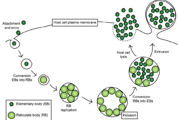
C. trachomatis, like all Chlamydiae, are obligate intracellular bacteria that have a distinctive biphasic
developmental cycle comprising the interconversion between two distinctive morphological forms, the elementary body (EB) and the reticulate body (RB) (Abdelrahman and Belland, 2005) (Figure 1.3). EBs are the infectious, non-replicative and extracellular form of the bacteria. They can temporally survive in the extracellular environment between infections, due to a highly crosslinked cell wall that confers resistance to osmotic and physical stress. These are the smaller form of the bacteria (~ 0.3 µm in diameter) and have a highly compact nucleoid. Although previously thought as being metabolically inactive, recent studies suggest that their metabolic and biosynthetic activity is high and depends on D-glucose as a source of energy (Omsland et al., 2014). The RBs (~ 1 µm in diameter) are the non-infectious but replicative form of the bacteria. They are more fragile than EBs due the lack of a cross-linked outer membrane. They have a more relaxed chromatin and are metabolically active as they are involved in nutrient acquisition and replication (Hackstadt et al., 1985; Barry et al., 1992; Abdelrahman and Belland, 2005; Omsland et al., 2014). C. trachomatis RBs can have an enlarged and aberrant morphology, designate as aberrant bodies (ABs). These are viable but non-infectious bacteria that result from Chlamydia entering in a state of persistence, where metabolism is slowed down and RB to EB conversion is hampered. This state is reversible and has been shown to be induced by several factors such as cytokines (IFN-), nutrient starvation and antibiotics (e.g., -lactams) (Beatty et al., 1994; Hogan
Figure 1.3 Transmission electron micrograph of different C. trachomatis morphologic forms. EB, elementary body. RB, reticulate body. IB, intermediate body, a conversion intermediate between EBs and RBs. Magnification x160,000. Reprinted form Philips et al. 1984
The developmental cycle of C. trachomatis is completed in 36 to 96 h (depending on the strains, e.g., for C. trachomatis LGV strains the cycle is about 48 h) (Miyairi et al., 2006). It comprises an entry phase, where bacteria attach to the extracellular side of the host cell plasma membrane and are subsequently internalized in a membrane-bound vacuole, normally designated as inclusion. Then, the inclusion migrates to the perinuclear region of the host cell (Clausen et al., 1997) and avoids fusion with lysosomes, as it mostly devoid of known markers of the endocytic pathway (Heinzen et al., 1996). EBs differentiate into RBs thus starting bacterial replication, which is supported by the acquisition of nutrients (lipids, amino acids and iron) from the host cell. Chlamydial replication has been presumed to occur by binary fission, however recent studies have reported different observations where one supports the hypothesis of replication by polarized cell division (Abdelrahman et al., 2016) and the other by binary fission (Lee et al., 2018). Those studies have been performed using different microscopy tecniques, and thus the mechanism of Chlamydia replication will require further clarification. Chlamydial replication is accompanied by the expansion of the inclusion and, later, RBs asynchronously convert into EBs. This process has been suggested to be regulated by the size of the RBs (Lee et al., 2018). Finally, EBs are released either by host cell lysis or extrusion of the inclusion and can undergo another round of infection on neighboring cells (Abdelrahman and Belland, 2005).
Throughout the developmental cycle, Chlamydia modulates host cellular functions (section 1.5.2) and one of the mechanisms used is the type III secretion system (T3SS; see Section 1.5.1), a mechanism of bacterial protein transport, that is likely crucial to the growth and survival of the bacteria within the host cell.
Figure 1.4 Schematic representation of the C. trachomatis developmental cycle. C. trachomatis
infection starts with the attachment of the infectious elementary bodies (EBs) to the surface of epithelial cells. After entry, bacteria become enclosed within a membrane-bound vacuole, named inclusion, where EBs differentiate in non-infectious reticulate bodies (RBs). Later in the cycle, RBs differentiate back into EBs and are then released,
either by host cell lysis or extrusion, to undergo another round of infection.
1.5.1 The type III secretion system of Chlamydia trachomatis
Protein secretion is an essential process for prokaryotic cells (and for all cells), allowing the transport of proteins to membrane compartments or to the extracellular space, whether this is the environment, another bacterium or a eukaryotic cell. To cross to the extracellular space, proteins from Gram-negative bacteria encounter a barrier composed by at least two phospholipid membranes and a peptidoglycan layer (Costa et al., 2015). Therefore, it is not surprising that these bacteria have evolved a variety of mechanisms of protein secretion, specific to their needs. The type III, type IV and type VI secretion systems (T3SS, T4SS, T6SS, respectively) allow the direct delivery of proteins and DNA, in a one-step process, through large multiprotein complexes, functioning as nanomachines, spanning both inner and outer bacterial membrane. Although these nanomachines appear similar and serve the same purpose, they have different evolutionary origins and functions. For instance, proteins of the T3SSs are evolutionary related to those of the bacterial flagellum and secrete proteins capable of manipulating eukaryotic host cells. The proteins of the T4SSs are evolutionary related to those of bacterial conjugation systems possessing a pilus-like structure. T4SS allow the transport of proteins, but its main physiological role is the transport of plasmid DNA by conjugation. Lastly, the proteins of T6SSs are homologous to those of phage tails. In fact, while T6SSs also transport proteins into eukaryotic cells, their most common function is the injection of proteins into other bacteria. They are important to bacterial communication/competition and interaction with the environment (Costa et al., 2015).
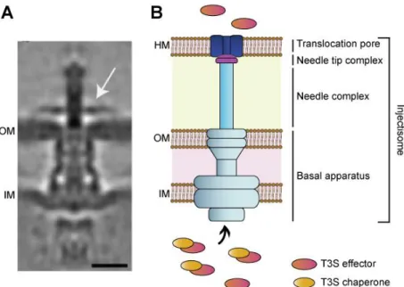
Non-flagellar T3SSs are exclusive of Gram-negative bacteria and are widely spread among bacterial pathogens of animals and plants, but are also involved in the establishment of symbiotic relationships between bacteria and eukaryotic hosts (Galán et al., 2014). These systems allow the direct delivery into the cytoplasm of eukaryotic cells of bacterial proteins, named type III secretion (T3S) effectors, which promote bacterial growth and survival by interfering with host cell processes. T3SSs comprise an injectisome, a multi-protein complex embedded in the two bacterial membranes consisting in a basal apparatus, a needle complex topped by a tip complex and a translocation pore inserted in a host membrane (the host plasma membrane and the inclusion membrane, in the case of Chlamydia). The injectisome forms a continuous channel that enables the one-step delivery of the T3S effectors, which can have specific T3S chaperones to aid their secretion (Figure 1.5) (Cornelis, 2006; Galán and Wolf-Watz, 2006).
Figure 1.5 The T3SS of C. trachomatis. (A) Cryo-electron tomogram of host-free T3SS of C. trachomatis; scale 15 nm; Reprinted from Nans et al. 2015 (B) Schematic representation of a prototypical T3SS. The injectisome basal apparatus is embedded in the bacterial outer (OM) and inner (IM) membranes and is topped by a needle-like structure. Upon activation of the T3SS, translocator proteins are secreted and inserted at the host membrane (HM), forming the translocation pore. T3S effectors in bacterial cytosol can interact with T3S chaperones that aid secretion through the injectisome.
Typically, T3SSs are encoded in a few operons, either in pathogenicity islands in the bacterial chromosome (e.g., Salmonella) or in virulence plasmids (e.g., Yersinia and Shigella) (Hueck, 1998). These systems have lost the motility genes of the flagellar apparatus and acquired components necessary for secretion and delivery of proteins across an eukaryotic cell membrane (Abby and Rocha, 2012). They are typically horizontally acquired and highly conserved even among bacteria with distinct evolutionary origins (Gophna et al., 2003).
Likely, the first observation of the chlamydial T3SS was made by Matsumoto and colleagues in 1973, which described the presence of rosette-like structures and projections on the surface of both EBs and RBs in different chlamydial species (Matsumoto, 1973). With the advent of DNA sequencing, genomic analysis confirmed the presence of T3S encoding genes in all sequenced Chlamydiaceae and
environmental Chlamydiae (Stephens, 1998; Kalman et al., 1999; Read et al., 2000; Read et al., 2003; Thomson et al., 2005; Azuma et al., 2006; Bertelli et al., 2010). Normally, T3SS structural components are clustered together; however, the genes encoding the T3SS in Chlamydia are in at least four clusters (Kalman et al., 1999) organized in multiple operons (Hefty and Stephens, 2007). Genes that code for T3SS have usually different G+C content (40-45%) when compared with the genome; however, this does not occur in Chlamydia indicating that recent gene integration did not occur and suggesting that chlamydial T3SS might represent a primordial system (Stephens, 1998; Abby and Rocha, 2012).
The chlamydial T3SS is thought to be activated in extracellular EBs upon contact with the host cell plasma membrane, resulting in the secretion of translocon proteins and followed by secretion of early effectors. Subsequently, within host cells, secretion is maintained in RBs by their association with the inclusion membrane which is accompanied by de novo expression of T3SS genes. When the transition of RBs to EBs occurs, RBs detach from the inclusion membrane and are thought to lose their protein secretion ability (Hansen-Wester and Hensel, 2001).
1.5.1.1 Chlamydial T3S effectors
Although the T3S injectisome is conserved among bacteria, normally T3S effectors do not have significant amino acid identity, but often have common structural features and perform similar functions. All effectors are thought to share a T3S signal localized at the N-terminus of the protein (approximately in the first 20 amino acid residues) (Sory et al., 1995; Lloyd et al., 2001). This signal is responsible to direct the protein for secretion and has been showed to be recognized by the T3SSs of different bacteria. The T3S signal is not cleavable, is unstructured and disordered, and enriched in serine, threonine, isoleucine and proline, but a clear consensus sequence is still unknown, despite of extensive studies (Wang et al., 2013). Upon secretion and delivery into the host cell, T3S effectors can perform a variety of functions that have been found to be crucial to the survival of many pathogenic bacteria.
In C. trachomatis, T3S effector genes have been predicted to represent approximately 5-10% of the coding capacity of the genome. Their initial identification and study mostly relied on the use of (i) complex bioinformatic tools to predict T3S signals (Arnold et al., 2009; Samudrala et al., 2009; Löwer and Schneider, 2009), (ii) use of heterologous T3SS such as Salmonella (Ho and Starnbach, 2005), Shigella (Subtil et al., 2005; Jewett et al., 2006; Pennini et al., 2010; Muschiol et al., 2011; Furtado et al., 2013), or Yersinia (Fields and Hackstadt, 2000; Clifton et al., 2004; Chellas-Géry et al., 2007; Hower et al., 2009; Pais et al., 2013; Hovis et al., 2013; da Cunha et al., 2014), and (iii) Saccharomyces cerevisiae to phenotypically characterize the effects of overexpression of candidate effectors (Sisko et al., 2006; Kumar et al., 2006). In addition, the identification of T3S effectors during C. trachomatis infection relied on the ability to produce specific antibodies against the proteins of interest and on their immunolocalization within host cells. More recently, the tools to genetically manipulate C. trachomatis strains have been used to study chlamydial T3S effectors.
1.5.1.1.1 Inc (inclusion membrane) proteins
The inclusion membrane (Inc) proteins are the most studied C. trachomatis T3S effectors. These proteins are characterized by a bilobed hydrophobic motif that likely mediates their insertion in the inclusion membrane (Rockey et al., 2002). Therefore, they can be identified using bioinformatics prediction tools and by immunofluorescence microscopy analysis of their presumed localization in the
C. trachomatis inclusion. Inc proteins, which are at the host-bacteria interface, have been shown to
modulate host cytoskeleton dynamics (Kokes et al., 2015; Mital et al., 2015; Dumoux et al., 2015; Wesolowski et al., 2017), vesicular and non-vesicular transport (Rzomp et al., 2006; Delevoye et al., 2008; Derré et al., 2011; Mirrashidi et al., 2015), release of the bacteria from host cells (Lutter et al., 2013; Nguyen et al., 2018) and inhibition of host cell death (Sixt et al., 2017; Weber et al., 2017). Moreover, Inc proteins have been hypothesized to contribute to the different tissue tropism and invasion characteristics of different C. trachomatis serovars (Lutter et al., 2012; Almeida et al., 2012).
1.5.1.1.2 Non-Inc T3S effector proteins
In addition to the Inc proteins, there are chlamydial T3S effectors that are delivered into the host cell and which do not possess the characteristic bilobed hydrophobic motif and mostly do not localize at the inclusion membrane. Their study is more challenging, as their primary structure normally lacks obvious distinguishable features.
CT456/TarP (translocated actin-recruitment phosphoprotein) is likely the most studied T3S effector of C. trachomatis. TarP is synthesized at the late stages of the developmental cycle, pre-packed into EBs but only delivered into the host cell within minutes after EBs attachment. Once the EBs enter host cell, TarP is readily phosphorylated and recruits actin to the entry sites. TarP comprises an actin-binding domain at its C-terminus that associates with both G- and F-actin and induces actin nucleation and polymerization independently of host cell factors. At the N-terminus, TarP contains tyrosine-repeated regions which have been shown to be phosphorylated by the host cell kinases Src, AblI and Syk family (Mehlitz et al., 2008; Jewett et al., 2008). Phosphorylated TarP interacts with guanidine exchange factors, Sos1/Eps8/Abi1, and Vav2, which activate the small GTPase Rac allowing WAVE2 and Arp2/3-mediated nucleation of actin at the host cell entry sites (Lane et al., 2008). Both actin nucleators, TarP and Arp2/3, act synergistically to enhance the rate of formation of actin filaments (Jiwani et al., 2012). TarP has been shown to interact with vinculin to mediate actin recruitment and assembly at the host cell plasma membrane, and with adaptor protein SHC1 to induce host cell survival early during chlamydial infection (Mehlitz et al., 2010). TarP has also been associated to the tropism of different C. trachomatis strains, as LGV strains contain higher number of tyrosine-rich repeat regions and less predicted actin binding domains, in contrast with TarP of the ocular strains (Lutter et al., 2010).
CT694/TmeA (translocated membrane-associated effector A) is a T3S effector important for infectivity and invasion of C. trachomatis. TmeA is delivered into the host cell in the early-steps of infection (Hower et al., 2009; McKuen et al., 2017). The C-terminus of TmeA interacts with human AHNAK affecting the formation of host cell actin stress fibers. However, this does not seem to be