Universidade de Lisboa/Faculdade de Ciências
Departamento de Biologia Animal
The Role of Notch Signaling in Thymic
Epithelium Development
Marta Sofia Carvalho Teles de Figueiredo
MESTRADO EM BIOLOGIA EVOLUTIVA E DO
DESENVOLVIMENTO
II
Universidade de Lisboa/Faculdade de Ciências
Departamento de Biologia Animal
The Role of Notch Signaling in Thymic
Epithelium Development
Marta Sofia Carvalho Teles de Figueiredo
MESTRADO EM BIOLOGIA EVOLUTIVA E DO
DESENVOLVIMENTO
Dissertação orientada por
Professora Dra. Rita Maria Pulido Garcia Zilhão (DBV/FCUL)
Professora Dra. Hélia Cristina de Oliveira Neves (FMUL)
III
Acknowledgments
Para que tudo isto fosse possível, e como ninguém “é sozinho”, houve uma série de pessoas que fizeram parte deste meu caminho e que deixaram a “sua marca”.
Quero agradecer, em primeiro lugar, à Hélia, que acreditou no meu potencial, desde que me conheceu numa rotação laboratorial do curso, e que me levou e guiou nesta jornada, não só intelectual como também psicológica. Deu-me a conhecer mais do mundo à minha volta (e do mundo da Ciência) mas também do mundo dentro de mim. Acreditou e investiu em mim, mesmo quando o cenário parecia negro, e deu-me força para continuar, e não desistir. Aceitou-me de braços abertos e fez por Aceitou-me pôr à vontade nesta nova fase da minha vida. Ganhei uma amiga. Pela sua paciência, dedicação, e exigência, um muito obrigada. Sem ela, e sem a sua paixão pela biologia e por este tema, este trabalho não seria possível.
Um obrigada à Prof. Rita Zilhão, por todo o seu conhecimento, apoio, e também pelo seu entusiasmo único, que é contagiante. Existem poucas pessoas assim, e fico contente por ter oportunidade de aprender e partilhar momentos com uma delas.
Queria agradecer também ao meu grupo da Unidade de Biologia da Hematopoiese do Instituto de Histologia e Biologia do Desenvolvimento da FMUL por me acolherem de braços abertos e por toda a ajuda que me ofereceram durante este trabalho. Em especial, à Isabel e ao Vítor, que estiveram mais perto, e que sempre me apoiaram e me ofereceram sorrisos.
Dentro do nosso grupo de trabalho, queria agradecer também ao Rafael, por toda a sua companhia, paciência, dedicação e gentileza, principalmente durante a fase inicial de integração no laboratório, que não teria sido tão agradável ou fácil como foi, se ele não tivesse estado presente. Um obrigada também pela amizade. Mais recentemente, a Carlota tornou-se um membro presente do grupo na recta final deste trabalho, e tal como o Rafael, a sua companhia e simpatia fizeram toda a diferença.
Queria deixar um agradecimento especial ao Filipe, da UBD, que nos sugeriu a estratégia de electroporação in vivo do tubo neural de embriões de galinha para testarmos as nossas construções. Obrigada pela sugestão que se revelou muito útil, por todo o ensinamento da técnica e paciência. Fica também um agradecimento à Joana Neves pela sua ajuda na técnica e pela simpatia. Quero agradecer também ao Domingos Henrique, por nos ceder as sondas do grupo de investigação dele, que foram também uma peça vital para o nosso estudo.
Um enorme obrigada a todos os amigos que acompanharam esta fase de mudança da minha vida, uns mais de perto do que outros (mas deixando, todos, o seu contributo). Aqui destaco somente alguns.
À Anita e à Marta, pelos bons momentos (cheios de sorrisos) passados dentro e fora de casa, e pelo apoio incondicional nas horas negras. Ao Diogo, que é quase o positivismo em pessoa, e que sempre tentou que eu acreditasse mais em mim própria (ou própia?). À Ana, que mesmo estando a km de distância, esteve sempre por perto. Principalmente, pelas palavras certas à hora certa; naqueles momentos em que mais precisava de força, lá estava ela, como se
IV
soubesse miraculosamente, a dizer-me as palavras que me faziam levantar outra vez. Obrigada por toda a força, pela transmissão da força que lhe é característica. Pela sua dedicação às coisas, pela esperança no futuro, e principalmente por acreditar em mim (mais do que eu própria). À Inês, presente desde os meus 4 meses de idade, amiga desde que tenho memória, até aos dias de hoje. Inseparáveis, caminhámos lado a lado desde o infantário até à universidade, e a verdade é que isso é algo raro. Obrigada pelo ombro amigo, pela força, pelos conselhos, e pela calma que me tenta transmitir quando eu não a sei ter.
Ao Marco, que entrou na minha vida como uma lufada de ar fresco, e que me ajudou nas fases mais complicadas desta jornada, tanto a nível pessoal, como profissional. Obrigada pela companhia, pelos sorrisos e gargalhadas, pela compreensão, pelo esforço e ajuda preciosa, pelo positivismo, por sonhar acordado, pela diferença (diferença essa que nos une e que me equilibra, de vez em quando), e por acreditar em mim.
Finalmente, aos meus pais, que são a razão pela qual estou no mundo. Foram eles que fizeram de mim quem sou. Um muito obrigada pela boa educação, pelo incentivo ao sorriso e à aprendizagem, pelo amor aos livros e ao conhecimento. Obrigada pelo apoio e amor incondicional. Por me incentivarem a procurar mais, a acreditar que é possível chegar mais longe e por acreditarem no meu potencial. Obrigada ao resto da família, que constitui, também, o ambiente que me tornou aquilo que sou hoje. Todos fizeram a sua parte ou deixaram a sua marca para sempre naquilo que me define.
Fica aqui um agradecimento eterno ao Marco, à Inês e à Carlota pelo apoio inacreditável da recta final, que só nós sabemos.
V
Abstract
The thymus generates central immune tolerance by producing restricted and self-tolerant T-cells as a result of interactions between developing thymocytes and thymic epithelial cells (TECs). While the functional importance of TECs is well established, the mechanisms that direct their embryonic development are unclear. The Notch pathway is a major signaling pathway involved in cell-fate determination. Recently, H. Neves group observed that, during chicken embryogenesis, Notch signaling-related molecules are expressed in the endoderm of the pharyngeal pouches, prior to their specification into TECs, suggesting the involvement of Notch signaling in this process.In this work we aimed to study the role of Notch signaling in early stages of thymic development in chicken.
To modulate Notch signaling in vivo, two new plasmids were generated with either the constitutively active form of Notch1 (intracellular domain of Notch1, ICN1) (pT2K-ICN1eGFP), or the dominant-negative (DN) form of Mastermind-like1 (MAML1) (transcriptional co-activator Notch signaling) (pT2K-DNMAML1eGFP). pT2K-ICN1eGFP and pT2K-DNMAML1eGFP were integrated in a “Tol2-mediated gene transfer” and “Tet-On” combined system of vectors developed by Y. Takahashi and collaborators, allowing the study of gain- and loss-of-function of Notch signaling, respectively. Our results show that pT2K-DNMAML1eGFP plasmid in this system, is capable of blocking Notch signaling. Conversely, further assays are required to confirm the functionality of pT2K-ICN1eGFP plasmid. Future studies of gain- and loss-of-function of Notch signaling in early thymic development will be performed by the genetic modification of isolated endodermal tissues of the presumptive territory of TECs with this system. The manipulated tissues will then be grafted into the body wall of a chicken embryo and thymic development followed by in situ observation.
In vitro assays of pharyngeal region explants of E3.5 and E4 chicken embryos were
performed to study the effect of Notch signaling inhibition during epithelial-mesenchymal interactions in early thymic development. Thymus and parathyroid glands development was assessed by Foxn1 and Gcm2 expression, respectively. We observed that Notch signaling inhibition by a γ-Secretase inhibitor (DAPT) interferes in Foxn1 expression in an apparent random fashion, making inconclusive its role in early thymic development. On the other hand,
Gcm2 expression is down-regulated when Notch signaling is inhibited. This data suggests that
Notch signaling is required in early stages of parathyroid development. Further studies are essential to unravel the role of Notch signaling in early thymic development.
VI
Resumo
O timo gera tolerância imunitária ao produzir células-T restritas” e “auto-tolerantes”, cuja geração depende das interacções das suas células precursoras, os timócitos, com células especializadas do nicho tímico, as células epiteliais tímicas (CET). Embora a importância funcional das CET esteja bem estabelecida, os mecanismos moleculares responsáveis pelo seu desenvolvimento embrionário são ainda desconhecidos. O desenvolvimento do timo é acompanhado de perto pelo desenvolvimento das glândulas paratireóides. Os domínios presuntivos do timo e das glândulas paratireóides foram identificados na endoderme da 3ª e 4ª bolsas faríngicas pela expressão de Foxn1 e Gcm2, respectivamente. A especificação das CET depende de interacções entre a endoderme da 3ª e 4ª bolsas faríngicas e o mesênquima circundante - interacções epitélio-mesenquimais. Algumas destas interacções epitélio-mesenquimais começam agora a ser reveladas, nomeadamente através do trabalho do grupo de H. Neves, que demonstra que factores de transcrição como o Bmp4 e o Fgf10 são expressos sequencialmente no mesênquima e são essenciais para a especificação da endoderme da 3ª e 4ª bolsas faríngicas em CET. Outras vias de sinalização têm sido sugeridas como prováveis intervenientes neste processo. A sinalização Notch é uma delas, sendo uma via altamente conservada no reino animal, envolvida nos processos de decisão do destino celular, no desenvolvimento embrionário e no adulto. Esta via de sinalização regula vários processos biológicos, incluindo a hematopoiese, miogénese, neurogénese, vasculogénese, desenvolvimento da pele e outros processos de organogénese.
Em 2001, o grupo de L. Parreira descreveu pela primeira vez, num contexto de um nicho estromal, a importância da formação de um microambiente Notch para a correcta especificação de progenitores hematopoiéticos nas diferentes linhagens linfóides. Este e outros grupos também observaram que os genes envolvidos na sinalização Notch são expressos de forma distinta nos diferentes territórios do timo adulto, reforçando a importância desta via de sinalização na função do mesmo. Recentemente, o grupo de H. Neves também observou, em embriões de galinha, que os genes envolvidos na sinalização Notch (receptores, ligandos e genes-alvo) estão diferencialmente expressos na endoderme das bolsas faríngicas, em estádios prévios à formação do rudimento tímico. Também observaram, usando um novo sistema in
vitro de associações heteroespecíficas de tecidos embrionários, que a sinalização Notch
interfere com a expressão normal de Foxn1 (marcador de CET) na endoderme das bolsas faríngicas. Este sistema de cultura permite uma análise funcional das moléculas envolvidas nas interacções epitélio-mesenquimais e também determinar a dinâmica temporal destas moléculas durante o desenvolvimento do timo. Este sistema recapitula in vitro os acontecimentos precoces do desenvolvimento tímico in vivo, uma vez que, quando enxertadas na membrana corioalantóide do embrião de galinha, estas associações de tecidos desenvolvem um timo funcional. Estes dados sugerem que a sinalização Notch está envolvida na especificação da endoderme das bolsas faríngicas em epitélio tímico.
Neste projecto, tivemos como objectivo estudar a sinalização Notch na especificação da endoderme das bolsas faríngicas em epitélio tímico. Para isso, desenvolvemos estratégias in
VII
abordagem in vivo, dois plasmídeos novos foram construídos, para expressar a forma constitutivamente activa de Notch1 (domínio intracelular de Notch1, ICN1) (pT2K-ICN1eGFP) ou a forma dominante-negativa de Mastermind-like1 (MAML1) (co-activador transcricional de sinalização Notch) (DNMAML1) (pT2K-DNMAML1eGFP). Estes plasmídeos foram integrados num sistema de vectores desenvolvido por Y. Takahashi e colaboradores, que combina a “transferência génica mediada por Tol2” e a “expressão condicional dependente de tetraciclina”. Assim, este sistema de vectores oferece uma abordagem experimental única para uma análise temporal, e específica de tecido, dos efeitos de ganho e perda-de-função da sinalização Notch durante a especificação/diferenciação das CET. Para realizar esses estudos, células endodérmicas do território gerador do epitélio tímico serão modificadas geneticamente com os plasmídeos pT2K-ICN1eGFP ou pT2K-DNMAML1eGFP (neste sistema de vectores) para activar ou bloquear a sinalização Notch, respectivamente, e serão enxertadas na parede do embrião de galinha, para acompanhar in situ o desenvolvimento do timo quimérico. Os nossos resultados mostram que o plasmídeo que expressa a forma DNMAML1, pT2K-DNMAML1eGFP, neste sistema de vectores, tem a capacidade de inibir a sinalização Notch. O trabalho poderá assim prosseguir para os estudos de perda-de-função da sinalização Notch na endoderme das bolsas faríngicas, antes da sua especificação em CET. Por outro lado, o plasmídeo que expressa o ICN1, pT2K-ICN1eGFP, neste sistema de vectores, necessita de mais experimentação para comprovar a sua funcionalidade.
A abordagem in vitro consistiu na realização de ensaios de cultura de explantes da região faríngica de embriões de galinha com 3.5 e 4 dias de desenvolvimento para estudar o efeito da inibição farmacológica da sinalização Notch (com um inibidor da γ-Secretase – DAPT) durante as interacções epitélio-mesenquimais no desenvolvimento inicial do timo. Tanto a especificação da endoderme faríngica em epitélio tímico como a manutenção do domínio das glândulas paratireóides nos explantes em cultura foram avaliadas através da expressão in situ de Foxn1 e Gcm2, respectivamente. Os nossos ensaios in vitro revelaram que a inibição da sinalização Notch, em fases iniciais do desenvolvimento tímico, interfere com a expressão de Foxn1 de forma aparentemente aleatória, mostrando elevada heterogeneidade de resultados. Assim, o papel da sinalização Notch em fases iniciais do desenvolvimento do timo continua por precisar. Por outro lado, a expressão de Gcm2 é bloqueada com a inibição da sinalização Notch nessas mesmas fases de desenvolvimento. Portanto, os nossos dados in vitro sugerem um papel da sinalização Notch no desenvolvimento das glândulas paratireóides, pelo menos em fases iniciais do desenvolvimento.
Novas experiências in vitro usando explantes da região faríngica de embriões de galinha em estádios de desenvolvimento diferentes (tanto mais precoces como mais tardios) daqueles estudados, e a realização dos estudos in vivo com o nosso sistema de vectores, serão essenciais para compreender estes resultados e também para estudar o papel da sinalização Notch na especificação/desenvolvimento das CET.
Com este projecto esperamos contribuir para o conhecimento do papel da sinalização Notch no desenvolvimento normal das células epiteliais tímicas, um passo fundamental na compreensão dos eventos responsáveis pela manutenção de um timo saudável ao longo da vida
VIII
e pela reparação da sua função em situações patológicas. Mais, o conhecimento de elementos chave envolvidos na especificação das CET poderá no futuro permitir a criação de novos sistemas in vitro para gerar células epiteliais tímicas, as quais, por sua vez, poderão abrir novas possibilidades de produção in vitro de repertórios de células-T e novas oportunidades para restaurar a função tímica em indivíduos atímicos ou imunodeficientes e, também, melhorar as terapias de transplantação de órgãos.
Palavras-chave: Notch, timo, células epiteliais tímicas, bolsa faríngica, endoderme, Foxn1
IX
Index
Acknowledgments ...III Abstract ... V Resumo ... VI Abbreviations ...1 I. Introduction ...2I.1. Thymus Development ...2
I.2. Notch signaling ...4
I.3. “Tol2-mediated gene transfer system” and “tetracycline-dependent conditional expression” system for the study of Notch signaling ...7
I.4. Chicken model for developmental studies...9
II. Objective ...10
III. Materials and Methods ...11
III.1. Molecular Biology procedures ...11
III.2. Cellular Biology procedures ...16
III.3. Developmental Biology procedures...18
IV. Results ...21
IV.1. In vivo modulation of Notch signaling in the 3/4PP endoderm ...21
IV.1.1. Production ...21
IV.1.2. Functional analysis ...23
IV.2. Expression of Notch signaling related genes in chicken embryos at stages of development prior to thymic epithelium specification ...27
IV.2.1. Expression of Notch1 and MAML1 in chick embryos at E3 and E4 ...27
IV.2.2. Expression of Notch signaling-related genes in the 3/4PP region (at E3 and E4) ...29
IV.3. In vitro assays: inhibition of Notch signaling in the pharyngeal region of the 3/4PP ...30
V. Discussion ...34
V.1. In vivo modulation of Notch signaling. Production of Notch Constructs: pT2K-ICN1eGFP (gain-of-function) and pT2K-DNMAML1eGFP (loss-of-function) ...34
V.2. Expression of Notch signaling related genes in chicken embryos at stages of development prior to thymic epithelium specification ...36
V.2.1. Expression of Notch1 and MAML1 in chick embryos at E3 and E4 ...36
X
V.3. In vitro assays: inhibition of Notch signaling in the pharyngeal region of the 3/4PP. ...37
VI. Conclusion ...39
VII. References ...40
APPENDIX I – BUFFERS, MEDIA AND OTHER SOLUTIONS ...48
APPENDIX II – PROTOCOLS ...51
1
Abbreviations
aa – amino acid bp – base pairs
DAPI – 4',6-diamidino-2-phenylindole
DAPT – N-[N-(3,5-Difluorophenacetyl-L-alanyl)]-S-phenylglycine t-Butyl Ester DMEM – Dulbecco's Modified Eagle Medium
DMSO – Dimethyl sulfoxide DN – Dominant Negative DNA – Deoxyribonucleic Acid E – Embryonic day
EDTA – Ethylenediaminetetraacetic Acid FACS – Fluorescence Activated Cell Sorter FBS – Fetal Bovine Serum
GFP – Green Fluorescence Protein
h, min, sec, ms – hour, minute, second, milisecond ICN – Intra-cellular domain of Notch
m/v – mass/volume
ng/mL – nanogram/milliliter o.n. – overnight
PA – Pharyngeal Arch
PBS – Phosphate Buffered Saline Pen/Strep – Penicillin/Streptomycin PFA – Paraformaldehyde
PP – Pharyngeal Pouch RNA – Ribonucleic Acid rpm – Revolutions per minute
RT-PCR – Reverse Transcriptase-Polymerase Chain Reaction SSC-H– Side light scatter-height
U/µL – units/microliter v/v – volume/volume
2
I. Introduction
I.1. Thymus Development
The thymus is a specialized organ of the adaptive immune system, responsible for the development of thymocytes into T lymphocytes. This organ is found in all jawed vertebrates and co-evolved with VDJ recombination mechanism which is used to somatically diversify antigen receptors (Thomas Boehm & Bleul, 2007). Although its existence was known for centuries, its immunologic function was only discovered in 1961 by Jacques Miller, when he observed a deficit in a specific type of lymphocytes (later called T-lymphocytes), after performing thymectomies in mice (Miller, 1961).
Thymus organogenesis is a highly dynamic process that is initiated during fetal life and can be divided in two main temporal phases; an initial thymocyte-independent phase, where cellular interactions between the endoderm and the surrounding mesenchyme direct thymic epithelial cells (TECs) specification, followed by a thymocyte-dependent phase. At this latter stage, the thymic enlagen depends on the colonization by lymphoid progenitor cells (LPCs) for further maturation of thymic epithelium into cortical (cTECs) and medullar (mTECs) compartments (Alves, Huntington, Rodewald, & Di Santo, 2009; G Anderson & E J Jenkinson, 2001; Auerbach, 1960; C Clare Blackburn & Nancy R Manley, 2004; N M Le Douarin, Dieterlen-Lièvre, & Oliver, 1984; Klug, Carter, Gimenez-Conti, & Richie, 2002; Nehls et al., 1996).
TECs are a specialized subset of thymic cells required for all stages of thymocyte differentiation (Anderson et al. 1993; Anderson, Owen, Moore, & Jenkinson, 1994; Oosterwegel et al. 1997; Klug et al. 1998; Ge & Chen, 2000; Bennett et al. 2002); specific TECs subtypes mediate particular aspects of thymopoiesis: cTECs are involved in positive selection (Cosgrove, Chan, Waltzinger, Benoist, & Mathis, 1992) and mTECs in tolerance induction (Gotter, Brors, Hergenhahn, & Bruno Kyewski, 2004).
In 1975, Le Douarin & Jotereau, using the chick–quail chimera system, showed that TECs derive from the pharyngeal pouches (PP) endoderm (N M Le Douarin & Jotereau, 1975). This single, endodermal germ layer origin, of the thymic epithelium was further supported by other studies in mouse (Bleul et al., 2006; Rossi, W. E. Jenkinson, Graham Anderson, & Eric J Jenkinson, 2006).
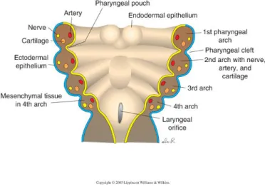
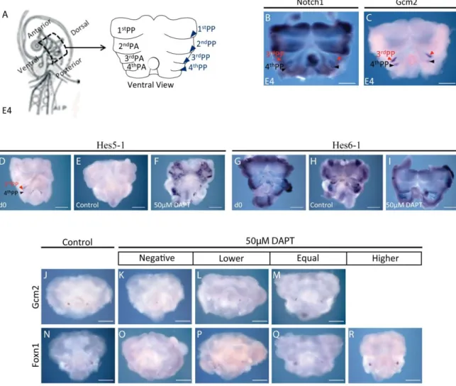
Thymic epithelium development is intimately linked to the development of the parathyroid glands, as their rudiments derive from the common embryonic structure of the PP [3rd and 4th PP (3/4PP) in chicken and humans, 3rd PP in mouse]. The pharyngeal pouches are bilateral transient structures that arise as outpocketings of the lateral foregut endoderm. The PP, along with the opposing pharyngeal clefts (PC) (invaginations of surface ectoderm) form the separation between pharyngeal arches (PA), the bilateral bulges that comprise the pharyngeal region (Fig. 1) (J. Gordon & N. R. Manley, 2011; Patel, Julie Gordon, Mahbub, C Clare Blackburn, & Nancy R Manley, 2006; Rodewald, 2008).
In the mouse, the prospective thymic epithelium was identified by the expression of
3
Manley, 2001), a product of the nude locus, which is required cell-autonomously for thymic epithelium differentiation and colonization by LPCs (Nehls et al., 1996; C C Blackburn et al., 1996; Bleul et al., 2006). The parathyroid rudiment was defined by the expression of Gcm2 (Glial Cell Missing 2) transcription factor (J Gordon et al., 2001); when Gcm2 is deleted, no parathyroid glands are formed (Günther et al., 2000; Z. Liu, Yu, & Nancy R Manley, 2007).
Figure 1. Scheme representing coronal section through the pharyngeal zone. Pharyngeal arches, consist of mesenchymal and mesodermal cells bounded by an outer layer of surface ectoderm (blue) and inner layer of pharyngeal endoderm (yellow). The ectoderm forms invaginations, the pharyngeal clefts, which separate the arches, whereas the endoderm forms the opposing outpocketings, the pharyngeal pouches.
In chicken, the transcription factors Foxn1 and Gcm2 are also expressed in the endodermal rudiments of the thymus and parathyroid glands, respectively. Specifically, Foxn1 domain of expression was identified as the emergent domain of thymic epithelium in the most dorsal portion of the 3/4 PP endoderm in chicken embryo at E4.5 (Fig. 2A and B) (Neves et al., 2011 in press). Conversely, parathyroid rudiments were identified in a more ventral position in the 3/4 PP by Gcm2 domain of expression (Fig. 2B) (Neves et al., 2011 in press).
The initial phase of thymic organogenesis is characterized by cellular interactions between the endoderm and the surrounding neural crest-derived mesenchyme to TECs specification (Rodewald, 2008). The importance of epithelial-mesenchymal interactions was demonstrated by Le Douarin, using the chick-quail chimera system. The 3/4PP endoderm isolated from early quail embryos was able to develop into thymic epithelium with the cooperation of a heterologous mesenchyme such as the somatopleure or splanchnopleure of E3 chicken embryos, which thus could be considered “permissive” to endoderm development. Furthermore, the grafted endoderm was capable of inducing the heterologous mesenchyme to participate in the formation of a fully developed thymus (N. Le Douarin, 1967; Le Douarin, N., Bussonnet, C., Chaumont, 1968; N. M. Le Douarin & Jotereau, 1975). In contrast, mesenchymal environments of the somite and limb bud were non-permissive to 3/4PP endoderm development (N. Le Douarin, 1967; Le Douarin, N., Bussonnet, C., Chaumont, 1968). These data provided the first evidence that epithelial-mesenchymal reciprocal interactions are essential for early thymic development; moreover, they revealed that some
4 Neves et al., 2011
B
A
heterologous mesenchymal tissues are able to mimic the role played by neural crest-derived mesenchyme during normal development of the thymus in the pharyngeal region.
Figure 2. Expression of Foxn1 and Gcm2 during thymic and parathyroid glands development in chick embryos. A) In situ hybridization showing Foxn1 expression in PP3/4 endoderm isolated by microsurgery in E4.5 chicken embryos. B) Schematic 3D-representation of Foxn1 and Gcm2 expression domains in the 3rd PP endoderm of E4.5 chicken embryos. (A, anterior; D, dorsal; c, chicken; P, posterior; PP, pharyngeal pouch; V, ventral).
Recently, H. Neves work unraveled some of the early molecular events occurring at the initial thymocyte-independent stage of thymic development. Her results showed that cellular interactions between the endoderm and adjacent mesenchyme involved a sequential expression of Bmp4 and Fgf10 in the mesenchymal compartment, fundamental for the development of the 3/4PP endoderm into of thymic and parathyroid glands epithelia. Also, a temporal regulation of Bmp4 expression in the mesenchymal compartment was observed, suggesting that the Bmp4 levels need to be tightly regulated in the developing pouches (Neves et al., 2011 in press).
Another signaling pathway known to be involved in epithelial cell development (dependent on mesenchymal interactions) in other organs like the skin and the gut is Notch signaling (Hu et al., 2010; T.-H. Kim, B.-M. Kim, Mao, Rowan, & Shivdasani, 2011).
I.2. Notch signaling
Notch signaling is a major signaling-pathway, highly conserved in the animal kingdom, which regulates biological processes in the development of the embryo and in the adult. Notch signaling has been shown to control hematopoiesis (Jaleco et al. 2001; Neves et al. 2006; Santos et al. 2007; Parreira et al. 2003; Eric J Jenkinson et al. 2006), myogenesis (Luo, Renault, & Rando, 2005), neurogenesis (Lasky & H. Wu, 2005), vasculogenesis (L. M. Anderson & Gibbons, 2007), skin development (Estrach, Cordes, Hozumi, Gossler, & Watt, 2008) and other aspects of organogenesis. Notch receptors (Notch1-4 in mammals, Notch1-2 in birds) and their ligands (Delta1, 3 and 4; Jagged1-2 in mammals, Serrate1-2 in birds) are evolutionary conserved transmembrane proteins that regulate cell-fates, cell numbers and cell position via
5
effects on proliferation and survival (Lai, 2004). These effects depend on dose, timing, and context of the Notch signal (Lai, 2004; Maillard, Fang, & Pear, 2005).
Notch-mediated transcriptional activation involves the activation of Notch receptors by their ligands that are expressed on neighboring cells. This receptor-ligand interaction will lead to the proteolytic cleavage of the intracellular domain of Notch (ICN), catalyzed by the γ-secretase complex. Thus, the ICN is released from the membrane and is translocated into the nucleus, where it cooperates with the DNA-binding protein CSL and co-activators to form the transcriptional activation complex, which will activate target gene expression (W. R. Gordon, Arnett, & Stephen C Blacklow, 2008). One important co-activator is Mastermind-like (MAML) protein (L Wu et al., 2000), which appears to function as a scaffold for the formation of a large multiprotein transcriptional activation complex (Jeffries, Robbins, & Capobianco, 2002) (Fig. 3). The Mastermind protein was originally identified in D. melanogaster as a neurogenic protein genetically linked to the Notch signaling pathway (Xu, Rebay, Fleming, Scottgale, & Artavanis-Tsakonas, 1990), and the mammalian homologues that were found afterwards were named Mastermind-like proteins.
Figure 3. Schematic representation of Notch signaling pathway in chicken. Ligand (Serrate1,3 or Delta1,3,4) binding between neighboring cells induces proteolytic cleavage of Notch receptors (Notch1,2), catalyzed by the γ-secretase complex, producing the free intracellular domain of Notch (ICN). ICN translocates to the nucleus and binds to transcription factors, MAML1 being one of them. The transcription of Notch target genes (ex: Hairy1, Hes5-1 or Hes5-1) is then activated.
It is known that, when a cell is forced to express the ICN, Notch signaling is constitutively active in a ligand independent manner (G Weinmaster, 1997). On the other hand,
in vitro experiments in mice have shown that truncated MAML1 proteins consisting of only the
N-terminal ICN-binding domain have potent dominant negative effects, presumably due to their inability to recruit other components of the Notch transcriptional activation complex (Fryer, Lamar, Turbachova, Kintner, & K. A. Jones, 2002; Weng et al., 2003). Thus, when a
6
cell is forced to express the dominant-negative (DN) form of MAML1 protein (DNMAML1), Notch signaling is profoundly blocked.
The activation of the Notch signaling induces a profound alteration of the cellular transcriptional program. The best-characterized Notch targets are the Hes genes. Hes genes are mammalian homologs of the Drosophila genes Hairy and Enhancer of split, characterized by basic helix–loop–helix proteins. Hes genes were shown to contribute to clocks that regulate somitogenesis, limb segmentation, and neural progenitor maintenance (Brend & Holley, 2009; Lewis, Hanisch, & Holder, 2009; Pascoal et al., 2007; Shimojo, Ohtsuka, & Ryoichiro Kageyama, 2008).
One of the first Hes genes described in vertebrates was a homologue of Drosophila
hairy in mouse, which was given the name of Hes1 (Sasai, R Kageyama, Tagawa, Shigemoto,
& Nakanishi, 1992). Hes1, in general, maintains cells in the undifferentiated progenitor state, influences progenitor cell proliferation and differentiation, specifically it inhibits neuronal and muscle differentiation ( Sasai et al., 1992; Ishibashi et al., 1994). Other Hes genes have been found over the time. Hes6 proteins are known to regulate neurogenesis by contributing to release differentiating neurons from Notch signaling (Vilas-Boas & Domingos Henrique, 2010). Also, Hes6 has been shown to regulate muscle differentiation (Cossins, Vernon, Zhang, Philpott, & P. H. Jones, 2002). Furthermore, Hes5-1 is known to be a direct target of Notch signaling in the developing nervous system; Hes5-1 transcription is severely reduced when Notch signaling is blocked (De La Pompa et al., 1997; Lütolf, Radtke, Aguet, Suter, & Taylor, 2002) and its promoter is directly regulated by Notch (Nishimura et al., 1998).
Previous work from L. Parreira and collaborators shown the importance of specific Notch microenvironments for the commitment of hematopoietic progenitors into different lymphoid lineages (Jaleco et al., 2001), for the expansion and lineage-differentiation of early-myeloid progenitors (Neves et al., 2006) and for the generation of plasma cells and the amount of antibodies secreted by them, in terminal B-cell maturation (Santos et al., 2007). They also observed that Notch-related genes are differentially expressed in the adult thymic microenvironments, stressing the importance of this signaling pathway in thymic function (Jenkinson et al., 2006; Parreira et al., 2003).
Notch signaling is known to play a role during late stages of thymic organogenesis. At the thymocyte-dependent phase, TECs provide Notch ligands to neighboring developing thymocytes, promoting their development (Alves, Goff, et al., 2009; Feyerabend et al., 2009; Hozumi et al., 2008; Koch et al., 2008; Tsukamoto, Itoi, Nishikawa, & Amagai, 2005). Specifically, Notch signaling activation mediated by Delta1 induced the appearance of a normal thymic architecture in murine fetal thymic organ cultures (Masuda et al., 2009). In contrast, only few evidences point to a role of Notch at early-phase of thymus formation.
Preliminary results of H. Neves and collaborators suggest a role of Notch signaling during chicken thymus development. They observed Notch signaling-related genes (receptors, ligands and target genes) expressed in the prospective thymic domain and surrounding mesenchyme. In particular, the expression of Hes1, in the endoderm of the 3/4PP at E3 and E4, suggests active Notch signaling in the prospective territory of TECs. Furthermore, using a
7
novel in vitro system (developed by Neves & Le Douarin, 2009) to study early-stages of thymus development, they observed that blocking Notch signaling interferes with the normal expression of Foxn1 in the endoderm of the pharyngeal-pouches. This in vitro culture system, with heterospecific associations of embryonic tissues, allows the functional analysis of molecules involved in epithelial-mesenchymal interactions and to determine the temporal dynamics of those molecules during thymus development. These heterospecific associations of embryonic tissues, when grafted in the chorioallantoic membrane of a chick embryo, develop into a functional thymus, showing the ability of this in vitro system to recapitulate the early events of thymic development in vivo. These observations suggest that Notch signaling is involved in the specification of TECs from the endoderm of 3/4PP.
To investigate how Notch signaling affects early stages of thymus organogenesis we aim to modify in a stable and cell-autonomous manner the quail endodermal tissues of the presumptive territory of TECs. This tissue will be genetically modified to either express the constitutively-active form of Notch1 , ICN1, or a dominant-negative form of MAML1, DNMAML1, using a “Tol2-mediated gene transfer” and “Tetracycline-dependent conditional expression” combined system of vectors.
I.3. “Tol2-mediated gene transfer system” and “tetracycline-dependent
conditional expression” system for the study of Notch signaling
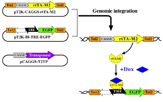
Y. Takahashi and collaborators developed a combined system of transposon-mediated transgene and tetracycline-induced conditional expression (Sato et al., 2007; Watanabe et al., 2007). This system has the advantage of allowing a stable integration and a conditional expression of a transgene in chicken embryos. The original system is composed of three plasmids: 1) pT2K-CAGGS-rtTA2SM2; 2) pT2K-BI-TREeGFP and 3) pCAGGS-T2TP (Fig. 1). Both 1 and 2 plasmids have a gene expression cassette surrounded by Tol2 transposable elements. After electroporation of the three plasmids, transient activity of transposase (plasmid 3) will induce the transposon construct containing either rtTA2S-M2 (plasmid 1) or TRE-eGFP (plasmid 2) to be integrated into the host genome. The reverse tet-controlled transcriptional activator (rtTA) (plasmid 1), ubiquitously expressed by the promoter-CAGGS, acts on the cis-element promoter, tetracycline responsive cis-element (TRE). rtTA binds to TRE only in the presence of doxycycline (an analog of tetracycline; Dox) and activates transcription of the TRE-driven gene (“Tet-On expression system”). pT2K-BI-TREeGFP has a bidirectional TRE with two minimal promoters of cytomegalovirus in both directions. In one direction, it has an eGFP sequence and on the other one a polylinker region where one can clone the transgenes of interest (Fig. 4). Thus, this system allows stable integration of transgenes in the avian genome, which can be conditionally expressed in specific time-points of development, upon administration of Dox. Cells expressing the transgene will be identified by GFP expression, therefore the correlation between the time of Dox administration and GFP expression will determine the exact moment of transgene expression.
As previously mentioned, the sequences coding for the ICN1, or for the DNMAML1, in fusion with GFP, were sub-clone into the pT2K-BI-TREeGFP plasmid, so that Notch signaling
8
can be constitutively activated or blocked, respectively, in a cell-autonomous manner. The reliability of the new constructs in activating or blocking Notch signaling was analyzed using the C2C12 myoblast differentiation assay.
Figure 4. “Tol2-mediated gene transfer system” and “tetracycline-dependent conditional expression” combined system. Transient activity of transposase (pCAGGS-T2TP) will induce the transposon construct containing either rtTA2S-M2 (pT2K-CAGGS-rtTA2SM2) or TRE-eGFP (pT2K-BI-TREeGFP) to be integrated into the host genome. Activation of transcription of the TRE-driven gene (“tet-on”) occurs only in the presence of doxycycline. Adapted from Sato et al., 2007.
Myoblast differentiation assay
The myoblast differentiation assay is a classical culture assay that uses differentiation of C2C12 murine myoblast cell line to evaluate the activation/inhibition of Notch signaling (R Kopan, Nye, & Weintraub, 1994; Lindsell, Shawber, Boulter, & Gerry Weinmaster, 1995). Under rich-serum conditions, the murine myoblast line C2C12 remains undifferentiated with Notch signaling activated. If deprived of serum, C2C12 cells start to differentiate, fuse to form myotubes and switch-off Notch signals (Jaleco et al., 2001; Nofziger, Miyamoto, Lyons, & G Weinmaster, 1999). C2C12 cells will be genetically modified to express either ICN1 or DNMAML1. We expect that C2C12 cells expressing ICN1 (Notch1 constitutively-active) will maintain their undifferentiated phenotype even in conditions that normally promote differentiation (low-serum conditions). Conversely, C2C12 cells expressing DNMAML1 (Notch signaling blocked) are expected to start to differentiate and form myotubes even in rich-serum conditions.
9
I.4. Chicken model for developmental studies
The chicken embryo has been a classic model in developmental biology since the time of comparative and experimental embryology, having the longest continuous history as an experimental model for studies in developmental biology. In the last 50 years, the chicken embryo has contributed to some of the most important general concepts in vertebrate developmental biology. The availability and low cost of fertilized eggs that can be incubated to specific, well characterized, stages of development; the easiness of tissue accessibility from pregastrulation throughout all developmental stages; the fact that chicken represents the model system which most resembles other higher vertebrates while still permitting experimental intervention in ovo, and the recent discovery that chicken share more than a half of its genes with human, stressing the fact that they share several biological processes, makes chicken embryo a powerful system for developmental biology (Bourikas & Stoeckli, 2003; Bronner-Fraser, 2008; Stern, 2004).
The chick-quail chimera system, developed by Le Douarin in the 1970s, have taken advantage of their difference in the heterochromatin structure, that could be exploited to follow easily the fate of grafted quail cells in chicken embryo. These chick-quail manipulations significantly contributed to some of developmental biology’s most important findings concerning induction of various tissues, fate mapping, patterning, cell lineage, and differentiation, because of the s. The use of chick-quail grafts was motivated by the need to selectively label define groups of cells in order to follow their pathways of migration and identify interactions during morphogenesis and organogenesis. The recent development of the transgenic technique by electroporation applied to the avian embryo has been an important advantage for this model in developmental biology. The fact that gene gain- or loss-of-function experiments can be combined with the chimeric technique brings about even more precision in the analysis of the developmental events under study, reinforcing the usefulness of the quail/chick model (Teillet, Ziller, & N M Le Douarin, 1999).
10
II. Objective
The main objective of this work was to study the role of Notch signaling in early stages of thymic development. For that, we developed in vivo and in vitro experimental approaches of gain- and loss-of-function of Notch signaling in chicken. To modulate in vivo Notch signaling in a stable and cell-autonomous manner, two new vectors were generated expressing either the constitutively active form of Notch1 (ICN1) or the dominant-negative form of MAML1 (DNMAML1). Future studies of gain- and loss-of-function of Notch signaling in thymic development will be performed using these two new vectors, integrated in a “Tol2-mediated gene transfer” and “Tetracycline-dependent conditional expression” combined system of vectors. Endodermal tissues of the presumptive territory of TECs will be genetically modified to either express ICN1 or DNMAML1; will be grafted into the body wall of a chick embryo and then thymic development will be followed by in situ observation.
In vitro assays of pharyngeal region explants were performed to study the effect of
Notch signaling inhibition during epithelial-mesenchymal interactions in early thymic development.
11
III. Materials and Methods
III.1. Molecular Biology procedures
Bacteria preparation and transformation
The DH5-α strain of E. coli was used for all transformations performed in this study. Before these cells can be transformed they need to go through a process that allows the intake of exogenous DNA. Non-competent bacterial cells from frozen glycerol stock were streak out onto LB plates, grown and one colony was selected for a starter culture. The next day a higher volume of LB was inoculated with 1/100 dilution of the starter culture and incubated until it reached 0.3 optical density at 600 nm (OD600). The cells were kept on ice and harvested by a
series of centrifugations in the presence of CaCl2 to generate chemically competent DH5-α
cells; the transformation of DH5-α cells was performed by heat shock treatment for 1 min. Detailed protocols for both preparation and transformation of DH5-α cells are shown in the Appendix1.
Plasmid DNA mini- and midi-preparation
For mini-preparation of plasmid DNA, single colonies of transformed bacteria were collected and inoculated into 5 mL of liquid LB medium supplemented with ampicillin (100 μg/mL) and incubated in a 37°C shaker (225 rpm) o.n.. The purification of plasmid DNA was carried out using the QIAprep® Spin Miniprep Kit (QIAGEN) according to the protocol recommended by the manufacturer.
For midi-preparation of plasmid DNA, single colonies of transformed bacteria were grown o.n. in 50 or 100 mL (high or low copy, respectively) of liquid LB medium supplemented with ampicillin (100 μg/mL) at 37°C (225 rpm) o.n.. The purification of plasmid DNA was carried out using the QIAfilter Plasmid Midi kit (QIAGEN) according to the protocol recommended by the manufacturer. DNA samples were stored at -20ºC.
Isolation of total RNA
Total RNA extraction from chicken embryos with 3 days of development was performed using High Pure RNA Isolation Kit (Roche) according to the manufacturer specifications. Embryos were cut in pieces and resuspended in 200 µL PBS. After adding 400 µL of Lysis/Binding Buffer and mixing 15 sec with a vortex, the samples were maintained at -20ºC until performing to the RNA extraction protocol. The RNA pellet was eluted in 50 µL of Elution Buffer). RNA samples were stored at -80ºC.
12
Complementary deoxyribonucleic acid (cDNA) synthesis
The synthesis of the first-strand cDNA from total RNA (previous section) was carried out using the SuperScript® III First-Strand Synthesis System for RT-PCR (Invitrogen), according to the manufacturer instructions. 2 μg of total RNA were used in each reaction. cDNAs were stored at -20ºC until needed.
PCR amplification
cDNA template synthesized from cE3 RNA (previous section) was amplified by PCR in a 25 µl reaction with a 0.5 µM final concentration of primers, using the Phusion™ Master Mix with HF Buffer (Finnzymes), according to instructions from the manufacturer. The cycling conditions were: 1 cycle of initial denaturation at 98ºC for 30 sec; 30 cycles of denaturation at 98ºC for 10 sec, annealing at optimal temperature (see below) for 30 sec, and extension at 72ºC for 15 sec per 1Kb; and 1cycle of final extension at 72ºC for 10 min. The optimal amplification conditions were obtained by testing several temperature gradients and the presence or absence of 3% of DMSO. The use of DMSO was suggested by Phusion™ Master Mix with HF Buffer manufacturer for situations of high %G/C, which was the case. The optimal conditions of each amplification were: ICN1 - annealing temperature = 64ºC, extension= 45 sec, PCR reaction with 3% of DMSO (2417 bp product); DNMAML1- annealing temperature = 63.4ºC, extension = 15 sec (205 bp product) and MAML1- annealing temperature = 60ºC, extension = 15 sec (989 bp product). Samples were stored at -20ºC. For the PCR reaction MyCycler™ Thermal Cycler (Bio-Rad) was used.
Primers selection for amplification of chicken ICN1, DNMAML1 and MAML1 sequences
ICN1 sequence: The start position of ICN1 sequence was identified in the valine 1792 in
chicken, when compared to the ICN1 start position in mouse (valine 1744) (Schroeter, Kisslinger, & Kopan, 1998). The comparison of ICN1 amino-acid (aa) sequences between Mus
musculus and Gallus gallus is shown below (First valine of ICN1 in bold/underlined):
Mus musculus ID: NP_032740.3
1690 AAFLGALASLGSLNIPYKIEAVKSEPVEPPLPSQLHLMYVAAAAFVLLFFVGCGVLLSRK 1749 AAFLGALASLG+LNIPYKIEAVKSE EP SQL+ MYV AA VLL F+G GVL+SRK 1738 AAFLGALASLGNLNIPYKIEAVKSETAEPARNSQLYPMYVVVAALVLLAFIGVGVLVSRK 1797 Gallus gallus ID: XP_415420.2
In the Gallus gallus Notch1 gene, the 1792 aa corresponds to the nucleotides 5376-78. The end of the ICN1 sequence coincides with the end of the Notch1 sequence (the 7770 nucleotide). Partial nucleotide sequence of Notch1 in Gallus gallus is shown below (the sequences chosen for primers construction are in bold, the ICN1 coding sequence is underlined and the stop codon that corresponds to the end of the Notch1 protein is in red):
13 Notch1 Gallus gallus Gene ID: XM_415420.2
…
5341 gcactggtct tgcttgcctt cattggagtg ggagtgctgg tgtcccgcaa gcggcgcagg 5401 gagcatggcc agctctggtt cccagagggc ttcaaagtga cggagtcgag caagaagaag 5461 cgccgggaac cacttgggga agattctgtt ggactgaaac ccctcaaaaa tgcttctgac (…)
7621 gaccacccct tcctcactcc ctctccggag tctccagacc agtggtcgag ctcctcgccc 7681 cactccaacg tgtccgactg gtccgagggc atctccagcc cccccaccag catgcagtcg 7741 cagatgggac acatccccga ggccttcaag tgagacccag tggggctcag ggactgcagc
…
To amplify the ICN1 sequence, and allow direct cloning and further expression of ICN1 the sequence of the 5’ primer was modified introducing a restriction site and a KOZAK sequence. The final primers were: forward 5’-GCTAGCCATGgtgctggtgtcccgcaag-3’ (the inserted sequences are in capital letter; the restriction sequence of NheI is underlined; the KOZAK sequence is in bold) and reverse 5’-ctgggtctcacttgaaggcctcg-3’.
DNMAML1 sequence: The dominant negative form of MAML1 was first identified in mouse
(Pear et al; Blood, 2004). This sequence (12-74 aa of MAML1) includes its exclusive binding domain to ICN1 and is sufficient to block Notch1-mediated transcriptional activation (Pear et al; Blood, 2004). Thus, we used this sequence to generate the DNMAML1 in chicken. The comparison of MAML1 aa sequences between Mus musculus and Gallus gallus is shown below (in bold the DNMAML sequence):
Mus musculus ID: NP_780543.2
1 MVLPTCPMAEFALPRHSAVMERLRRRIELCRRHHSTCEARYEAVSPERLELERQHTFALH 60 MVLP CPMA +PRHSAVMER +RIELCRRHHS CE+ RY+ AVSPERLELERQ TFALH 1 MVLPPCPMAHLVVPRHSAVMERPFQRIELCRRHHSACESRYQAVSPERLELERQQTFALH 60 Gallus gallus Gene ID: XP_414607.2
Mus musculus ID: NP_780543.2
61 QRCIQAKAKRAGKHRQPPAAATAP---VAAPAPASAPAAARLDAADGPEHGR--P 110 QRC+QAKAKRAGKHRQPP A P VA A S +AA G +HGR
61 QRCLQAKAKRAGKHRQPPPAPPPPAPPPPAAAVAGSAERSGANGLDGEAASGEQHGRSST 120 Gallus gallus ID: XP_414607.2
In the Gallus gallus MAML1 gene the 12 aa corresponds the 36-38 nucleotides and the 74 aa corresponds to the 222-224 nucleotides. The nucleotide sequence of MAML1 Gallus
gallus is shown below (the sequences chosen for primers are in bold, the DNMAML1 sequence
14 MAML1 Gallus gallus ID: XM_414607.2
1 atggtgctgc ccccctgccc catggcccat ttagtggtgc cgcggcacag cgcggtgatg 61 gagcggccct ttcagcgcat cgagctctgc cggcggcacc acagcgcctg cgagtcccgc 121 taccaggccg tgtccccgga gcgcctggag ctggagcgcc agcaaacctt cgccctgcac 181 cagcgctgcc tgcaggccaa ggccaagcgg gccggcaagc accgccagcc gcccccggcc …
To amplify the DNMAML1 sequence, and allow direct cloning and further expression of DNMAML1 the sequence of the 5’ primer was modified introducing a restriction site and a KOZAK sequence. The final primers were: forward 5’- GCTAGCCATGgtggtgccgcggcacagc-3’ (the inserted sequences are in capital letter; the restriction sequence of NheI is underlined; the KOZAK sequence is in bold) and reverse 5’- TCATCAgtgcttgccggcccgcttgg-3’ (the inserted sequence of two stop codons is in capital letter).
MAML1 sequence: To generate a 989 bp riboprobe for chicken MAML1 gene (GenBank
database sequence ID: XM_414607.2), the following primers were used: forward 5’-cctgtgaggacaagcagtca- 3’ and reverse 5’-aacaggtgcaaaggaaatgg-3’. The product amplified corresponds to the sequence of the gene containing nucleotides 907-1895.
Restrictions Digestions
Enzymatic restriction of DNA was performed for approximately 2 h using commercially available restriction enzymes and respective buffers (Promega, New England Biolabs). The volume of reaction depended on the quantity of DNA (as a rule final volume should be 10x higher (in µL) than the quantity of DNA (in µg)). The volume of enzyme used in each reaction never exceeded 10% of the total reaction volume. In all cases the temperature of reaction was 37ºC.
TOPO II PCR cloning
PCR products were cloned in TOPO II PCR vector using Zero Blunt® TOPO® PCR Cloning Kit (Invitrogen) according to manufacturer instructions. Afterwards, the mix was used to transform competent DH5α bacteria. Then 250 μL of S.O.C. medium (TOPO® PCR Cloning Kit) was added and transformed cells were incubated in a 37°C shaker for 45 min (200 rpm). Bacteria were plated (20 μL and 100 μL) on solid LB agar medium supplemented with ampicillin (50µg/mL) (Sigma) and 30 μL of X-Gal (50mg/mL) (Promega) and were incubated. at 37ºC o.n.. Plasmid DNA was extracted as described (section Plasmid DNA Mini Preparation and Midi Preparation). Restriction analysis and DNA sequencing was performed to confirm the correct construction and correct sequence of the insert (PCR amplification product).
15
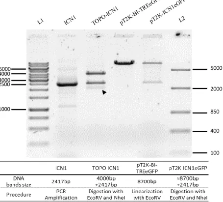
Cloning of ICN1 and DNMAML1 into the BI-TREeGFP to generate pT2K-ICN1eGFP and pT2K-DNMAML1eGFP
A second cloning reaction was performed to create the final recombinant plasmids. TOPO-ICN1, TOPO-DNMAML1 and pT2K-BI-TREeGFP were digested with EcoRV and
NheI restriction enzimes. 3 μg of TOPO-ICN1 and 8 μg of TOPO-DNMAML1 were digested,
each in a total reaction volume of 80 μL. For pT2K-BI-TREeGFP vector, 2 μg of DNA was digested in a total reaction volume of 30 μL. Afterwards, reaction products were loaded on an agarose gel (see section Agarose gel electrophoresis) and DNA fragments of interest were recovered and purified (see section Qiaquick gel extraction kit).
The ligation reaction of the linearized vector and insert (with the correct termini) was performed according to the formula: μg of vector/vector’s dimension = 10x (μg of insert/insert’s dimension). For the ligation reactions we used 50 ng of pT2K-BI-TREeGFP (8.7kb). For the ICN1 construct we used 150 ng of the insert (ICN1 insert with 2417bp) in a total volume of 20 μL and for the DNMAML1 construct we used 12.5 ng of the insert (DNMAML1 insert with 205bp) in a total volume of 15 μL. The reactions were made in 1X Buffer for T4 DNA Ligase (Biolabs), with 1-2 μL T4 DNA ligase (Biolabs 400U/μL)at room temperature (22ºC) o.n.. The reaction product was directly used to transform DH5-α competent cells. Plasmid DNA was extracted and isolated. The correct orientation of the inserts (ICN1 and DNMAML1) into the vector (pT2K-BI-TREeGFP) was confirmed using single and double digestion with EcoRV and KpnI. Sequencing analysis was performed to verify the insert sequences.
Qiaquick gel extraction kit
DNA extraction from agarose gel was carried out using the QIAquick Gel Extraction Kit (QIAGEN), according to manufacturer instructions.DNA was eluted with 50 μL of sterile distilled water.
DNA and RNA quantification
The concentration of nuclear acids was determined by spectrophotometry using the NanoDrop® ND-1000 Spectrophotometer (Thermo Scientific). One A260 unit corresponds to 50
μg/ml of double-stranded DNA and to 40 μg/ml of single-stranded RNA. Samples purity was evaluated based on A260/A280 ratio (pure preparations of DNA and RNA, i.e., without
significant amounts of proteins or phenol contaminants, show ratio values of 1.8 and 20, respectively).
Preparation of riboprobes for in situ hybridization
To generate the antisense and sense transcripts, TOPO II PCR plasmids containing the sequence of interest, were linearized with the appropriate restriction enzyme (see appendix I). The digestion reaction was performed in a total volume of 150 μL containing 20 μg of DNA, 15
16
μL of 10X enzyme buffer, 5 μL of restriction enzyme (6 U-20 U/μL) and RNase-free water. After digestion, linearized plasmid DNA was purified using phenol:chloroform extraction and ethanol precipitation (see appendix II). The synthesis of digoxigenin (DIG) -labelled antisense and sense RNA probes was carried out by in vitro transcription at 37°C for 2h. The reaction contained 8 μL of Transcription Optimized 5X Buffer (Promega), 4 μL of 0.1M DTT (Promega), 2 μL of each rGTP, rATP, rCTP (10 mM) (Roche), 1,3 μL of rUTP (10 mM) (Roche), 0,7 μL of Digoxigenin-11-UTP (10 mM) (Roche), 2 μL of RNasin® Ribonuclease Inhibitor (Promega) and 8 μL of RNAse free water. To this mixture solution and 2 μL of the appropriate RNA polymerase (see appendix I), 2 μL (2g) of the linearized templates were added. After incubation for riboprobe synthesis, the sample was treated with 6 μL of DNase I recombinant RNase-free (10 U/μL) (Roche) at 37°C for 15 min. Purification of the probe was performed using illustra MicroSpin G-50 Columns (GE Healthcare) according to manufacturer instructions. To check for probe quality and success of transcription reaction, 2 μL of reaction product were analyzed by agarose gel electrophoresis (see below).The samples were stored at -80ºC.
Agarose gel electrophoresis
Agarose gel electrophoresis was used to confirm PCR amplification products and the complete digestions with restriction enzymes, to recover and purify specific DNA fragments using extraction kit and to check riboprobe and DNA samples integrity. UltraPureTM Agarose (Invitrogen) was dissolved by heating in 1X TAE buffer (composition provided in Appendix I) to a final concentration of 0.8-1.5% (according to the required resolution for DNA fragment). To check for the presence of nuclear acids, GelRedTM Nucleic Acid Gel Stain (Biotium) or SYBR® Safe DNA Gel Stain (Invitrogen) were added to this solution in a 1:10 proportion. Samples were mixed with 6X MassRuler™ DNA Loading Dye (Fermentas) in 6:1 proportion and were loaded into the gel. Electrophoresis was performed in 1X TAE at 5-10 V/cm of gel lenght. Samples were observed under UV light and images acquired with AlphaImager HP (Alpha Innotech). The size of the fragments was estimated by comparison with the DNA ladders (FastRuler™ Low Range DNA Ladder, FastRuler™ Middle Range DNA Ladder or O'GeneRuler™ 1 kb DNA Ladder, Fermentas) run along with DNA samples.
III.2. Cellular Biology procedures
Maintenance of C2C12 murine cell line
The murine C2C12 cell line (provided by Christel Brou from Alain Israel’s group,
Unité de Biologie Moléculaire de l’Expression Génique, Institut Pasteur, Paris, France) was
maintained undifferentiated in medium containing DMEM (Invitrogen) supplemented with 20% of FBS (Invitrogen) and 1x Pen/Strep (Invitrogen). The cells were cultured on T25, T75 or T175 flasks (Nunc) using a media volume to surface area ratio of 0.1-0.2 mL/cm2. All cultures
17
were grown in a humidified incubator (Heraeus® HERAcell®) at 37ºC with 5% of CO2.
C2C12 cell line aliquotes were taken from the liquid azote and put in a 37ºC bath, without immerging, long enough to start to thaw. Immediately after, the cells were resuspended in maintenance medium and plated onto a T75 flask. The medium was changed regularly until cells reached 20% confluence (higher cell confluence induces differentiation). For this, the media was removed and the cells were washed with PBS. In order to detach the cells, pre-warmed 1-2 mL of 0.25% Trypsin/EDTA (Sigma) was added and the cells were incubated at 37ºC for about 5 min. When cells detached, trypsin was inactivated by addition of medium. For dilutions the appropriate volume was taken and plated.
Differentiation assay
C2C12 cell differentiation was induced with DMEM medium (Invitrogen) containing 2% of Horse Serum (Invitrogen) and 1x Pen/Strep (Invitrogen). Cells from two T175 flasks with 10% confluence of C2C12 were trypsinized, recovered by centrifugation at 1200 rpm for 5 min using Sorvall RT7 Plus RTH-750. Cells from each flask were resuspended in 4 mL of medium (cells from one flask resuspended in maintenance medium and cells from the other flask in differentiation medium). For each medium condition, 2 mL of cell suspension was plated in one well of a 6-well, flat-bottomed plate (TPP) covered with10x10 mm cover slips pretreated with 0.1% gelatin (m/v) in water (Sigma). After 24h, Troponin T expression was assessed by immunocytochemistry in cultured cells (see next section).
Immunocytochemistry for Troponin-t
To perform the immunocytochemistry for Troponin-T, C2C12 cells were fixed in 3.7% PFA/PBS, o.n.. Cells were then permeabilized with 0.5% TritonX-100 (Sigma) in PBS for 15 min, and washed 3 times with PBT (composition in appendix I) for 5 min. Then, the primary antibody (Monoclonal Mouse anti Troponin-t, CT3 - Developmental Studies Hybridoma Bank, 1:10 in PBS) was added and incubated in a humidity chamber at 37ºC for 30 min. Three washes of 5 min with PBT were followed by the addition and incubation of the secondary antibody (Policlonal Goat Anti-Mouse IgG + IgM (Jackson Immunoresearch Labs)) in a humidity chamber at 37ºC for 30 min. Three final washes of 5 min with PBT were performed and the cover slips were plated upside-down in slides with 15µL of VECTASHIELD® Mounting Medium (VectorLabs) (containing DAPI). The cover slips were sealed with nail polish and stored protected from light. The immunofluorescence data was acquired with Leica DM5000B Widefield Fluorescence Microscope.
Electroporation of C2C12
C2C12 cells were electroporated with the pT2K-CAGGS-EGFP and pCAGGS-T2TP vectors (Sato et al., 2007) to assess the best electroporation conditions and efficiency rate of the procedure. Electroporation of C2C12 was performed using Gene Pulser® II Electroporation System device (Bio-Rad). To assess the best electroporation conditions, several conditions from 1 to 3 pulses of 500 V to 1650 V were tried. Cells were harvested from the flasks by trypsinization and centrifugation (procedures mentioned above), and resuspended in a proper
18
volume of PBS (800 µL/2 mm electroporation cuvette). Cell numbers from 1x10⁵ to 1x10⁶ and 20 µg of each vector were added slowly through the wall of each cuvettes avoiding bubble formation. The cuvette walls were dried and placed in contact with the electrodes. After, the pulsed cells were plated in T75 flasks with maintenance medium. The next day the medium was replaced to remove dead cells and a preliminary quantification of GFP positive C2C12 cells was obtained by eye observation using Leica DMIL inverted microscope. At all times a negative control was employed, using the same conditions without DNA. The percentage of GFP positive C2C12 cells was obtained by Flow Cytometry. To do so, ≥1x10⁴
cells were added to a cytometry tube and the fluorescence data was acquired in the FACSCalibur flow cytometer (Becton Dickinson).
Transfection of C2C12
C2C12 cells were transfected with the pT2K-CAGGS-EGFP and pCAGGS-T2TP vectors (Sato et al., 2007) using FuGENE 6 Transfection Reagent (Roche) according to manufacturer instructions. C2C12 cells grown until 20% confluence were trypsinized and 1x10⁵ cells were suspended in a 2 mL of maintenance medium and plated in a 6-well, flat-bottomed plate (TPP). After 24 h of culture and 3 h prior to transfection, the media was changed to 1 mL of DMEM (medium without serum and antibiotics). Three concentrations of FuGENE reagent (3, 6 and 9 µL) were tested, as well as two DNA concentrations (1 and 2 µg of pT2K-CAGGS-EGFP and pCAGGS-T2TP each). After further 16h, the supernatant of the transfection mix (DMEM with DNA and FuGENE) was removed and the cells from each well were trypsinized, resuspended in maintenance medium and plated into T75 flasks (Nunc). After a fewdays in culture (with replating to maintain cell confluence ≤20%) GFP positive cells were quantified by Flow Cytometry, using the same method as mentioned in the previous section.
III.3. Developmental Biology procedures
Chicken embryo manipulation
Fertilized chicken eggs were obtained from Sociedade Agrícola Quinta da Freiria, S.A., Portugal, stored at 16ºC and incubated at 38ºC to initiate development. Embryos were staged according to Hamburger and Hamilton (Hamburger & Hamilton, 1951). At specific stages of development, embryos were dissected from the eggs, and the extra-embryonic membranes removed. Embryos were further processed differently depending on the method used subsequently: isolation of RNA, whole mount in situ hybridization or in vitro culture of branchial arch explants.
In ovo electroporation of neural tube
The neural tube of E2 chicken embryos was first injected with different combination of vectors (Fig. 5A), then electroporated and finally administrated with doxycycline to induce GFP and Notch construct expression in the vector system (Fig. 5B). Control conditions were performed by electroporating embryos with a mix of the four vectors:
pT2K-CAGGS-19
A B
rtTA2SM2, pT2K-BI-TREeGFP, pCAGGS-T2TP (Sato et al., 2007) and pCAG-CherryNLS plasmid (encoding a nuclear form of a red fluorescent protein driven by the constitutive CAG promoter (Vilas-Boas, Fior, Swedlow, Storey, & Domingos Henrique, 2011). Plasmid pT2K-BI-TREeGFP and pCAGGS-T2TP were at around 2 µg/µL; however pT2K-CAGGS-rtTA2SM2 and pT2K-BI-TREeGFP were at 1:2 proportion as recommended by Watanabe and collaborators (Watanabe et al., 2007). The same proportions were used for all plasmids when injecting recombinant plasmids pT2K-ICN1eGFP and pT2K-DNMAML1eGFP (instead of pT2K-BI-TREeGFP). pCAG-CherryNLS plasmid was at a concentration of 0,1 µg/µL and was used as a control vector to assess the electroporation efficiency. Fast Green was used in the plasmid mix a 1:10 proportion to dye the DNA mix to visualize of the injection site and to follow the distribution of the injected solution in the embryo.
DNA solution was injected into the lumen of the neural tube of E2 chicken embryos with a microinjection capillary glass (Harvard Apparatus), using the Inject + Matic Microinjector (Inject + Matic) (Fig. 5A). Platinum electrodes (Nepagene), distanced 4 mm apart were placed parallel to the neural tube on the surface of the embryo, along the anteroposterior axis (Fig. 5A), and some drops of PBS with 1x Pen/Strep were added to the surface of the embryo. Using an ElectroSquare Porator ECM830 (BTX), 4 pulses of 25 V for 50 ms, spaced by 100 ms were applied twice. The side of the neural tube closer to the positive electrode was the experimental side (having directional entry of DNA into those cells), while the other was the control side (Fig. 5B). Immediately after electroporation, 500 µL of 0.1mg/mL doxycycline hyclate (doxycycline Sigma) in Hank’s Buffered Salt Solution (HBSS, Invitrogen) (Sato et al., 2007) were injected between the embryo and the yolk, using a 1 mL syringe and without penetrating the area opaca (Watanabe et al., 2007). For each experimental condition, embryos were electroporated with either the ICN1 or DNMAML1 DNA mix of Figure 5. In ovo electroporation of the neural tube of chick embryos. A) Schematic representation of an HH13 embryo (E2) (adapted from Hamburguer & Hamilton,1951) injected with DNA solution into the neural tube’s lumen previous to electroporation. Electrodes position and polarity are shown by bars and by minus (-) and plus (+) signs, respectively. B) Schematic representation of the electroporation of a neural tube injected with DNA solution (blue) followed by DOX administration to induce gene expression. GFP (green) and CherryNLS (red) expression are expected in the right side of the neural tube, according to electrodes position; n:neural tube, n:notochord.