i
FACULDADE DE FARMÁCIA
The role of high S-adenosylhomocysteine levels
and histone hypomethylation in cardiovascular
disease
Dora Sofia Chan Roseira Dias
Dissertação orientada por:
Doutora Rita Castro
Doutora Isabel Rivera
Mestrado em Ciências Biofarmacêuticas
ii
FACULDADE DE FARMÁCIA
The role of high S-adenosylhomocysteine levels
and histone hypomethylation in cardiovascular
disease
Dora Sofia Chan Roseira Dias
Dissertação orientada por:
Doutora Rita Castro
Doutora Isabel Rivera
Mestrado em Ciências Biofarmacêuticas
ii
The studies presented in this thesis were performed within the Metabolic & Genetics (Met&Gen) research group, at the Research Institute for Medicines (iMed.ULisboa), Faculty of Pharmacy, Universidade de Lisboa, under the supervision of Rita Castro, Ph.D, and Isabel Rivera, Ph.D. This work was supported by grant PEstOE/SAU/UI403 by FCT.
iii
As doenças cardiovasculares são a principal causa de morte nos países desenvolvidos. Assim sendo, é importante estudar os mecanismos moleculares causadores destas patologias, descobrindo novos mecanismos moleculares subjacentes e factores de risco, bem como novas formas de prevenção e terapias mais eficazes.
A desregulação do metabolismo da homocisteína, que leva ao aumento dos seus níveis circulantes no plasma (designada por hiperhomocisteinémia), está associada às doenças cardiovasculares. No entanto, os mecanismos inerentes à toxicidade vascular da homocisteína permanecem ainda por elucidar. Recentemente foi colocada a hipótese de que a acumulação do precursor da homocisteína, a S-adenosil-homocisteína (SAH), que ocorre em situações de hiperhomocisteinémia, poder conduzir à hipometilação celular, explicando assim a toxicidade vascular associada à hiperhomocisteinémia. A SAH funciona como inibidor da actividade enzimática da maioria das metiltransferases celulares que usam S-adenosilmetionina (SAM), o precursor metabólico da SAH, como dador de grupos metilo para as reacções de transmetilação que actuam sobre diversos compostos como DNA e proteínas, incluindo as histonas. A metilação do DNA e das histonas constituem mecanismos epigenéticos que regulam a expressão genética.
A Enhancer of Zeste Homolog 2 (EZH2) estabelece a trimetilação da lisina 27 da histona H3 (H3K27me3), uma marca epigenética envolvida na repressão de genes, através do aumento da condensação da cromatina. De entre os genes cuja transcrição é reprimida no endotélio, incluem-se aqueles que têm um papel na promoção da aterosclerose. A EZH2 é uma histona metiltransferase dependente da SAM e, consequentemente, a acumulação de SAH poderá afectar negativamente o funcionamento da EZH2. Assim sendo, se os níveis intracelulares de SAH aumentarem, o conteúdo de H3K27me3 celular poderá diminuir, com a consequente activação de genes pró-aterogénicos, o que contribuirá para o estabelecimento da aterosclerose e da patologia cardiovascular. Esta possibilidade constitui a hipótese de trabalho investigada no presente trabalho.
Para confirmar esta hipótese avaliámos se a acumulação intracelular de SAH levaria à diminuição de H3K27me3, quer in vitro, em células endoteliais humanas, quer in vivo usando células mononucleares do sangue periférico obtidas voluntariamente de doentes cardiovasculares e de controlos. Os nossos resultados indicam que, in vitro, a acumulação intracelular de SAH diminui a marca H3K27me3. Nas células endoteliais, a quantificação dos níveis intracelulares de SAM e SAH revelou uma acentuada diminuição da sua razão,
iv
células controlo. Relativamente aos estudos in vivo, os resultados obtidos demonstraram que o conteúdo de H3K27me3 não diferiu de forma significativa nos doentes cardiovasculares e a na população controlo, apesar da razão SAM/SAH intracelular estar diminuída nos doentes cardiovasculares em relação aos controlos.
Embora os resultados in vitro corroborem a nossa hipótese, fomos incapazes de evidenciar in vivo, um relação inversa entre o ambiente de hipometilação celular e o conteúdo da marca epigenética dependente da EZH2 em células mononucleares do sangue periférico de doentes cardiovasculares. Assim, serão necessários estudos futuros, numa população-alvo mais alargada, para confirmar os resultados obtidos in vivo. Idealmente, estes estudos futuros deveriam incidir em doentes cardiovasculares que não estejam submetidos a nenhum tipo de medicação. Isto porque os resultados da avaliação de mediadores da aterosclerose, como o colesterol por exemplo, foram nitidamente influenciados pela terapia a que os doentes cardiovasculares estavam sujeitos aquando da colheita de sangue. Por outro lado, estes resultados foram, ainda assim, interessantes, comprovando a eficácia da medicação aconselhada aos doentes cardiovasculares, uma vez que estes revelaram, no geral, níveis de lípidos circulantes significativamente mais baixos do que os indivíduos controlo.
Adicionalmente, investigações futuras deverão alargar o estudo a outro tipo de células, como por exemplo células do tecido vascular. Com efeito, o conteúdo da marca epigenética H3K27me3 das células estudadas in vivo poderá não representar o conteúdo das células endoteliais. Só assim poderemos avaliar inequivocamente se a marca H3K27me3 está relacionada com o aparecimento e/ou desenvolvimento de um fenótipo endotelial pró-aterogénico. Deste modo poderemos também clarificar se a desregulação da actividade da EZH2 induzida pela SAH contribui efectivamente para o aparecimento da aterosclerose e doença cardiovascular.
Palavras-chave: doença cardiovascular, Enhancer of Zeste Homolog 2, epigenética,
hipometilação, homocisteína.
v
Cardiovascular diseases (CVD) are the main cause of death in developed countries. Thus, it is important to study the mechanism that causes disease progression, uncover new risk factors, and new ways of effective prevention and therapeutic approaches.
Increased levels of circulating homocysteine are associated with cardiovascular disease. However, the mechanisms underlying this association remain elusive. Cellular hypomethylation caused by accumulations of the homocysteine precursor, S-adenosylhomocysteine (SAH), may explain homocysteine’s toxicity. SAH inhibits the enzymatic activity of most cellular methyltransferases, which use S-adenosylhomocisteine (SAM), SAH’s precursor, as a methyl group donor, acting upon several important compounds like DNA and proteins, including histones. Methylation of DNA and histones are epigenetic mechanisms that regulate gene expression and whose deregulation may cause diseases.
Enhancer of Zeste Homolog 2 (EZH2) is a histone methyltransferase that establishes trimethylation of lysine 27 on histone H3 (H3K27me3), an epigenetic mark associated with repression of pro-atherogenic genes in endothelial cells. EZH2 activity depends on SAM, therefore SAH accumulation may negatively affect EZH2 methyltransferase activity diminishing H3K27me3 content, and leading to the expression these pro-atherogenic genes, and to CVD. This possible mechanism constitutes the basis of our work.
To study this hypothesis, we evaluated whether SAH accumulation leads to H3K27me3 decrease either in vitro, in human endothelial cells, or in vivo, in peripheral mononuclear blood cells (PMBC) of CVD patients. Our in vitro results demonstrate that H3K27me3 content decreases if SAH accumulates in the cells. Yet, our in vivo results show no significant difference in the H3K27me3 content in PBMC from CVD patients and controls. Although our
in vitro results corroborate our hypothesis, additional studies with a larger cohort of patients
and with other cellular material, namely vascular tissue, are necessary in order to ascertain whether SAH-induced deregulation of EZH2, and consequent H3K27me3 endothelial content, contributes to atherosclerosis and CVD.
Key words: cardiovascular disease, DNA hypomethylation, Enhancer of Zeste Homolog 2,
vi
Às minhas orientadoras, professora Rita Castro e professora Isabel Rivera, que me ajudaram e apoiaram durante estes dois anos e pouco, e que me permitiram crescer tanto científica como pessoalmente. Esta tese não seria a mesma sem a vossa presença, críticas, sugestões e observações. Obrigada por tudo o que me ensinaram!
Às professoras Isabel Tavares de Almeida e Paula Leandro por terem dado a oportunidade de realizar esta tese no grupo Met&Gen. A todas as pessoas do grupo Met&Gen, que de alguma forma me ajudaram e contribuíram de alguma forma para a execução desta tese, principalmente à Madalena Barroso, Marco Moedas, Hana Pereira e Cristina Florindo. E à Inês Vieira da Silva, pela companhia e simpatia. À professora Maria João Silva pela ajuda e disponibilidade.
Ao Hospital de Santa Maria, cuja colaboração foi imprescindível para a realização desta tese.
A todos os que participaram neste estudo de forma voluntária. Sem essa colaboração, esta dissertação seria completamente diferente. Muito obrigada!
À Dra. Elisa Alves que colheu o sangue do grupo controlo. Ao Tiago Leite, que levou as amostras para serem analisadas no laboratório Dr. Joaquim Chaves e que me ajudou com algumas informações do grupo de estudo.
À Maria Carlos Nunes, pelos brainstormings, piadas (nerds), conversas filosóficas e palmadinhas nas costas durante este tempo. És tão grande Maria! Nunca duvides disso! Algo de grande te espera. E espero que encontres o teu caminho, sem medos. E esse teu percurso se cruze com o meu (if you know what a mean…).
À Eli, Eleonora Scarpa, que mesmo no curto período de tempo que esteve presente no CPM, se tornou numa amiga especial. Por me ter permitido desvanecer quando mais precisei. Espero que a luz continue a brilhar, Eli!
Às poderosas, Rita Leones e Shikha Raikundalia, que durante todo o mestrado tornaram os meus dias muito mais alegres. Por estarem sempre disponíveis e bem-dispostas, mesmo que mais de longe durante este ano de tese. À terceira poderosa, Carla Ferreira Mendes, que juntamente com a Rita e a Shikha, aturou os meus desvaneios, disparates e crises existencias (incluindo as laboratoriais). Mas a ti, Carla, agradeço-te especialmente por teres contribuído para a minha felicidade e pela companhia constante no CPM. Gosto tanto, tanto, de vocês! Pelo que são, pela amizade que têm por mim e por terem partilhado o vosso tempo comigo. Foram cruciais neste meu percurso…
vii
Dionísio, Peter, pelas dicas, sugestões e disponibilidade.
Aos meus biólogos, Ana Sofia Cruz, Ana Sofia Oliveira, Verónica Mixão e Bruno Gonçalves, que estiveram presentes ao longo da minha licenciatura. Ao Pedro Quina, cujas piadas nunca se esgotam, e que está sempre pronto a animar o dia, seja de que forma for.
À Rita Dias, ao Tiago Baeta, meus afilhados de curso, cuja amizade e presença me são indispensáveis. Não consigo expressar o quão importante é o vosso apoio, sorriso, abraço e sentido crítico (científico ou não). Mas vocês sabem… Obrigada, do fundo do coração.
Ao Timon, Pedro Timóteo, meu padrinho de curso. Um obrigado gigante! Por seres único, por teres sido um pilar durante a minha licenciatura. Obrigada por continuares a cuidar de mim, mesmo longe. Quando for grande quero ser como tu!!! E contar piadas como só tu fazes!
À Ana Soares, pelas conversas de café e apoio. Ao Martim Rodrigues, por todas as conversas e pela ligação cósmica. À Rita Raposeiro pelos abracinhos.
Ao Sasha Fonseca, que se revelou um grande amigo. Obrigada por teres ouvido os meus desabafos durante estes anos. Por todos os conselhos e dicas informáticas, pelos jantares, pelos cafés…
Ao Miguel Mendes, cuja amizade, apesar dos seus altos e baixos, já dura há 11 anos. Obrigada por teres crescido comigo e de me teres mostrado diferentes perspectivas de vida. Ao Daniel Azevedo, por ter também ter crescido comigo.
À Sara Pereira, minha colega de casa, que aturou com muitos sorrisos e paciência, os humores que esta tese suscitou.
Ao “Sr. Doutor” Ricardo Viegas por ter sempre uma palavra de apoio. E também ao Marcos Nascimento, pelas conversas, sempre interessantes. Ao meu professor de matemática, Zé Carlos, que me encorajou a seguir os meus sonhos. Um grande obrigada, onde quer que esteja!
À Madalena Calisto, Sónia Tomás, Inês Pinto, Ana Teresa Pepe e tantas outras que aprenderam e partilharam (talvez ainda o partilhem) o grande lema de vida que a patinagem nos ensinou, mas que faz mais sentido dito em inglês: “if you are going to land, it might as be on your feet.”.
viii Sr. Américo pelas luzes.
Aos meus pais, por nunca me ter faltado nada. Por, desde muito cedo, terem estimulado a minha curiosidade e a minha procura pelo saber. Por terem apoiado os meus sonhos e as minhas pequenas conquistas. Por me terem ajudado a caminhar pelo meu próprio pé. Nada disto seria possível sem vocês! Esta tese, que exigiu muito trabalho e dedicação, dedico-a, em retorno, a vocês!
Ao Rui Pinheiro, por tudo o que fez por mim. Por todo o amor e carinho. Por tudo que me ensinou. Por todos os momentos e desafios que me proporcionou. Por todo apoio e paciência. Por nunca ter desistido de me fazer sorrir, mesmo quando estava extremamente ocupado. Por ter cuidado tão bem de mim e por continuar a fazê-lo. Por não me ter faltado, por me ter dado colo sempre que precisei (e mesmo quando não precisei)… Por mais outras N coisas, que se as fosse escrever todas, teria que escrever outra tese… Mas acima de tudo, por me fazer verdadeiramente feliz.
No fundo, a todas as pessoas, que se cruzaram no meu caminho e que de alguma forma, fizeram de mim o que sou hoje: “Everyone you will ever meet knows something you don’t.” – Bill Nye.
ix
Resumo ... iii
Abstract ... v
Agradecimentos ... vi
Index of Tables ... xi
Index of Figures... xii
Abbreviations ... xiii
I – INTRODUCTION ... 1
1. Cardiovascular Disease ... 1
1.1. Endothelium dysfunction, atherosclerosis, and CVD ... 2
2. Homocysteine Metabolism ... 5
2.1. Homocysteine and link to disease ... 7
2.2. Homocysteine and cell hypomethylation... 8
3. Epigenetics ... 9
3.1. DNA methylation ... 9
3.2. Histone methylation ...11
3.2.1 Enhancer of Zeste Homolog 2 ...12
II - OBJECTIVES ...15
III – METHODS AND MATERIALS ...17
A. In vitro studies: incubation procedure in endothelial cells...17
A.1. Cell culture and treatments ...17
A.2. Sample preparation ...17
A.3. SAM and SAH analysis ...18
A.4. Western blot analysis ...18
A.4.1. Histone extraction ...18
A.4.2. Histone quantification ...18
A.4.3. Western blotting ...19
A.4.4. Image and Statistical analysis ...20
B. In vivo studies: characterization of CVD patients and healthy individuals ...20
B.1. Participants and sample collection ...21
B.2. Preparation of biological samples ...21
B.2.1. PBMC isolation ...21
B.2.2. Isolation of histones and RNA ...23
x
B.3. Biochemical analysis ...24
B.3.1. Statistical analysis ...24
B.4. Western blot analysis and statistical analysis ...24
B.5. RT-qPCR ...25
B.5.1. Statistical analysis ...26
IV – RESULTS AND DISCUSSION ...27
A. In vitro studies: incubation procedure in endothelial cells ...27
A.1. Influence of ADA in H3K27me3 on HUVEC cells ...27
B. In vivo studies: characterization of CVD patients and healthy individuals ...29
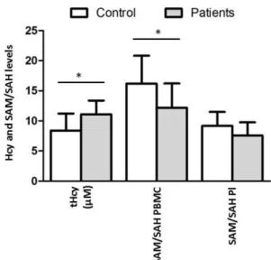
B.1. Quantification of Hcy levels and SAM/SAH ratio ...29
B.2. Evaluation of cardiovascular risk parameters in the population ...33
B.2.1. Evaluation of pro-atherogenic indicators in circulating plasma ...33
B.2.2. Evaluation of CVD risk in the population ...36
B.2.3. Quantification of pro-inflammatory indicators ...37
B.2.3. Evaluation of circulating pro-atherogenic mediators ...39
B.3. H3K27me3 detection in the PBMC of the population ...41
B.4. Determination of EZH2 and IL-1β expression ...42
V – CONCLUSION ...45
xi
TABLE 1.LIST OF PRIMARY AND SECONDARY ANTIBODIES USED IN IMMUNOBLOT ASSAYS. ...20
xii
FIGURE 1.PERCENTAGE OF DEATHS DUE TO CVD IN RELATION TO TOTAL DEATHS IN 2013. ... 1
FIGURE 2.SCHEMATIC REPRESENTATION OF THE ATHEROSCLEROTIC PROCESS. ... 3
FIGURE 3:HOMOCYSTEINE METABOLIC PATHWAY. ... 6
FIGURE 4:POTENTIAL MECHANISM(S) BY WHICH SAH CAUSES ENDOTHELIAL ACTIVATION. ...13
FIGURE 5.ISOLATION OF LEUKOCYTES FROM EDTA-BLOOD USING FICOLL-PAQUE™ DENSITY GRADIENT CENTRIFUGATION. ...22
FIGURE 6.SIMPLIFIED SCHEME OF RNA AND HISTONE EXTRACTIONS EXECUTED FROM THE SAME SAMPLE OF PBMC. ...23
FIGURE 7.INTRACELLULAR SAM/SAH RATIO IN HUVEC INCUBATED WITH ADA0 ΜM AND 20 ΜM FOR 24 H AND 48 H. ...27
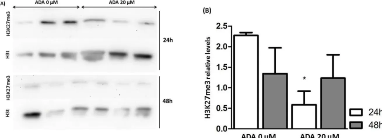
FIGURE 8.EFFECT OF EXCESS OF SAH ON H3K27ME3 MARK IN HUVEC. ...28
FIGURE 9.EVALUATION OF THCY LEVELS BY GENDER IN THE PLASMA. ...30
FIGURE 10.EVALUATION OF THCY,SAM/SAH RATIO IN PBMC AND IN PLASMA IN THE STUDIED POPULATION. ...30
FIGURE 11.PLOT OF INDIVIDUAL VALUES OF PLASMA THCY VS SAM/SAH RATIO. ...31
FIGURE 12.PLOT OF INDIVIDUAL VALUES OF SAM/SAH RATIOS IN THE PLASMA VS SAM/SAH RATIO IN THE PBMC. ...32
FIGURE 13.EVALUATION OF SAH LEVELS IN THE STUDIED POPULATION. ...33
FIGURE 14.LEVELS OF PLASMA CIRCULATING LIPIDS IN THE STUDIED POPULATION. ...33
FIGURE 15.CVD RISK ANALYSIS IN THE STUDIED POPULATION. ...37
FIGURE 16.INFLAMMATORY PARAMETERS IN THE STUDIED POPULATION. ...38
FIGURE 17.EVALUATION OF ENDOTHELIAL DYSFUNCTION INDICATORS IN THE STUDIED POPULATION. ...40
FIGURE 18.H3K27ME3 RELATIVE LEVELS IN THE STUDIED POPULATION. ...42
xiii
Adenosine-2,3-dialdehyde ADA
Angiotensin Converting Enzyme ACE
Apoliprotein A-I apoA-I
Apoliprotein B apoB
Cardiovascular Diseases CVD
C-reactive Protein CRP
Cytosine preceding Guanine CpG
Diethylpyrocarbonate DEPC
DNA methyltransferases DNMT
Dr. Joaquim Chaves Laboratories JCLab
Endothelial Cells EC
Endothelial Dysfunction ED
Enhancer of Zeste Homolog 2 EZH2
High-density Lipoprotein HDL
High-density Lipoprotein cholesterol HDL-c
Histone Methyltransferases HMT
Homocysteine Hcy
Human Coronary Artery Endothelial Cells HCAEC
Human Umbilical Endothelium Cells HUVEC
Hyperhomocysteinemia HHcy
Hypotonic Lysis Buffer HLB
Intercellular Adhesion Molecule-1 ICAM-1
Low-density Lipoprotein LDL
Low-density Lipoprotein cholesterol LDL-c
Methionine Met
Nitric Oxide NO
Nuclear Factor-KappaB NF-κB
Núcleo de Prestação de Serviços de Bioquímica, FFULisboa
NPSB
Oxidized LDL ox-LDL
Peripheral Blood Mononuclear Cells PBMC
Polycomb Repressor Complex 2 PCR2
Reactive Oxygen Species ROS
xiv
S-adenosylhomocysteine SAH
S-adenosylhomocysteine Hydrolase SAHH
S-adenosylmethionine SAM
Soluble Intercellular Adhesion Molecule-1 sICAM-1
Soluble Vascular Cell Adhesion Molecule-1 sVCAM-1
Total Homocysteine tHcy
Trimethylation of histone H3 at lysine 27 H3K27me3
Vascular Cell Adhesion Molecule-1 VCAM-1
1
I – INTRODUCTION
1. Cardiovascular Disease
Cardiovascular diseases (CVD) are a group of diseases resulting from dysfunctional settings of arteries, veins, and heart1. Within this context, CVD morbidity and mortality are usually caused by ischemic heart disease, stroke, and congestive heart failure2, and the amount of deaths rises with age3.
CVD is considered the leading cause of death in developed countries4, causing 17.1 million deaths every year5, and this number is expected to grow up to 23.6 million by the year of 20306. In Portugal, the percentage of deaths attributed to CVD by gender is higher in women than in men, as illustrated in Figure 1, where the percentage of death due CVD in 2013 is shown.
Results worldwide correspond to CVD death percentage in relation to total deaths at a national level. The color bar scheme indicates the percentage of CVD death rate, from the lowest, represented in dark blue, to the highest, represented in red. Results of Portugal correspond to CVD death percentage in relation to total deaths per gender.
Abbreviation: CVD, cardiovascular disease. Data source collected from references7,8.
In approximately ten per cent of cases CVD are an heritable condition9, but there are numerous non-heritable risk factors contributing to CVD, such as hypertension, smoking, diabetes, bacterial infections, inappropriate vitamin consumption, abnormal blood cholesterol, high blood pressure, lack of exercise, obesity or overweight, aging, and high plasma levels of homocysteine (Hcy)1,6,10–15. Air pollution has also shown to increase the risk for CVD morbidity and mortality, even though it depends on the individuals’ ethnicity16.
2
CVD develops after a prolonged asymptomatic phase17 and negative cardiovascular events may occur in the absence of notorious risk factors18, therefore, it is important to find new ways to diagnose and prevent these group of diseases.
The cardiovascular systems is composed by the heart, connected to a network of arteries, veins, and capillaries19. Different cell types constitute the vessel wall, such as smooth muscle cells, and endothelial cells (EC)20. The endothelium is composed by a monolayer of EC, coating the entire vascular system21. In normal conditions, the endothelium is in a quiescent state, with a turnover rate from months to years. It conducts the blood ensuring the nutritional support for organs, and regulates vascular tone, platelet activity, and blood vessel formation and growth. The endothelium also plays a role on innate and adaptive immunity, by regulating leukocyte adhesion and hemostasis22–25. It also limits clot formation, by maintaining blood’s fluidity, and functions as a barrier between blood and surrounding tissues24.
1.1. Endothelium dysfunction, atherosclerosis, and CVD
The homeostasis of the endothelium may be disturbed by metabolites, hormones23 and by microbiological, immunological or mechanical insults, resulting in vascular injury26. Upon injury, the endothelium will undertake major changes27, causing the reshaping of the vessel wall, both in size and composition28. In this process, the endothelial barrier permeability is disrupted29, leading to arterial stiffness, and to endothelium dysfunction (ED)19. ED is characterized by vasoconstriction, vascular inflammation, upregulation of cytokines and adhesion molecules, and decreased bioavailability of nitric oxide (NO)30,31. NO is an important molecule with vasodilatory properties and cardioprotective effects31. The reduction of NO bioavailability occurs due to NO synthase disruption32, as NO is synthesized from ʟ-arginine via endothelial NO synthase (eNOS), which has anti-atherogenic functions33. ED is linked with eNOS dysfunction34.
ED is the initial stage of atherosclerosis, which is the leading cause of CVD24,35. Atherosclerosis is a multifactorial disease that progresses with age, usually begins in youth and continues asymptomatic until a later point in life36,37. It develops in the intima of middle and large size arteries, mainly in vessel bifurcations38 and inner curvatures39, mostly in areas of disturbed blood flow40. Alterations like vascular remodeling, calcification, oxidative stress, cell death, and in lipoprotein metabolism occur during atherosclerosis development36. Ages over 45 years for men and 55 years for women are a risk factor41. Aged vessels are more susceptible to develop vascular injuries and impaired angiogenesis, therefore they can easily develop atherosclerosis19. Until today, there is no way to reverse atherosclerosis by medical therapeutics42.
3
The accumulation of low-density lipoprotein (LDL) in the subendothelial matrix is an initial step of atherosclerosis43. LDL can diffuse passively through EC junctions, and its interaction with apoliprotein B (apoB) seems to be important for its retention in the vessel wall43, and consequent progression of atherosclerosis. ApoB is the principal apolipoprotein of LDL and the largest isoform present in the body35, allowing the internalization of LDL particles by the cells, so they can absorb cholesterol44. LDL and other apoB-containing lipoproteins can accumulate in the intima43. LDL can undertake modifications like oxidization. Oxidized LDL (ox-LDL) can activate EC, monocytes and macrophages38.
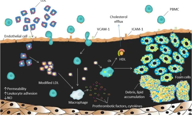
Figure 2. Schematic representation of the atherosclerotic process.
The structure of a normal artery is represented with the endothelial cell layer at the top, followed by the intima (exaggerated in this figure), and smooth muscle cells at the bottom. Changes in the blood flow affect EC, influencing their permeability and gene expression, leading to a decreased NO bioavailability. The retention of LDL and other lipoproteins are a key initiator event. LDL undergoes oxidative modification, which are, inhibited by HDL. Oxidized LDL stimulates the production of adhesion molecules (like ICAM-1 and VCAM-1) by EC, resulting in PBMC recruitment into the vessel wall. Oxidized LDL forms aggregates that are recognized by macrophages (derived from monocytes), that forms foam cells. When they die, foam cells contribute to a growing mass of debris and lipids that accumulate over time.
Abbreviations: LDL, low-density lipoprotein, NO, nitric oxide; ICAM-1, intercellular adhesion molecule-1; VCAM-1, vascular cell adhesion molecule-1; PBMC, peripheral blood mononuclear cells; Ch, cholesterol; HDL, high-density lipoprotein; EC, endothelial cells.
Adapted from references43,45.
The upregulation of adhesion molecules, such as vascular cell adhesion molecule-1 (VCAM-1) and intercellular adhesion molecule-1 (ICAM-1), will favor the adhesion of peripheral blood mononuclear cells (PMBC - namely lymphocyte and monocytes) to the endothelium31, as shown in Figure 2. Moreover, the dysfunctional EC phenotype will increase the expression of pro-inflammatory cytokines, promoting the entrance and retention of
4
cholesterol containing LDL in the artery wall and modifications of LDL structure can increase the expression of adhesion molecules in EC46–48. LDL is composed by phospholipids and free cholesterol, and acts as a cholesterol transporter44.
These accumulations can occur over time42, as lipoprotein retention seems to be a self-perpetuating process35, maintained by the dysfunctional EC phenotype46. This process results in EC activation, and acts as a chemoattractant for monocytes38, and can also be an indicator of oxidative stress18.
Atherosclerosis is characterized by high plasma concentrations of cholesterol49, that leads to the formation and growth of the atherosclerotic plaque41. These plaques are formed by accumulation of fibrous materials, cell debris, minerals, and lipids50; infiltration of immune cells leads to the establishment of a collagen fibrous cap45. Proliferation and migration of vascular smooth muscle cells (VSMC) can occur, as well as accumulation of collagen and proteoglycans, resulting in blood vessel obstruction28.
In opposition to LDL, high-density lipoprotein (HDL) has a protective function against atherosclerosis. HDL is responsible for the cholesterol efflux from macrophages, through the action of apolipoprotein A-I (apoA-I), transporting the cholesterol back to the liver51. From there, HDL go to the intestine where apoA-I is liberated from lipids, so it can be attached to new HDL particles, or it is degraded in the kidneys52. HDL may have a protective role upon ED, by enhancing NO release, preventing apoptosis, and reducing oxidative damage29,51. Nevertheless, HDL seems to function as a chameleon-like lipoprotein, having anti-inflammatory functions in the absence of an acute phase response or systemic inflammation, and promoting inflammation in the reverse case scenario53.
With the progression of atherosclerosis, deposits of C-reactive protein (CRP), an innate immune response protein, can also be found in the intima54,55. CRP may affect the expression of endothelial cell adhesion molecules, chemokine production and contributes for EC’s apoptosis21. CRP levels may depend on the individuals genetic variation56.
Besides CRP, fibrinogen is also associated with atherosclerosis development15. High levels of plasma fibrinogen are found in patients with acute thrombosis, and acute coronary syndrome57. Fibrinogen is a complex hexamer composed by α, β and γ chains58, and it is the major plasma coagulation factor59. The isoform γ’ is associated with atherothrombotic events60.
As the plaques grow, EC proliferate43 and plaques can extend beyond into the arterial lumen61, being able to form a necrotic core45. The lesions will grow continuously until a critical point, leading to the narrowing of the lumen (stenosis). At some point, the dilation of the
5
artery can no longer compensate stenosis49, macrophages will become apoptotic42 and will destabilize the atherosclerotic plaque62. The plaques can grow enough to block the blood flow, leading to vein occlusion and eventually resulting in adverse vascular events as myocardial infarction (MI) or stroke43. Alternatively, the degradation of the fibrous cap may lead to plaque rupture43,63, releasing prothrombotic factors, promoting thrombosis38,41. The beginning and development of CVD complications depend on the interplay between inflammation response and oxidative stress64. If the inflammatory response continues, the artery wall will thicken and harden41, and the number of macrophages and lymphocytes will increase, leading to the release of cytokines, chemokines, growth factors, and hydrolytic enzymes. Over time, this could lead to focal necrosis49.
Elevated shear stress can lead to platelet aggregation and further plaque rupture. On the other hand, a low shear stress permits the formation and development of atherosclerotic plaques10. Activated EC will also interact with leukocyte through the same adhesion molecules and through pro-inflammatory cytokines, like IL-6 and -829. Monocytes produce IL-1β, a powerful cytokine, and high levels of IL-1β seems to contribute for atherosclerosis development65.
2. Homocysteine Metabolism
Homocysteine (Hcy) is formed during the metabolism of methionine (Met)66, an essential amino acid found in foods with high protein content1.
Met is transformed in S-adenosylmethionine (SAM), a reaction catalyzed by methionine adenosyltransferase (MAT, EC 2.5.1.6), which has two isoenzymes: one distributed along all tissues and another present in the liver67. SAM is the universal methyl donor in the cell, and specific methyltransferases transfer SAM’s methyl group to various molecules like DNA, RNA, proteins or lipids68,69. After the transfer of the methyl group, S-adenosylhomocysteine (SAH) is formed. SAH is a strong competitive inhibitor of the catalytic action of most SAM-dependent methyltransferases67,70. As such, high levels of SAH may decrease the cellular methylation status71. SAM/SAH ratio is used as an indicator of cellular methylation potential, and decreased SAM/SAH values indicate that the cell is hypomethylated72.
SAH is further hydrolyzed into Hcy and adenosine by SAH hydrolase (SAHH, EC 3.2.2.9) through a reversible reaction that favors SAH synthesis. However, in physiological conditions, Hcy is quickly metabolized, favoring SAH hydrolysis66,67,72.
Following its production, Hcy can be catabolized by the transsulphuration pathway, in which Hcy and serine are condensed into cystathionine, by the action of cystationine-β-synthase (CBS,EC 4.2.1.22), that uses pyridoxal phosphate (PLP), the active form of vitamin
6
B6, as co-factor67. Cystathionine is converted into cysteine which will be further oxidized into
urinary sulphates that are eliminated from the body73, as illustrated in Figure 3. Interestingly, the transsulphuration pathway is absent in vascular and myocardial cells due to the lack of CBS, and for this reason the cardiovascular system is highly susceptible to Hcy accumulations and resulting toxicity32.
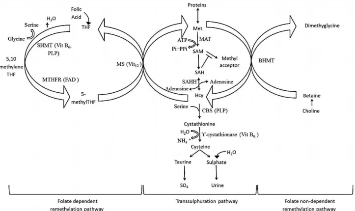
Figure 3: Homocysteine metabolic pathway.
Abbreviations: Met, methionine: MAT, ATP-ι-methionine S-adenosyltranferase; SAM, S-adenosylmethionine; SAH, S-adenosylhomocysteine; SAHH, S-adenosylhomocysteine hydrolase; Hcy, homocysteine; THF, tetrahydrofolate; MS, methionine synthase; BHMT, betaine-homocysteine methyltransferase; MTHFR, 5,10-methylenetetrahydrofolate reductase; SHMT, serine hydroxymethyltransferase; CBS, cystathionine β-synthase; PLP, pyridoxal phosphate; Vit, vitamin.
Adapted from reference67.
Met consumption only provides a portion of the body’s needs and its synthesis requires a lot of energy; so, the organism depends on optimal Hcy recycling1. Hcy can be remethylated into Met through two ways: a folate-dependent pathway or a folate-independent pathway68. In the folate-dependent pathway, methionine synthase (MS, EC 1.16.1.8) is responsible for the conversion of Hcy into Met74. 5-Methyl tetrahydrofolate (5-methylTHF) supplies the methyl group used by MS for Hcy remethylation66. MS requires vitamin B12 as a co-factor and
produces also tetrahydrofolate (THF)75. THF is further reduced of 5,10-methylenetetrahydrofolate (5,10-methyleneTHF), and then converted into 5-methylTHF, by the action of methylenetetrahydrofolate reductase (MTHFR, EC 1.5.1.20)75. Mutated MTHFR can lead to Hcy accumulation76.
7
The betaine-homocysteine pathway is a folate-independent remethylation cycle, in which Met is produced from Hcy by a reaction catalyzed by betaine-homocysteine methyltransferase (BHMT, EC 2.1.1.5) that uses betaine as a methyl donor group. Betaine is then converted into dimethylglycine67,68.
Adequate levels of B vitamins are mandatory to ensure a regular metabolism of Hcy77. Accordingly, deficiencies in B vitamins can lead to an increase Hcy circulating levels74. On the other hand, treatments with folic acid can lower the levels of accumulated Hcy78.
Women tend to have lower Hcy circulating levels than men, probably due to the influence of sex hormones1. Also, Hcy levels increase progressively with age79.
2.1. Homocysteine and link to disease
High Hcy plasma levels are associated with atherosclerosis69, and Hcy is considered to be an independent cardiovascular risk factor70. Meta-analyses have shown that increases of 3 - 5 μmol/L of Hcy in the plasma can enhance 10 % - 27 % the risk for thromboembolism, myocardial infarction, or stroke80.
The increase of plasma Hcy levels (hyperhomocysteinemia or HHcy) have been linked to impaired endothelial vasodilation81, platelets aggregation, vascular inflammation32, and ED82. A large cohort study, performed in Italy, demonstrated that high plasma levels of Hcy were linked to coronary atherosclerosis32. Subjects presenting HHcy also exhibited high levels of circulating SAH in the plasma and alterations in DNA methylation pattern in the lymphocytes67.
Several mechanisms have been proposed to explain the vascular toxicity of Hcy. Various studies showed that increased levels of plasma Hcy are important for the initiation and progression of vascular disease, including ED, inhibition of methylation due to SAH accumulation, protein modification at a post-translational level, decreased NO levels, and increased oxidant stress83.
The beneficial effects of folate supplementation as an Hcy-lowering therapy aiming to diminish CVD risk are still controversial. Oral administration of folic acid, either alone or in combination with vitamin B12 has shown to lower Hcy levels84, and folic acid supplementation
in HHcy subjects can ameliorate endothelial-dependent dilatation32. Also, vitamin B ingestion may lead to carotid plaque decay, even in patients with normal Hcy levels85. Other studies also confirmed the benefits of folate. For example, a Canadian cohort study revealed that low folate levels in the serum were linked to a higher coronary heart disease. Also, the US Physician Health Study showed that low folate levels were linked to a higher probability of acute myocardial infarction86.
8
However, the NORVIT trial showed that treatments with folic acid and with vitamin B6,
although effective in diminishing plasma Hcy levels, did not lowered CVD risk78. Other studies also confirmed these negative results. For example, the SEARCH study, a large Hcy-lowering trial could not show the advantage of vitamin B consumption on reducing the CVD risk84. Another example is the VISP study, which did not show benefits of high-dose vitamin therapy on the outcome of stroke, coronary heart disease or death85.
These controversial studies lead to an impasse in the scientific field of Hcy research. In fact, several authors claimed that it would be expected that treatments aiming to lower Hcy levels should reduce the associated cardiovascular risk47. Nonetheless, current views hold that several factors may explain these contradictory results. For example, most studies had a less than 5 years period of follow-up, an insufficient period of time to allow accurate conclusions about CVD risk decline, when atherosclerosis is a chronic condition that takes years to develop87. Importantly, SAH, and not Hcy, is now considered the main culprit for vascular toxicity associated to HHcy. In accordance, a study revealed that vitamin B supplementation, which can reduce Hcy plasma levels, did not decreased SAH levels82.
2.2. Homocysteine and cell hypomethylation
Currently SAH, the Hcy precursor, which accumulates in the setting of HHcy, is considered a more accurate measure of CVD risk than Hcy82,88. As discussed before, under normal conditions, the reaction catalyzed by SAHH favors Hcy rather than SAH synthesis67. Nevertheless, if Hcy accumulates (due to genetic or nutritional causes), SAH will increase as well82. As referred above, SAH inhibits almost all SAM-dependent methyltransferases70. In fact, this inhibitory effect of excess SAH may explain the vascular toxicity attributed to HHcy. Thus, excess SAH may promote hypomethylation of DNA, proteins, and other small molecules. Several observations support this possibility. For example, in cultured cells, elevated SAH levels leads to gene-specific DNA hypomethylation80. Additionally, increased levels of SAH (and low SAM/SAH ratios) were observed in plasma and in erythrocytes from HHcy subjects with occlusive vascular disease74. Furthermore, the Met&Gen Group have demonstrated that intracellular accumulation of SAH results in DNA and protein hypomethylation. Moreover, a SAH-induced hypomethylating environment promotes ED and a pro-atherogenic phenotype of EC89.
The methylation status of DNA and histone-proteins constitute epigenetic marks that modulate gene expression. As such, these observations support the hypothesis that SAH-induced epigenetic deregulation may underlie HHcy-related CVD.
9
3. Epigenetics
Epigenetic changes are heritable changes in gene expression that do not involve any alteration in the underlying DNA sequence90 and that may be mediated through complex genome-environment interactions91. The environment can induce stable modification in the cell function that persist until adulthood, leading to a panoply of phenotypes, including disease risk92. Epigenetic changes affect gene function by altering the packing of chromatin (chromosomal DNA associated with histone proteins) and, consequently, the genome stability and the accessibility of regulatory proteins69,71,93. Epigenetic changes can also result in inappropriate gene silencing or expression17,94.
Numerous types of human diseases, including CVD, have been linked to epigenetic alterations28. Environmental factors, like diet, can trigger epigenetic modifications associated with CVD, as exemplified by the Dutch Hunger Winter case, where it was observed that food restriction during pregnancy had long-lasting consequences for adult health, including increased risk for coronary heart disease69. Besides diet, other habits like smoking, exercise, and endocrine exposures may lead to transgenerational nongenomic modifications92.
There are various types of epigenetic modifications and these include histone and DNA modifications95, which are linked to Hcy metabolism.
3.1. DNA methylation
DNA methylation has an important role in various biological processes including development, inactivation of the X chromosome in females, imprinting, and suppression of DNA sequences that are considered as parasitic96. Methylation in repetitive gene sequences, like satellite DNA, are a necessary step for normal gene regulation, as it enables spatial positioning of the chromosomes97. DNA methylation is a powerful way to silence gene expression93 and can also influence transcriptional elongation process, alternative splicing98, and cellular growth, differentiation, survival and senescence99.
In mammalian genomes, methylation deposition normally occurs in 5’ cytosine (C) that precedes guanine (G), or CpG dinucleotides. High density of CpG dinucleotides form the so-called CpG islands100, the majority located in gene promoters, including housekeeping genes, tissue-specific genes and developmental regulatory genes96. DNA methylation generally leads to increased chromatin condensation and inhibition of normal DNA-protein binding, which leads to gene silencing97,100.
DNA methyltransferases (DNMT) are SAM-dependent enzymes that establish and maintain DNA methylation patterns. Mammals possess different types of DNMT: DNMT1, and DNMT3A and DNTM3B. DNMT1 functions at a post DNA-replication level, maintaining the methylation pattern100 as it copies the methylation pattern during mitosis92, although it can
10
also have de novo methylation capacities40. DNMT1 activity seems to be lower in EC exposed to Hcy71, and in advanced stages of atherosclerosis, DNMT1 expression decreases significantly, causing a global reduction of DNA methylation101. Also, DNMT1 expression can be induced by disturbed blood flow102.
DNMT3A and DNMT3B are associated with de novo methylation processes93,100 which can
occur in 3 - 5 % of adult somatic cells during mitosis17. In peripheral blood mononuclear cells (PBMC), low DNMT3A/B levels are associated with high SAH levels. Nevertheless, this relation between increased Hcy levels and decreased DNMT-mediated hypomethylation is not widely accepted103.
It has been observed that in vivo inhibition of DNMT led to low levels of atherosclerotic plaque development102. In fact, alterations in the DNA methylation capacity leading to hypomethylation have been reported in CpG islands of patients with heart failure69. Also, DNA methylation was found to play an important role in atherosclerosis development72, as DNA hypomethylation was reported in VSMC of advanced atherosclerotic plaques in human patients17. Also, DNMT expression seems to respond to disturbed blood flow, which in turn, regulates endothelial gene expression and function, which could lead to atherosclerosis46.
Experiments with animals models demonstrated that DNA methylation has great implications in the development of atherosclerosis, as mice without DNMT genes showed DNA hypomethylation in their lymphocytes and an increase in inflammatory mediators17.
DNA methylation patterns can be modified by various factors: nutrition, chromatin accessibility, HHcy state, oxidative stress, inflammation, aging, or SAM and SAH availability93. SAH has the ability to bind to DNMT with more affinity than SAM does, thus inhibiting their activity and subsequent DNA methylation reactions. This explains why patients with HHcy can show alterations in DNA methylation pattern of their PBMC72. Alteration of the DNA methylation pattern induced by SAH results in hypomethylation and enhances DNA’s sensitivity to SAH toxicity82.
Furthermore, hyper-mutability of distinct cardiac genes can be attributed to disturbed DNA methylation patterns104. Atherosclerotic patients showed DNA strands breaks and chromosomal damage in VSMCs and other circulating cells105. In coronary arteries of patients with advance atherosclerotic plaques, hypomethylation of genes involved in inflammation and immune responses was reported106.
New sequencing chemistries have the advantage to reveal direct DNA methylation measures107. Recently, the epigenome-wide survey of human candidate genes that partake in atherogenesis revealed, in atherosclerosis patients, a genome-wide increase in DNA
11
methylation levels, contrary to expectations108,109. Large genome sequencing studies also reported an increase of global DNA methylation levels as atherosclerosis evolves9. Although, genome-wide and age-related studies showed that DNA hypomethylation was predominant9. Differences in the methylation pattern in different studies can be explained by the response of different cell types upon atherogenesis106.
Lastly, DNA methylation can enable histone modifications but on the other hand, histone methylation can facilitate DNA methylation101, as methylated intragenic regions have a strong effect on histone modifications98. Histone modification can act as docking sites, recruiting chromatin-modifying enzymes, regulating specific transcriptional gene’s states110
.
3.2. Histone methylation
Histones are nuclear proteins that form globular octamers of H2A, H2B, H3 and H4 assemblies, around which 146 base pairs of DNA segments are wrapped. These assemblies form the nucleosomes, which are the basic units of chromatin111–113. Nucleosome structure changes according to histone charges, which affect the chromatin structure by interfering with the contact between histones and DNA and between different histones114,115. Nucleosome disposition can block the access of transcription factors and polymerases111, thereby blocking gene transcription93,116.
Changes of histone charges usually occur in amino acid residues present in their tails117. Histones are susceptible to different modifications, including methylation115.
The effects of histone methylation on transcription depend on the specific residues that are methylated. For example, methylation of lysines 4, 36 and 79 in histone H3 are mainly associated with active transcription110, while methylation at lysines 9 and 27 are associated with gene repression. Furthermore, the methylation status of a single lysine residue can be determinant for gene expression112. In fact, lysines can be mono-, di-, or trimethylated115, these marks are associated with both active and inactive chromatin, depending on the methylation degree and the position within the nucleosome and gene112. Trimethylated histone H3 lysine 4 (H3K4me3) is a mark of active promoters110, while di- and trimethylated histone H3 lysine 9 (H3K9me2/3) residues are strongly associated with transcriptional repression118.
Additionally, one of the most studied histone modifications is the epigenetic mark H3K27me3 (trimethylation of histone H3 at lysine 27), known for heritable gene silencing expression110,114. DNA hypomethylation and global alterations in histone tail can lead to an increase of H3K27me3 levels119.
12
Histone methyltransferases (HMT) are enzymes with the ability to methylate histones and depend on SAM as the methyl donor compound. Although there are other HMT100, we will focus on the one responsible for the catalytic activity towards lysine residues112: the Enhancer of Zeste Homolog 2 (EZH2).
3.2.1 Enhancer of Zeste Homolog 2
Polycomb group proteins (PcG) are widely expressed120 and represent an evolutionary conserved multiprotein family that include the polycomb repressor complex 2 (PCR2)121. PcG are epigenetic regulators of transcription, leading to repression of gene expression97. EZH2 is a SET-domain (Su(var)3-9; E(z); Trithorax)122,123 HMT, that represents the catalytic core of PCR299,124, which epigenetically silences gene expression125. Also, EZH2 relies on SAM as a methyl-donor compound99. EZH2 can associate with DNMT as they both might function with a similar mechanism for silencing gene expression. The interaction between EZH2 and DNMT seems to facilitate the binding to promoters of genes that are targets of EZH2126.
PCR2 is known to respond to high nucleosome density, leading to a higher level of chromatin condensation, and therefore, gene repression127. In fact, H3K27me3 in repressed genes will recruit PCR2 to copy the methylation pattern, during DNA replication. This way, the methylation pattern will be conserved in both DNA strands even after cell division, assuring the cell’s identity as it controls the gene expression profile121.
H3K27me3-enriched repressive chromatin structure is crucial for stem cells to maintain their quiescent and undifferentiated status128–130. In a wide variety of cancer cells EZH2 is overexpressed97,131. In contrast to studies in cancer cells, little is known concerning EZH2 role in differentiated cells. Recent studies revealed that EZH2 epigenetically regulates EC proliferation, migration, and communication, and angiogenesis125,132. Additionally, EZH2 target genes consist of several mediators implicated in ED, including pro-inflammatory cytokines that induce endothelial cell expression of adhesion molecules125. The same phenotype that Met&Gen Group reports had attributed to excess SAH89. As such, these observations raise the possibility that deregulation of epigenetic control mediated by EZH2 may contribute to ED and CVD125. In support, a reduction in global histone H3K27me3 in atherosclerotic plaques was just reported133.
In addition, the Met&Gen Group recently reported that, in cultured human endothelial cells, SAH decreases EZH2 expression and reduces the content of the epigenetic mark H3K27me3. Furthermore, EZH2 knockdown recapitulated the effects of excess SAH on endothelial activation, i.e., it induced up-regulation of adhesion molecules and cytokines.
13
These findings suggest that suppression of EZH2 activity by excess SAH may contribute to the vascular toxicity of HHcy89. This constitutes our working hypothesis that is illustrated in Figure 4.
Figure 4: Potential mechanism(s) by which SAH causes endothelial activation.
SAH can inhibit EZH2 activity leading to a decrease in repressive histone mark H3K27me3, which suppresses the expression of various genes, including IL-1β. Exposure to excess of SAH activates the transcription of cytokines (such as IL-1β) and adhesion molecules (such as ICAM-1 and VCAM-1), contributing to endothelial cell activation, which leads to endothelial dysfunction.
Abbreviations: SAH, S-adenosylhomocysteine; EZH2, Enhancer of zeste homolog 2, H3K27me3, trimethylation of lysine 27 in histone H3; IL-1β, interleukin 1-β; VCAM-1, vascular adhesion molecule-1; ICAM-1, intercellular adhesion molecule-1.
15
II - OBJECTIVES
We postulate that hypomethylating stress induced by high levels of SAH will suppress histone H3 specific methylation, therefore contributing to a pro-atherogenic endothelial phenotype. We believe that a decreased content of the repressive epigenetic mark H3K27me3 induced by excess SAH will promote atherogenesis and CVD. Recently, Loscalzo and colleagues134 validated the use of peripheral blood cells in the study of the pathophysiology of atherosclerosis-related diseases. Thus, and considering our working hypothesis, the objectives of this work are:
to confirm, in vitro, the ability of SAH to decrease the H3K27me3 mark
to determine the content of H3K27me3 mark in PMBC of healthy individuals and CVD patients
to evaluate whether H3K27me3 mark is associated with plasma and PMBC markers of hypomethylation and atherosclerosis, and whether they correlate with CVD
17
III – METHODS AND MATERIALS
A. In vitro studies: incubation procedure in endothelial cells
Human umbilical endothelium cells (HUVEC)135 are a cellular model widely used to study the atherosclerosis process136, since their phenotypic changes are similar to those of the adult vascular endothelium137. To test the ability of SAH to diminish the endothelial H3K27me3 epigenetic content we performed in vitro studies in HUVEC.
A.1. Cell culture and treatments
HUVEC from pooled donors (Lonza) in passage 2 were cultured in T75 flasks with filter cap
(Orange Scientific, Belgium), at 37 °C with 5 % CO2, in a Hera Cell 150 incubator (Thermo
Scientific by Thermo Fisher Scientic, Waltham, MA, USA). Cells were grown in EGM™-2 Medium containing SingleQuot™ Kit supplements, including antibiotic and antimitotic (Lonza) until 70 - 80 % confluency and were further harvested with a 1:3 split. In this process, the medium was removed, cells were washed twice with DPBS (Lonza) without calcium and magnesium, and 0.5 % trypsin – EDTA (Gibco™ by ThermoFisher Scientific, Waltham, MA, USA) diluted in DPBS (1:10) was added. After 2 min at 37 oC, the cells were collected and grown in fresh culture medium at 37 oC, which was replaced 12 h after tripsinization, and further, every 48 h.
Both DPBS and EGM™-2 Medium were filtered before usage, with the help of a stericup® and steritop™ vacuum-driven filtration TM PLUS system with 0.22 μm connected to a chemical duty pump, 220 V/50 Hz WP6122050 pore (Merck Millipore, Darmstadt, Germany).
Experiments were performed between passages five and eight and with cells 70 to 80 % confluent. Adenosine-2,3-dialdehyde (ADA), a SAHH inhibitor, was used to increase the intracellular levels of SAH135. Treatments with ADA were performed for 24 or 48 h at 20 μM. A sterile solution of ADA was used, which was obtained using a 5 mL Terumo® sterile syringe (Terumo Corporation, Japan) and a sterile syringe-driven 0.22 μm filter (Millex® from Merck Millipore, Darmstadt, Germany). Cells incubated in unsupplemented medium were used as control.
All cell manipulations were executed in a Holten Laminar flow hood HVR 2640 (Thermo Scientific).
A.2. Sample preparation
After the incubation period, the culture medium was removed and the cells were washed twice with DPBS (Gibco™ by ThermoFisher Scientific
).
Cell detachment was executed in 500 μL of DPBS using a 23 cm cell scrapper (Orange Scientific, Belgium); a process18
repeated two more times. All manipulations were performed on ice, outside the flow chamber. The cells were collected into a 2 mL sterile microtube (Eppendorf, Hamburg, Germany) and centrifuged at 4000 rpm for 2 min in a Hermle Z233M-2 centrifuge (Labnet International, Inc, Edison, USA). Part of the pelleted cells was immediately deproteinized by adding 100 μL of 10 % perchloric acid (v/v) and stored at -80 o
C in an ultra-low temperature freezer (Panasonic Biomedical Sales, Japan) until SAH and SAM analysis. The remaining cells were also stored at -80 oC until further Western blot analysis.
A.3. SAM and SAH analysis
Deproteinized cell extracts were sent to the Metabool Laboratory at VU Medisch Centrum, in Amsterdam, The Netherlands, to measure SAM and SAH content by tandem mass spectrometry, as previously described138.
A.4. Western blot analysis
A.4.1. Histone extraction
Histone can be extracted following nuclei isolation139. With this purpose, the cells were lysed by hypotonic lysis buffer (HLB: 10 mM Tris-HCl pH 8.8; 1 mM KCl; 1.5 mM MgCl2; 1
mM diothiothreitol; 25x complete protease inhibitor; in diethylpyrocarbonate (DEPC) treated water) followed by 30 min incubation on ice in a Rocker 25 agitator (Labnet International, Inc). Protein extraction is usually executed at 4 oC, as they can easily denaturate once released from cells140.
After the incubation with HLB, the microtubes containing the cell lysates were centrifuge at 10000 g for 10 min at 4 oC. To the obtained pellets, 50 μL of 0.25 M H2SO4 were added and
lefted incubating on ice in a Rocker 25 agitator for at least 2 h. At this point, most of nuclear proteins had precipitated139. Then 200 μL of 100 % ethanol was added and a centrifugation at 1000 g for 10 min at 4 oC was performed. The obtained pellet (histones) was washed 3 times, each with 500 μL of cold PBS (Sigma-Aldrich, St. Louis, Missouri, USA) followed by centrifugation at 1000 g at 4 oC for 10 min. Lastly histones were resuspended in 100 μL of 0.1 % PBS-Triton and stored at -20 oC until quantification.
A.4.2. Histone quantification
Before subsequent analyses, samples were sonicated for 50 cycles, with 100 % amplitude using a UP100H Ultrasonic Processor (Hielscher Ultrasound Technology, Teltow, Germany).
Histones were quantified using the Pierce™ BCA Protein Assay Kit (Thermo Fisher Scientific, Waltham, USA) according the manufacturer’s instructions. This method is based on the reduction of Cu2+ to Cu1+ by proteins followed by colorimetric detection of Cu1+ by
19
BCA141. BCA readings were executed using a microplate reader ASYS Expert PLUS (LaboControle, Oeiras, Portugal).
A.4.3. Western blotting
Histone samples were diluted in loading buffer were denatured at 95 oC for 7.5 min in the thermocycler Swift™ Maxi® (ESCO®, Singapore). Subsequently, histone samples were loaded in a 20 % (w/v) SDS/polyacrylamide gel (Nzytech Company, Lisbon, Portugal) in a Mini-Protean® Tetra Vertical Electrophoresis Cell (Bio-Rad, California, USA), and ran under reducing conditions, according to the method described by Laemmli142. Electrophoresis conditions were maintained at 15 mA, per gel, connected to a PowerPac™ Basic (Bio-Rad).
Histones were then electrotransferred onto a 0.45 μm pore membrane (Amersham Hybond™-P PVDF; GE Healthcare, Buckinghamshire, UK), which was stirred in 100 % methanol for about 20 seconds prior to use.
The transference process was undertaken using a Mini Trans-Blot® Cell Kit (Bio-Rad), with cooled transfer buffer circulating at 600 rpm in a Heidolph MR 3001 agitator (Heidolph, Schwabach, Germany), at 124 mA for 45 min, and connected to a PowerPac™ Basic (Bio-Rad). The transfer buffer consisted in 25 mM Tris-HCl pH 8.2 - 8.7, 192 mM glycine and 20 % (v/v) methanol.
Further, membranes were incubated in a 5 % blocking solution (1 % (w/v) milk fatty acids-free diluted in TBS (14.2 % NaCl (w/v), 10 % Tris-HCl 1 M pH 7.5, (v/v)) with 0.1 % Tween® 20 (v/v) (Sigma-Aldrich, St. Louis, USA), for 2 h in a Mini LabRoller™ Dual Format Rotator at RT (room temperature). TBS with 0.1 % Tween® 20 (v/v) was used to wash the membranes. Then, the membranes were incubated overnight at 4 oC with anti-rabbit Histone H3K27me3 antibody (pAb) (1:500, Active Motif, Carlsbad, California, USA) using a Mini LabRoller™ Dual Format Rotator.
After another washing with TBS with 0.1 % (v/v) Tween® 20, the membrane was incubated with anti-rabbit immunoglobulin G, HRP-linked antibody (1:2000, Cell Signaling Technology®, Danvers, MA, USA) for 1 h 45 min at RT in a Mini LabRoller™ Dual Format Rotator followed by washings (4 times) with TBS with 0.1 % Tween® 20 (v/v), in a Rocker 25 agitator at 100 rpm, for 5 min each. Immunocomplexes were revealed using the Amersham™ ECL™ Prime Western Blotting Detection Reagent (GE Healthcare) in 1:1 concentration, using the Chemidoc™ XRS+ System (Bio-Rad).
The membranes were stripped off their antibodies in a Rocker 25 agitator at 100 rpm. Each step takes 5 min, involving a washing step with water, incubation with the stripping solution (25 % acetic acid (v/v), 0.5 % Tween® 20 (v/v), 0.05 % sodium dodecyl sulfate (w/v),
20
0.75 % glycine (w/v)), two series of washings with water, and one last washing with TBS with 0.1% Tween® 20 (v/v).
The membrane was blocked again and washed, as previously described above and was further incubated with anti-mouse Histone H3t antibody (mAb) (1:20000, Active Motif) overnight at 4 oC. After proper washing and incubation with peroxidase-conjugated anti-mouse secondary antibody IgG+IgM (H+L) (1:4000, Jackson ImmunoResearch, West Grove, PA, USA), immunocomplexes were detected as previously described.
All antibodies specified in this protocol are described in Table 1.
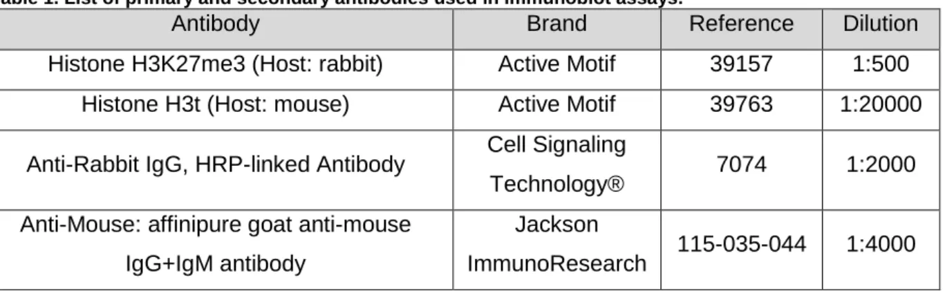
Table 1. List of primary and secondary antibodies used in immunoblot assays.
Antibody Brand Reference Dilution
Histone H3K27me3 (Host: rabbit) Active Motif 39157 1:500
Histone H3t (Host: mouse) Active Motif 39763 1:20000
Anti-Rabbit IgG, HRP-linked Antibody Cell Signaling
Technology® 7074 1:2000
Anti-Mouse: affinipure goat anti-mouse IgG+IgM antibody
Jackson
ImmunoResearch 115-035-044 1:4000
A.4.4. Image and Statistical analysis
All images were captured using Quantity One® 4.6 software for Windows (Bio-Rad), exposing the membrane in the Chemidoc XRS+ System, using the chemiluminescent option for incubations with Histone H3t antibody, and the chemiluminescent high-sensitive option for incubations with Histone H3K27me3 antibody. Membrane expositions where stopped when the signal reached saturation point.
Images where processed and analyzed by using ImageJ 1.48v software from National Institutes of Health, (USA); converted into 8-bit format. The percentage of H3K27me3 was normalized to H3t percentage. One way ANOVA followed by post hoc Bonferroni’s multiple comparison test was performed in GraphPad Prism version 5.00 for Windows, GraphPad Software, San Diego, California, USA, “www.graphpad.com”.
B. In vivo studies: characterization of CVD patients and healthy individuals
To determine whether EZH2 suppression and decrease of H3K27me3 content were associated with markers of hypomethylation and CVD, we studied plasma and PBMC from CVD patients and controls.
21
B.1. Participants and sample collection
We studied 14 atherosclerotic vascular patients [mean age 56.08 ± SD 10.56 years] and 15 healthy controls [mean age 52.63 ± SD 6 years]. The patients group was constituted by 4 women and 10 men; the control group was constituted by 8 women and 7 men. Cases were recruited by Prof. Doutor Fausto Pinto’s team (Centro Hospitalar Lisboa Norte, Lisboa, Portugal) amongst patients who had been admitted to Hospital de Santa Maria, Lisbon, Portugal with a diagnosis of acute coronary syndrome; samples were collected at least 6 months after the occurrence. Controls were selected among staff of the Faculty of Pharmacy from the Universidade de Lisboa whose lifestyle details (i.e., alcohol consumption, medication, smoking, physical exercise, and personal, and family histories) were established by use of standardized questionnaires and protocols. The criteria for inclusion in the control group were: normal hematology and liver/renal function tests and no history of vascular pathology. Exclusion criteria for both groups were metabolic, hepatic, or renal pathology; cancer; alcohol or drug abuse. All patients, but none of the controls, were under therapy with the following drugs: non-steroidal anti-inflammatory + antiplatelet + angiotensin converting enzyme (ACE) inhibitors + statins. Written informed consent was obtained from all participants, and the study was approved by Local Ethical Committees.
B.2. Preparation of biological samples
A total of 50 mL of EDTA-blood samples were collected after a fast of 12 h; plasma and PBMCs were isolated no more than 4 h after the blood collection66. We used 10 mL of blood for plasma separation by centrifugation at 1800 rpm at 8 oC for 10 min, using an Eppendorf 5810R centrifuge (Eppendorf). From the obtained plasma, 750 μL were immediately prepared to preserve SAH and SAM by adding 750 μL of 10 % perchloric acid (v/v). The remaining plasma was further used to perform the biochemical analysis detailed in section B.3. All preparations were stored at −80 oC until use. From the remaining blood, PBMC were
immediately isolated using density gradient centrifugation (section B.2.1). The obtained cells were further used to measure intracellular SAM/SAH levels (section A.3) and to isolate histones and RNA (section B.2.2) for the studies detailed in B.3 and B.4, respectively.
B.2.1. PBMC isolation
PBMC were isolated from the blood samples by density gradient centrifugation143, using LEUCOSEP™ tubes (Greiner Bio-One GmbH, Kremsmünster, Austria), that were put on ice and filled up to the filter with Ficoll-Paque™ PLUS (GE Healthcare, Buckinghamshire, UK). After homogenizing carefully, the EDTA-blood was layered on the LEUCOSEP™ tubes and centrifuged at 2700 rpm for 20 min, with no brake. Differential migration during centrifugation resulted in the formation of layers, containing different cell types143, as illustrated in Figure 5.
22
The bottom layer contains red blood cells and granulocytes, and the layer above contains the Ficoll-Paque™ PLUS solution. Because of their lower density, PBMC were found at the interface between the plasma and the Ficoll-Paque™ PLUS solution143.
The PBMC were recovered with a plastic sterile Pasteur pipette (FL Medical, Torreglia, Italy) and were divided into two 15 mL Cellstar® tubes (Greiner Bio-One GmbH). The tubes are centrifuged at 4000 rpm for 7 min at 8 oC. The supernatant was discarded and 3 mL of Hanks Solution (Sigma-Aldrich) was added to the PBMC pellets. The tubes were centrifuge again, in the same conditions as previously described, and the supernatant was discarded. All centrifugations up to this point were executed in an Eppendorf 5810R centrifuge (Eppendorf).
Figure 5. Isolation of leukocytes from EDTA-blood using Ficoll-Paque™ density gradient centrifugation.
Abbreviations: PBMC, peripheral blood mononuclear cells. Adapted from reference144.
The PBMCs’ pellets were washed twice with 750 μL of Hanks Solution, and then transferred into a 2 mL sterile microtube (Eppendorf). After a centrifugation at 12000 rpm for 20 seconds, the supernatant discarded and 750 μL of cold DEPC-treated water were added to ressuspend the pellet, and another 750 μl of Hanks solution were added to the microtubes, which were centrifuged again in the same conditions. These last two centrifugations were executed in a Hermle Z233M-2 centrifuge (Labnet International, Inc).
100 μL of cold PBS were added to one of the microtubes, which was immediately frozen at -80 oC until further SAM/SAH determinations (after immediate deproteinization with 200 μL of 10 % perchloric acid (v/v)). To the other microtube, 1000 μL of RNAlater® solution (Ambion, ThermoFisher Scientific, Waltham, USA) was added, and followed by an overnight incubation at 4 oC. The microtube was stored at -80 oC, until further histone and RNA extractions, as detailed in section B.2.2.1 and B.2.2.2.