Review
Current pathophysiological concepts and management of pulmonary hypertension
André P. Lourenço, Dulce Fontoura, Tiago Henriques-Coelho, Adelino F. Leite-Moreira
⁎
Department of Physiology and Cardiothoracic Surgery, Faculty of Medicine, University of Porto, Porto, Portugal
a b s t r a c t
a r t i c l e i n f o
Article history:
Received 11 October 2010
Received in revised form 14 February 2011 Accepted 13 May 2011
Keywords:
Pulmonary hypertension Pulmonary arterial hypertension Pathophysiology
Treatment
Pulmonary hypertension (PH), increasingly recognized as a major health burden, remains underdiagnosed due mainly to the unspecific symptoms. Pulmonary arterial hypertension (PAH) has been extensively investigated. Pathophysiological knowledge derives mostly from experimental models. Paradoxically, common non-PAH PH forms remain largely unexplored. Drugs targeting lung vascular tonus became available during the last two decades, notwithstanding the disease progresses in many patients. The aim of this review is to summarize recent advances in epidemiology, pathophysiology and management with particular focus on associated myocardial and systemic compromise and experimental therapeutic possibilities. PAH, currently viewed as a panvasculopathy, is due to a crosstalk between endothelial and smooth muscle cells, inflammatory activation and altered subcellular pathways. Cardiac cachexia and right ventricular compromise are fundamental determinants of PH prognosis. Combined vasodilator therapy is already mainstay for refractory cases, but drugs directed at these new pathophysiological pathways may constitute a significant advance.
© 2011 Elsevier Ireland Ltd. All rights reserved.
1. Introduction
Pulmonary hypertension (PH), is de
fined by mean pulmonary
arterial (PA) pressure (mPAP) elevation above 25 mm Hg at rest
[1]
. In
most cases, PH accompanies cardio-respiratory conditions and does
not involve the pulmonary vasculature. However, more rarely it may
present itself as pulmonary arterial hypertension (PAH), de
fined
additionally by normal left ventricular (LV)
filling pressure
[2]
. PAH is
viewed as a vasoproliferative disease with characteristic pathological
abnormalities, such as arteriolar plexiform lesions, as found in most of
cases. Initial symptoms, mainly fatigue and dyspnea, are usually vague
and insidious, thus most cases are diagnosed when cardiac output
(CO) is already low
[3]
. Right ventricular (RV) failure due to PH is an
important cause of death
[4]
whose complex pathophysiological
Abbreviations: 5-HT, 5-Hydroxytryptamin, serotonin; 5-HT2A, serotonin type 2A receptor; 6MWT, 6-minute walk test; AC, adenylate cyclase; AS, atrial septostomy; BMP, bone
morphogenetic protein; BMPR1, bone morphogenetic protein receptor 1; BMPR2, bone morphogenetic protein receptor 2; BNP, type B natriuretic peptide; CaL, L-type Ca2+
-channel; CC, cardiac cachexia; CCB, Ca2+
-channel blocker; CCR2, chemokine receptor 2; CCR5, chemokine receptor 5; CDK, cyclin-dependent kinase; cGMP, cyclic guanosine monophosphate; CHD, congenital heart disease; CO, cardiac output; CO–A/R, co-repressors or activators; COPD, chronic obstructive pulmonary disease; CT, computerized tomography; CTD, connective tissue disease; CTEPH, chronic thromboembolic pulmonary hypertension; CVC, central venous catheter; CX3CR1, chemokine receptor 1; CXCR4,α-chemokine receptor; DCA, dichloroacetate; DLCO, carbon monoxide diffusion; e-, electron; ECM, extracellular matrix; EF, ejection fraction; EGFR, epidermal growth factor receptor; EPC, endothelial
progenitor cells; ERA, endothelin-1 receptor antagonists; ET-1, endothelin-1; ETA, endothelin-1 type A receptor; ETC, electron transport chain; FDA, Food and Drug Administration;
fPAH, familial pulmonary arterial hypertension; GC, guanylate cyclase; Gq, protein Gq; HF, heart failure; HIF-1α, hypoxia-inducible factor-1α; HIV, human immunodeficiency virus;
HLT, heart-lung transplantation; Id, inhibitor of DNA binding proteins; IL-6, Interleukin-6; IP, prostaglandin receptor; IP3, inositol 3-phosphate; iPAH, idiopathic pulmonary arterial
hypertension; iv, intravenous; Kv1.5, O2-sensitive K+-channels; LHD, left-heart disease; LV, left ventricular; LT, lung transplantation; MCP-1, monocyte chemotactic protein-1; MLC,
myosin light-chain; MLCK (−P), myosin light-chain kinase, and respective phosphorylated form; MMP, matrix metalloproteinases; mPAP, mean pulmonary artery pressure; NFAT, nuclear factor of activated T lymphocytes; NIH, National Institutes of Health; NO, nitric oxide; NRCT, non-randomized clinical trial; O2. -, superoxide anion; PA, pulmonary arterial;
PAP, pulmonary artery pressure; PAH, pulmonary arterial hypertension; PASMC, pulmonary artery smooth muscle cell; PCH, pulmonary capillary haemangiomatosis; PCWP, pulmonary capillary wedge pressure; PEA, pulmonary endarterectomy; PDE, phosphodiesterases; PDE5, type 5 phosphodiesterase; PDEi, phosphodiesterase inhibitors; PDGF,
platelet derived growth factor; PDGFR, platelet derived growth factor receptor; PDH (−P), pyruvate dehydrogenase, and respective phosphorylated form; PDK, pyruvate dehydrogenase kinase; PGI2, prostacyclin; PH, pulmonary hypertension; PKA, protein-kinase A; PKG, protein-kinase G; PPH, portopulmonary hypertension; PTE, pulmonary
thromboembolism; PVOD, pulmonary veno-occlusive disease; PVR, pulmonary vascular resistance; QOL, quality of life; RANTES, regulated upon activation, normal T expressed and secreted; RCT, randomized clinical trials; RHC, right-heart catheterisation; ROS, reactive oxygen species; RV, right ventricular; RVAD, right ventricular assist device; sc, subcutaneous; SDF-1, stromal cell-derived factor-1; SLE, systemic lupus erythematosus; SOD, superoxyde dismutase; SPAP, systolic pulmonary artery pressure; SR, sarcoendoplasmic reticulum; TGF-β, transforming growth factor -β; TGF-βR, transforming growth factor -β receptor; TP, ThromboxaneA2receptor; TnC, tenascin C; TNF-α, tumor necrosis factor-α; trp, transient
receptor potential; TTCW, time to clinical worsening; TxA2, thromboxane A2; VEGF, vascular endothelial growth factor; VEGR, vascular endothelial growth factor receptor; WHO,
World Health Organization.
⁎ Corresponding author at: Department of Physiology and Cardiothoracic Surgery, Faculty of Medicine, University Hospital São João, Alameda Professor Hernâni Monteiro, 4200– 319 Porto, Portugal. Tel.: + 351 225513644; fax: + 351 225513646.
E-mail address:amoreira@med.up.pt(A.F. Leite-Moreira).
0167-5273/$– see front matter © 2011 Elsevier Ireland Ltd. All rights reserved. doi:10.1016/j.ijcard.2011.05.066
Contents lists available at
ScienceDirect
International Journal of Cardiology
mechanisms are just beginning to be understood. The last decades
have been proli
fic in experimental and clinical studies in both PAH
and PH. Several new drugs have become available
[3]
. Nevertheless,
the prognosis remains poor, and many patients require
transplanta-tion
[5]
. The present review aims to summarize the most recent
concepts on the epidemiology, pathophysiology, diagnosis and
man-agement of PH
[6,7]
.
2. Aetiology and classi
fication
Several conferences on PH have been fostered by the World Health
Organization (WHO). A classi
fication was proposed in 1973 and then
modi
fied at Evian in 1988 to better reproduce pathophysiology and
clinical presentation. At Venice in 2003, the term primary PH was
substituted for idiopathic PAH (iPAH) and pulmonary veno-occlusive
disease (PVOD) and pulmonary capillary haemangiomatosis (PCH)
were grouped under a single PAH subcategory. In 2008, the 4th World
Symposium held in Dana Point (
Table 1
) endorsed the expression
“non-PAH PH” to address categories other than PAH. Additionally,
left-heart disease PH was subdivided in systolic heart failure (HF),
diastolic HF and valvular heart disease, and schistosomiasis was
included as a new class of disease-associated PAH.
3. Diagnosis
During the 4th Conference (
Table 2
) exercise values were excluded
as a criteria for diagnosis since the increase in mPAP during exercise
frequently exceeds 30 mm Hg among the elderly
[8]
. Additionally,
non-invasive echocardiographic criteria of systolic tricuspid
regur-gitant velocity were contemplated
[9]
. Nevertheless, transpulmonary
flow and pulmonary venous pressure are not reliably measured by
echocardiography thus right-heart catheterisation (RHC) remains the
gold standard while echocardiography is usually a screening exam.
RHC is mandatory in every patient, allowing the selection of patients
that may bene
fit from Ca
2+-channel blockers (CCB), the positive
responders during vasoreactivity test, those in whom mPAP drops
more than 10 mm Hg or to values bellow 40 mm Hg with normal or
increased CO, after administration of a short-acting vasodilator, such
as nitric oxide (NO)
[10]
, epoprostenol or adenosine
[7]
.
4. Epidemiology
The incidence and prevalence of PAH were estimated to be 2.4
–
7.6 cases/million/year and 15
–26 cases/million, respectively, in large
population studies
[11,12]
. Worldwide prevalence is hard to appraise,
but it is surely underdiagnosed
[13]
and its onus is likely greater than
recognized, given the newly revealed associations with haemodialysis
[14]
, the metabolic syndrome
[15]
, and developing world diseases,
such as human immunode
ficiency virus (HIV) infection,
schistosomi-asis, and sickle cell disease
[16]
. Apart from iPAH no precise estimates
of incidence or prevalence are available. Nevertheless, non-PAH PH is
increasingly recognized as a major health burden. HF is the most
common cause of pulmonary hypertension (PH). Not only up to 60% of
patients with severe LV systolic dysfunction but also 70% of those with
HF and normal ejection fraction (EF)
[17]
develop PH
[18,19]
. Moreover,
PH af
flicts 70% of patients with rheumatic heart disease
[20]
. Many
patients develop chronic thromboembolic PH (CTEPH) after pulmonary
thromboembolism (PTE)
[20]
or PH during the progression of chronic
obstructive pulmonary disease (COPD). Prevalence ranges from 35 to
90% according to stage
[21,22]
. Systolic PAP (SPAP) is mostly limited to
values ranging from 25 to 35 mm Hg, and severe PH is uncommon in
advanced COPD
[23]
. Nevertheless, some patients develop
dispropor-tionate PH. These warrant particular attention
[21]
, but even modest PH
has a strong impact on quality of life (QOL) and survival
[22]
. Right HF,
its most severe complication, is responsible for 10
–30% of admissions
due to decompensated HF
[24]
. Presently COPD is already responsible
for 84% of cor pulmonale cases and, due to smoking, will be the 3 rd cause
of death by 2020
[23]
. Portopulmonary hypertension (PPH) is a
pulmonary-hepatic vascular disorder that af
flicts approximately 5–6%
of patients referred for liver transplantation due to advanced liver
disease. It is an underrecognized complication that adversely affects
survival, after liver transplantation but presumably also in the early
stages of liver disease
[32,33]
.
5. Clinical presentation and workup
Severe disease may present with chest pain, palpitation, oedema,
ascites, and syncope
[9]
but earlier treatment, at reversible stages, is
fundamental. Diagnosis is challenging, a delay of 2 to 3 years is common
and a high suspicion level is needed
[13]
. The clinician may
find RV
hypertrophy on the electrocardiogram and hilar PA prominence on the
chest X-ray. Echocardiography, generally undertaken after a suspicion,
may show increased SPAP, estimated by the velocity of tricuspid
Table 1
New classification for pulmonary hypertension (PH) from the 4th World Symposium on PH (Dana Point, 2008). Pulmonary arterial hypertension
(PAH)
Non-PAH pulmonary hypertension (PH)
Well defined cause Unclear or multifactorial
PAH (1) Left-heart disease (2) Unclear/multifactorial mechanisms (5)
Idiopathic Systolic dysfunction Haematologic disorders
Hereditary Diastolic dysfunction Myeloproliferative disorders, etc.
Drug/toxin induced Valvular disease Systemic disorders
Disease associated Lung diseases/hypoxia (3) Vasculitis, sarcoidosis, neurofibromatosis, etc.
CTD COPD Metabolic disorders
HIV infection Interstitial lung disease Glycogen storage disease, thyroid disorders, etc.
Portal hypertension Sleep-disordered breathing Congenital heart disease
Systemic-pulmonary shunts Chronic exposure to high altitude (Other than systemic-pulmonary shunt)
Schistosomiasis Broncho pulmonary dysplasia Other
Chronic haemolytic anaemia Developmental abnormalities Fibrosing mediastinitis, chronic renal failure on dialysis, etc.
Subclass of PAH (1′) CTEPH (4)
PVOD and PCH
Classes are presented between parentheses. CTD, connective tissue disease; HIV, human immunodeficiency virus; PVOD, pulmonary veno-occlusive disease; PCH, pulmonary capillary angiomatosis; COPD, chronic obstructive pulmonary disease; CTEPH, chronic thromboembolic PH.
Table 2
New diagnostic criteria for pulmonary hypertension (PH) from the 4th World Symposium on PH (Dana Point, 2008).
Method Normal Borderline Clear
mPAP (mm Hg) b21 21–25 N25
systolic tricuspid regurgitation (m.s− 1) b2.5 2.5–2.8 N2.8
regurgitation jet, and/or increased RV out
flow tract acceleration time. It
is fundamental to evaluate valve or primary myocardial disease, as well
as the degree of RV hypertrophy and dysfunction
[9]
. Comprehensive
echocardiographic evaluations of RV function have been proposed as
useful approaches to risk strati
fication in PAH
[25]
, although magnetic
resonance imaging techniques have also been used
[26]
. Regarding
differential diagnosis, patients with suspicion of PTE should undergo the
highly sensitive ventilation-perfusion (V-Q) scan. Staging and
opera-bility also relies on chest computerized tomography (CT) and
angiography. High-resolution CT is useful to assess PVOD or PCH and
to diagnose interstitial lung or connective tissue disease (CTD)
[7,9]
.
Finally antinuclear antibodies, autoimmune disease markers, HIV and
viral hepatitis screening, coagulation disorder markers (eg, protein S
and C, lupus anticoagulants, von Willebrand factor) and type B
natriuretic peptide (BNP) may be carried out for differential diagnosis
[7,9]
. The key feature differentiating PH resulting from left-heart disease
(LHD) is elevated pulmonary capillary wedge pressure (PCWP), which is
absent in PAH
[27]
. To establish the diagnosis of PPH patients must
present with portal hypertension and not only haemodynamic criteria
for PH, in the absence of other causes, but also increased pulmonary
vascular resistance (PVR)
[28]
. Functional respiratory evaluation relies
on spirometry and carbon monoxide diffusion (DL
CO). Spirometry may
be markedly altered in lung disease, whereas minor changes are found
in iPAH. DL
COimpairment correlates with lung vascular surface area and
PAH severity
[29]
. The 6-minute walk test (6MWT), a common clinical
trial end-point that evaluates moderate to severe heart or lung disease,
is an easily performable and reproducible test originally developed as a
surrogate of peak O
2consumption (
Table 3
). It correlates well with CO,
PVR, O
2consumption, QOL, and predicts mortality in PAH
[30]
.
Nevertheless, since it depends on many individual variables, it is not a
reliable marker of disease progression
[7]
. Additionally, its validity has
been questioned for CTD
[31]
. Cardiopulmonary exercise testing,
regarded by most as gold-standard in exercise capacity evaluation and
still a cornerstone in PAH functional evaluation, also assesses PH
prognosis
[32]
, but requires an experienced laboratory
[33,34]
.
6. Pathophysiology
Although no animal model completely recapitulates human PAH,
combining multiple insults, according to the multiple-hit hypothesis,
yielded severe phenotypes that closely mimic it
[35]
. Pathophysiological
knowledge, derived mostly from these animal studies, once viewed PH
as an imbalance between pulmonary vasoconstrictors and vasodilators
[36]
. While prostacyclin (PGI
2) and NO normally govern vascular tone,
endothelin-1 (ET-1), thromboxane A
2(TxA
2) and serotonin (5-HT) take
over in PH. Not surprisingly, lung arteries vasodilators have been the
mainstay of therapy (
Fig. 1
)
[3]
. Nevertheless, recent research showed
this view to be highly incomplete.
6.1. PAH as panvasculopathy
PAH is currently viewed as a panvasculopathy, accompanied by
histological features as intimal hyperplasia, medial hypertrophy, and
arteriolar occlusion by thrombosis, in
filtration by inflammatory cells or
angioproliferative plexiform lesions (
Fig. 1
)
[7]
. Apoptosis may generate
apoptosis-resistant endothelial cell phenotypes that cross-talk with PA
smooth muscle cell (PASMC) through growth factors such as
transform-ing growth factor-
β (TGF-β), that are involved in endothelial cell and
fibroblast transdifferentiation and PASMC proliferation
[37]
.
Metallo-proteinase activation leads to the disruption of the basement membrane
enabling in
flammatory cell recruitment and further generation of
mitogenic peptides
[38]
. The main mechanisms involved in in
flamma-tion, endothelial progenitor cell (EPC) recruitment, growth factor
activity and extracellular matrix remodeling are summarized in
Fig. 1
.
PAH shares a mitochondrial-metabolic abnormality with cancer, the
“Warburg phenotype”, a shift from oxidative phosphorylation to
glycolysis (despite adequate O
2supply) that enhances proliferation
and prevents apoptosis (
Fig. 2
). Hyperpolarization of the mitochondrial
membrane, reduced production of reactive oxygen species (ROS),
normoxic-activation of hypoxia inducible factor-1
α, overexpression of
pyruvate dehydrogenase kinase (PDK) and decreased expression of O
2-sensitive K
+channels (Kv1.5) have been postulated to underlie changes
in mitochondrial O
2sensing
[39]
. PDK activation suppresses aerobic
glucose metabolism and decreased Kv1.5 conductance depolarizes the
membrane. Dichloroacetate (DCA), a mitochondrial PDK inhibitor and
Kv1.5 channel opener, improved PAH
[39]
both by activating pyruvate
dehydrogenase (PDH) and aerobic metabolism and by restoring
membrane potential and ROS production
[40]
.
6.2. Genetics of PAH
Mutations in bone morphogenetic protein (BMP) receptor-2
(BMPR2), a constitutively active receptor responsive to TGF-
β
super-family (including BMP), are seen in more than 80% of familial PAH
(fPAH) cases, leading to loss of smad signalling (
Fig. 3
) and therefore to
increased proliferation and decreased differentiation of PASMC
[41,42]
.
Still, penetrance is low and the mutation is seen only in 10 to 20% of
non-fPAH
[43]
. Other genetic mechanisms predispose to PAH, namely
single-nucleotide polymorphisms of Kv1.5
[18]
, transient receptor potential
(trp) channels
[13]
, and serotonin transporters
[44]
. Trp channels
regulate contractility and cell proliferation by intracellular Ca
2+[45]
.
Elevated 5-HT levels and 5-HT transport have been implicated in PAH
pathogenesis
[44]
.
Table 3
The 6-minute walk test (6MWT) in pulmonary hypertension (PH). Features
Submaximal exercise test
Correlates well with activities of daily living (useful for moderately severe functional impairment)
Non-specific (evaluates the response of all systems)
Well tolerated (nevertheless, appropriate response to an emergency should be available)
Measurements
Dyspnoea and fatigue self-rating at the beginning and end (according to the Borg scale, see legend)
Distance walked
Demographic and anthropometric determinants Gender, age and ethnicity
Height and weight Advantages
Practical and inexpensive to perform (no equipment or specially trained technicians needed)
Reproducible (estimated coefficient of variability of 8%) Ongoing monitoring of cardiopulmonary disease progression Evaluation of response to therapy
Limitations
Merely a rough estimate of the general functional status (does not discard specific assessment tools)
Lack of validation for connective tissue disease associated PAH (musculoskeletal involvement)
“Ceiling effect” for patients with better baseline capacity Biases: disturbed cognition, motivational factors, test repetition, musculoskeletal limitations, etc.
A comprehensive perspective on the 6MWT including indications, contraindications, safety precautions, technical aspects, biases, can be found in the guidelines from the American Thoracic Society[134]. The 6MWT measures the distance that a patient can walk on aflat surface in a period of 6 min, patients are allowed to stop and rest. The normal walked distance for healthy 60 year-old men and women of average constitution is approximately 630 and 550 m, respectively[135], whereas idiopathic PAH patients on World Health Organization functional class IV usually walk less than 200 m[30]. A clinically important improvement in walking distance for the PAH patient is generally 44–76 m[103].
Borg scale: (0) nothing at all, (0.5) just noticeable, (1) very slight, (2) slight, (3) moderate, (4) somewhat severe, (5) severe, (7) very severe, and,finally, (10) maximal[136].
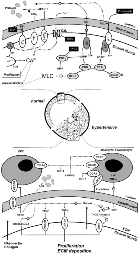
Fig. 1. Pulmonary artery smooth muscle cell (PASMC) constriction and proliferation mechanisms. The major sites of action of lung vasodilator drug classes are shown in the upper panel, namely Ca2+
-channel blockers (CCB), endothelin-1 (ET-1) receptor antagonists (ERA), phosphodiesterase inhibitors (PDEi) and prostanoids. Myosin light-chain (MLC) kinase (MLCK) is inactivated upon phosphorylation (MLCK-P). Other mechanisms are presented in the lower panel. Several cytokines, beyond interleukin-6 (IL-6) and tumor necrosis factor-α (TNF-α), mostly produced byfibroblasts, such as stromal cell-derived factor-1 (SDF-1), monocyte chemotactic protein-1 (MCP-1), fractalkine, RANTES (regulated upon activation, normal T expressed and secreted), and vascular endothelial growth factor (VEGF) are upregulated and induce PASMC proliferation and monocyte recruitment, while monocytes upregulate the α-chemokine receptor (CXCR4) and chemokine receptors 1, 2 and 5 (CX3CR1, CCR2 and CCR5, respectively)[46]. Elastase, early activated in PH, triggers growth factors release from the extracellular matrix (ECM) and induces tenascin C (TnC) through the activation of matrix metalloproteinases (MMP). When TnC binds surface integrins on PASMCs cell-survival signals are generated and growth factor receptors are further activated. Serotonin (5-HT) induces proliferation of PASMCs by stimulation of S100A4/Mts1, a S100 Ca2+
-binding protein family member with metastasis-inducing ability[3]. Endothelial progenitor cells (EPC) may participate in vessel repair, but on the other hand also take part in plexiform lesions[146]. Other abbreviations: TxA2, thromboxane A2; Gq, protein Gq; IP3, inositol 3-phosphate; SR, sarcoendoplasmic reticulum; ETA, ET-1 type A receptor; TP, TxA2receptor; 5-HT2A, 5-HT type 2A receptor; CaL, Type L
Ca2+-channel; GC, guanylate cyclase; AC, adenylate cyclase; IP, prostacyclin receptor; PDE
5, phosphodiesterase type 5; PKG, protein-kinase G; PKA, protein-kinase A; VEGFR, VEGF
6.3. In
flammation
The in
flammatory state of the vessel wall has recently gained
interest as primary event, rather than mere consequence of the disease
[46]
. Autoantibodies and in
filtration by inflammatory cells are
common in PAH associated with CTD but are also seen in iPAH
[46]
.
Increased levels of cytokines and their receptors have been
demon-strated, particularly in iPAH patients
[47]
, who also present heightened
expression of in
flammatory cell-associated nuclear factor of activated
T lymphocytes (NFAT)
[48]
. Cytokines involved in the pathogenesis of
chronic in
flammatory diseases and cancer, such as tumor necrosis
factor-
α (TNF-α) and IL-6, may play a role in PAH vasculopathy
[49]
.
Our group has tested an anti-in
flammatory approach in experimental
models of PH with variable success
[50,51]
. In
flammatory activation
may also underlie systemic manifestations, for instance cardiac
cachexia (CC). CC is characterized not only by neuroendocrine and
in
flammatory activation but also by suppressed appetite and
nutri-tional derangements
[52]
and poses a signi
ficant prognostic burden on
HF patients
[53]
. CC accompanies the progression of PH, indeed,
patients with severe PH have exaggerated and early post-prandial
satiety hormone response
[54]
.
6.4. The RV in PH
The RV effectively serves as a thin, compliant reservoir for blood
returning to the LV whose primary function of is to deliver deoxygenated
blood to the lungs, while maintaining low-pressure perfusion
[55]
. It is
thus best suited for volume work and unable to suddenly withstand high
PAP. Sudden afterload decreases stroke volume and dilates the RV
[56]
,
whereas progressive overload allows gradual hypertrophy, remodeling
and substantial increases in mPAP. Curiously, although RV response
partly determines the outcome
[26,57]
, despite the fact that the RV was
shown to be an independent therapeutic target in experimental PH
[58]
,
and even though RV remodeling is potentially reversible, as seen after
lung transplantation (LT)
[59]
, little is known about the mechanisms
underlying RV dysfunction
[55]
. Many have shown neuroendocrine
activation can contribute to RV hypertrophy
[60,61]
,
fibrosis and
apoptosis, as well as to oxidative stress, and activation of in
flammatory
cytokines and growth factors
[55,62]
. A state of myocardial hibernation
has been proposed based on systolic
flow impediment to coronary
arteries which is proportional to RV pressure and mass
[63]
. In contrast to
the normal
flexible metabolism, in RV hypertrophy the myocardium
relies solely on anaerobic glucose metabolism partly due to PDK
activation
[64]
and possibly impaired mitochondrial energy-producing
ability
[65]
. Moreover, changes in cardiomyocyte redox state can
underlie electrophysiological instability and remodeling, by mechanisms
similar to those already described for pulmonary vessels
[66]
.
Experi-mental
findings and clinical observations suggest that elevated mPAP
cannot be the single driver for RV failure
[62]
, therefore, targeting the RV
may be a promising approach
[6]
. As for the LV myocardium,
echocardiography shows compromised LV function in various
aetiol-ogies of PH
[67]
, mainly due to ventricular interdependence and
impaired
filling
[68]
. Nevertheless, myocardial abnormalities partly
underlie LV dysfunction. Indeed, despite immediate restoration of LV
geometry and RV function, LV
filling is only normalized 1 year after
single-LT in severe PH
[69]
, and combined heart-lung transplantation
(HLT) is favored if LV function is impaired because the LV may not
recover after LT alone
[70]
. We have con
firmed intrinsic LV myocardial
dysfunction and neuroendocrine activation experimentally
[61,71]
.
6.5. Pathophysiology of non-PAH PH
Contrarily to PAH, and paradoxically, few data are available on the
pathophysiology of the far more common non-PAH PH. Regarding
chronic pulmonary disease, several mechanisms are potentially
responsible. Hypoxia, such as is found at high altitude, is known to
Fig. 2. Reactive oxygen species (ROS), disturbed O2sensing, and mitochondrial dysfunction in pulmonary arterial hypertension (PAH). During oxidative phosphorylation, electrons (e−)
are conveyed by the electron transport chain (ETC) from donors (NADH and FAH) to O2, but minor side reactions also generate by-products, as superoxide anion (O2.−) that must be
detoxified to H2O2by superoxyde dismutase (SOD)[147]. Under normoxia H2O2constitutively opens plasma membrane O2-sensitive K+-channels (Kv1.5) and inhibits hypoxia-inducible
factor-1α (HIF-1α) activity, whereas during hypoxic vasoconstriction, ROS and H2O2production are decreased, Kv1.5 channels close, the plasma membrane depolarizes, Ca2+enters the
cell and myocytes contract. In PAH, mitochondrial abnormalities, most notably pyruvate dehydrogenase kinase (PDK) activation, shift metabolism toward anaerobic glycolysis and impair the ETC. Reduced ROS production, nuclear translocation of HIF-1α, and decreased expression of Kv1.5 ultimately lead to sustained membrane depolarization, L-type Ca2+
-channel (CaL) activation and hypertrophy by Ca2+-calcineurin-dependent activation of the nuclear factor of activated T lymphocytes (NFAT)[146]. PDH, pyruvate dehydrogenase, and respective
induce PH, but low arterial O
2is not an independent predictor of
mPAP, therefore after the Evian Conference COPD-associated PH was
no longer classi
fied as ‘associated with hypoxemia’
[29]
. Pulmonary
vessels in COPD consistently develop intimal
fibro-elastic thickening
and overall muscularisation but this does also not provide a consistent
explanation
[72]
. Endothelial dysfunction and in
flammation, are
currently viewed as the key to vascular remodeling
[73]
. Findings
strongly suggest an involvement of vasoactive mediators and cytokines
[72]
. Plasma IL-6 correlates with mPAP and certain IL-6 genotypes are
associated with PH development in COPD
[74]
. Indeed, vascular
remodelling and endothelial dysfunction can be observed in mild
COPD without hypoxaemia and in ordinary smokers
[75]
. Symptomatic
CTEPH affects 3.8% of patients within 2 years of initial PTE
[76]
, but up to
5.1% of patients may develop de
finite CTEPH
[77]
. Unlike PAH, CTEPH is
mainly associated with obstructions in larger vessels. Its
pathophysiol-ogy remains obscure, while most argue that it results from recurrent
pulmonary embolism, it has also been suggested that endothelial
dysfunction could lead to thrombus formation in situ, and, in fact many
patients do not have a clear history of embolism
[78]
. Variable degrees of
small vessel disease, a PAH-like vasculopathy, accompany CTEPH and
the mechanisms that underlie them are probably common to PAH,
namely endothelial dysfunction
[79]
. As for LHD, two major
mecha-nisms underlie PH, an hydrostatic and a vasoreactive. Increased
filling
pressures are transmitted to the pulmonary circulation and generate,
initially, pulmonary venous hypertension, but, later on, also PVR
increase. SPAP correlates tightly and is roughly twice the PCWP
[80]
.
When the compensatory mechanisms of the highly distensible
pulmonary vasculature are surpassed PA pressure increases
first on
exertion and later on also at rest. Endothelial dysfunction,
sympathetic-adrenergic stimulation and disturbances of 5-HT, TxA
2and
angiotensin-II production further aggravate PH
[81]
, contributing to structural
changes at the capillary level, namely swelling of the endothelial cells,
thickening of the basal lamina, and proliferation of reticular and elastic
fibrils. These changes participate in increasing PVR, decreasing
permeability of the vascular bed, and lower the possibility of developing
pulmonary edema, but ultimately lead to increased likelihood of right
ventricular failure
[82]
. These changes are initially reversible if cardiac
filling pressures are reduced, but on the long term become irreversible
and pose a relative contraindication to cardiac transplantation
[83]
.
6.6. Prognosis
Although PAH has been most extensively studied, its rarity, diverse
etiology and changing therapeutics preclude an estimation of yearly
mortality rates. An early registry followed 194 patients with iPAH from
1981 to 1985 and estimated a median survival of 2.8 years, with 1-, 3-,
and 5-year survival rates of 68, 48, and 34%, respectively
[84]
.
Present-day registries, however, reveal a better prognosis, with 1 year survival
ranging 83 to 88% and 3 year survival 58 to 72%
[85]
. A risk-prediction
equation could be derived from multivariate analysis, including gender,
6MWT, and CO at diagnosis as covariates
[86]
. Four variables were
associated with increased 1-year survival: WHO functional class I,
6MWT
≥440 m, BNPb50 pg/mL, and DL
CO≥80% of predicted
[86]
.
Recently, echocardiographic evaluation of RV function has also been
successfully used for risk strati
fication in PAH
[25]
. The progression in
non-PAH PH is generally slower and the overall prognosis is better. Still,
there is a substantial impact on QOL and survival
[22,27]
. The level of
PAP is a good indicator of prognosis in COPD and a 50% 5-year survival
rate has been reported with PH
[87]
. Regarding CTEPH, survival changed
dramatically. Before the advent of pulmonary endarterectomy (PEA)
patients who had mPAP higher than 30 mm Hg steadily progressed to
PH and 2 year-survival was lower than 20% after it reached 50 mm Hg
[88]
. Currently, in experienced centres and carefully selected patients,
PEA provides remarkable haemodynamic and clinical improvement
with low procedural mortality rate
[5]
. In severe HF, the EF of the RV is
the most important determinant of short-term prognosis among
hemodynamic variables
[89]
. Although increased mPAP is frequently
coupled with reduced RV function, exceptions must be taken in account
during prognostic strati
fication
[19]
.
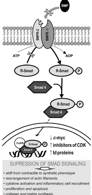
Fig. 3. Growth-promoting pathways in bone morphogenetic protein (BMP) receptor type 2 (BMPR-2) mutations. Bone morphogenetic protein (BMP) receptor type 1 (BMPR-1) and -2 dimerize upon activation by BMP and initiate a cytosolic receptor-activated Smad protein-signaling cascade. Smads (homology with drosophila's mothers against decapentaplegic– MAD – and Caenorhabditis elegans' small phenotype – sma – proteins) ultimately complex with common partner Smad4 and translocate to the nucleus. The weak Smad–DNA interaction requires co-repressors or activators (Co–A/R)[148]. Signal disrupting mutations in BMPRII can be found in the ligand-binding domain, in the kinase domain or in the cytoplasmic tail. Suppression of Smad signalling partly underlies the hypertrophic and proliferative phenotype of pulmonary artery smooth muscle cells (PASMC) [149]. In normal PASMC, BMP stimulates transcriptional activation of cyclin-dependent kinase (CDK) inhibitors and repression of c-myc. CDK inhibitor prevents progression in cell cycle, while c-myc encodes a transcriptional activator responsible for growth and proliferation[150]. Inhibitor of DNA (Id) binding proteins, a family deprived of DNA-binding domain that acts by inhibition of transcription factors, are also major targets. Failure to induce Id genes makes PASMC unresponsive to the growth suppressive effects of BMPs [151]. Prostanoids, by cyclic adenosine monophosphate and a direct effect on the Id promoter, drive the expression of Id proteins[148].
7. Therapeutics
Treatment of PAH has evolved considerably over the past decade,
many treatment algorithms have been proposed, mainly based on
studies conducted in patients with iPAH and PAH associated with
CTDs. In
Table 4
we summarize the major therapeutic studies on PAH.
Several general measures can be recommended. Regarding
exercise practice, patients may practice low level aerobic exercise,
such as walking, whereas exertion that may lead to breathlessness,
dizziness or chest pain should be avoided. Some patients may not
tolerate high altitudes, for instance during airplane
flights, and
therefore require in-
flight O
2administration. It is currently
recom-mended for patients either in WHO classes III and IV or whose arterial
O
2pressure is below 60 mm Hg. Dietary sodium restriction can be
advised particularly in RV failure (
≤2.4 g.d
− 1), but current European
Society of Cardiology guidelines do not recommend it. Immunization
against common respiratory pathogens is recommended
[7]
. PAH is a
contraindication to pregnancy due to the high mortality rate
[90]
.
Despite the lack of randomized controlled trials (RCT), the initial
therapeutic, and largely supportive, approaches to the treatment of
PAH were anticoagulation, diuretics, O
2therapy and digoxin.
Obser-vational studies suggested improved survival after anticoagulation in
patients with iPAH, therefore most experts recommend
anticoagula-tion in iPAH, heritable PAH, and PAH due to use of anorexigens
(titrated to international normalized ratio of 2.0
–3.0). As for non-iPAH,
anticoagulation may be advised for severe cases
[7]
. Diuretics are used
to manage HF symptoms. O
2therapy in hypoxemia is employed strictly
to avoid vasoconstriction. Based on a short-term effect study, digoxin
may be used in patients with low CO, but its use is clearly only
recommended in patients with supraventricular tachyarrhythmias
Table 4
Summary of randomized clinical trials (RCT) on pulmonary arterial hypertension (PAH).
Class Drug Year Author Study type Sample Patients Follow-up Positive outcomes
Prostanoid Epoprostenol 1996 Barst[96] RCT (not blind) iPAH (WHO III–IV) 81 12 weeks Haemodynamics, QOL, WHO class, survival Treprostinil (sc) 2002 Simmoneau[98] RCT iPAH, CTD and CHD
(WHO II-IV)
470 12 weeks Haemodynamics, 6MWT,
clinical evaluation Iloprost (inh) 2002 Olschewski[99]
(AIR)
RCT iPAH, CTD and CTEPH
(WHO III-IV)
203 12 weeks Haemodynamics, 6MWT,
QOL, WHO class
Beraprost 2002 Galiè[137]
(ALPHABET)
RCT iPAH, CTD, CHD, portal hypertension and HIV (WHO II-III)
130 12 weeks Exercise tolerance, 6MWT, clinical evaluation
2003 Barst[138] RCT iPAH, CTD and CHD
(WHO II-III)
116 1 year Exercise tolerance, 6 MWT,
TTCW,
ERA Bosentan 2002 Rubin[103]
(BREATHE1)
RCT iPAH, CTD or SLE
(WHO III-IV)
213 16–28 weeks Exercise tolerance, 6MWT, WHO class, TTCW; 2005 McLaughlin[139] RCT (not blind) iPAH (WHO III-IV) 169 3 years Survival (NIH prediction) 2006 Galiè[140]
(BREATHE5)
RCT CHD (WHO III) 54 16 weeks Haemodynamics, 6 MWT
2008 Galiè[106] (EARLY) RCT iPAH, CTD, CHD, HIV (WHO II) 185 6 months Haemodynamics, NT-pro-BNP and TTCW 2008 Jais[123] (BENEFIT)
RCT CTEPH (WHO II–IV) 157 16 weeks Haemodynamics
Ambrisentan 2005 Galiè[141] RCT (dose ranging) iPAH, CTD, HIV and anorexigen (WHO II–III) 64 12 + 12 weeks (not-blind) Haemodynamics, 6 MWT, clinical evaluation 2008 Galiè[142] (ARIES 1 and 2)
RCT iPAH, CTD, HIV and
anorexigen
202+192 (parts 1 and 2)
12 weeks 6MWT, WHO class, QOL, TTCW, NT-pro-BNP 2009 Oudiz[108]
(ARIES 1, 2 and E)
RCT iPAH, CTD, HIV and
anorexigen
383 2 years 6MWT, TTCW and survival
(combined outcome) Sitaxsentan 2004 Barst[143] (STRIDE1) RCT iPAH, CTD and CHD (WHO II–IV) 178 12 weeks Haemodynamics, 6MWT, WHO class 2006 Barst[144] (STRIDE2) RCT iPAH, CTD and CHD (WHO II–IV)
245 18 weeks 6MWT, WHO class
PDEi Sildenafil 2005 Galiè[110]
(SUPER1) RCT iPAH, CTD and CHD (WHO II–IV) 278 12 weeks Haemodynamics, 6MWT, WHO class Tadalafil 2009 Gàlie[145] (PHIRST)
RCT (dose ranging) iPAH, CTD, CHD, HIV and anorexigen
405 16 weeks Haemodynamics, WHO
class, 6MWT, TTCW, QOL Combined Bosentan+Iloprost (inh) 2006 McLaughlin[114]
(STEP)
RCT iPAH, APAH (WHO III) 67 12 weeks Haemodynamics, WHO
class, TTCW Epoprostenol +
sildenafil
2008 Simmoneau
[115](PACES)
RCT iPAH and CTD 267 16 weeks Haemodynamics, exercise
tolerance, QOL, TTCW Bosentan or sildenafil+
treprostinil (inh)
2010 McLaughlin[100]
(TRIUMPH I)
RCT iPAH, CTD, HIV and
anorexigen (WHO III–IV)
255 12 weeks QOL, NT-pro-BNP
RCT on PAH therapeutics are summarized according to drug class, drug type and publication date. Study acronyms are presented when applicable. Studies enrolling less than 50 patients as well as studies involving specific PAH groups, namely HIV-related and portal hypertension-related were excluded. iPAH, idiopathic pulmonary arterial hypertension; WHO, World Health Organization; QOL, quality of life; 6MWT, 6-minute walk test; NIH, National Institute of Health; APAH, disease associated pulmonary arterial hypertension; CTD, connective tissue disease APAH; CHD, congenital heart disease APAH; CTEPH, chronic thromboembolic pulmonary hypertension; HIV, human immunodeficiency virus APAH; TTCW, time to clinical worsening; SLE, systemic lupus erythematosus APAH; NT-pro-BNP, N terminal fragment of pro-type B natriuretic peptide. Study acronyms stand for: AIR, Aerosolized Iloprost Randomized; ALPHABET, Arterial Pulmonary Hypertension and Beraprost European Study Group; BREATHE, Bosentan Randomized trial of Endothelin Antagonist THErapy Study Group; EARLY, Endothelin Antagonist tRial in miLdlY symptomatic PAH patients; BENEFiT, Bosentan Effects in iNopErable Forms of chronic Thromboembolic pulmonary hypertension; ARIES, Ambrisentan in pulmonary hypertension, randomized, double blinded, placebo controlled, multicenter, efficacy studies); STRIDE, Sitaxsentan To Relieve ImpaireD Exercise; SUPER, Sildenafil Use in Pulmonary Arterial Hypertension Study Group; PHIRST, Pulmonary Arterial Hypertension and Response to Tadalafil; STEP, Safety and pilot efficacy Trial in combination with bosentan for evaluation in pulmonary arterial hypertension; PACES, pulmonary Arterial hypertension Combination study of epoprostenol and sildenafil; TRIUMPH I, efficacy and tolerability of inhaled Treprostinil sodium in patients with severe pulmonary arterial hypertension.
[91]
. During the last two decades substantial RCT and pharmacological
research have yielded several new and more effective alternatives to
treat PH. The main pharmacological classes will be brie
fly presented.
Most of the studies are small scaled and short-termed, not suitable for
survival analysis, but a recent meta-analysis found an overall bene
fit in
mortality
[92]
. Nevertheless, most therapies reduce mPAP by only 10
–
20%, with the exception of strong responders to CCB. Despite all the
advancement, many patients still remain symptomatic, with a
suboptimal QOL and warrant combined therapy or even invasive or
surgical procedures.
7.1. Ca
2+channel blockers
A marked improvement in survival rates was shown with long-term
high-dose CCB therapy for patients with iPAH and a positive
vasoreactivity test
[93]
. Long acting nifedipine, diltiazem, or amlodipine
are more commonly used. If there is no recovery to functional classes I or
II patients are deemed as non-responders and should discontinue CCB.
True responders are rare in non-iPAH
[94]
. Indiscriminate use is not
recommended, due to systemic vasodilation and negative inotropic
effects
[95]
.
7.2. Prostanoids
There are presently several commercially available prostanoid
formulations. Intravenous (iv) epoprostenol was the
first shown to
improve functional class, hemodynamics and survival in a 12-week
follow-up period in patients with iPAH of classes III and IV
[96]
. These
bene
ficial effects were reproduced in long-term observational
compar-isons with historical controls
[97]
. Moreover, epoprostenol was also
evaluated in CTD associated PAH and other forms of non iPAH with
favourable outcomes. Presently, because of the complex administration
and cumbersome follow-up, epoprostenol use is mainly con
fined to
highly experienced centres. Patients must keep a central venous catheter
(CVC) and handle drug preparation and infusion. Dosing must be
carefully titrated. Most patients do well with an initial in-hospital dose of
2 ng.kg
-1.min
-1and a dose range between 25 and 40 ng.kg
−1.min
−1.
Unfortunately, substantial side-effects have been reported, namely
flushing, headache, and sudden death after abrupt discontinuation, as
well as risk of infection related to CVC
[7]
. Treprostinil, a longer half-life
prostanoid, amenable to administration by subcutaneous (sc) route,
circumventing the need for CVC, showed minor bene
ficial effects in
patients with functional classes II
–IV of idiopathic, CTD and (CHD)
associated PAH
[98]
. The Food and Drug Administration (FDA) approved
it for functional classes II
–IV also by iv route, when the sc route is not
tolerated due to pain or erythema. It is currently not approved by the
European Medicines Agency (EMA). On another attempt to facilitate
administration, iloprost was developed for inhalation by aerosol device.
After a 12-week administration, iloprost improved the 6MWT and
functional class in a multicentre RCT enrolling patients with PAH of
different aetiologies
[99]
. Treprostinil is now also available by inhalation
[100]
, and trials of oral formulations have been iniciated (FREEDOM,
Trial of Oral Treprostinil in Pulmonary Arterial Hypertension).
7.3. Endothelin receptor antagonists
We have previously reviewed the role of ET-1 and its antagonists
(ERA) in cardiovascular pathophysiology
[101]
. Brie
fly, after a small
magnitude RCT had shown improvement in the 6MWT, mPAP and CO
with the non-selective ERA bosentan
[102]
, a larger scale study
conducted in patients with idiopathic or CTD associated PAH,
reproduced these
findings and reported improvement in time to clinical
worsening (TTCW), a secondary endpoint de
fined as a composite of
mortality, LT, hospitalization, discontinuation due to lack of recovery or
need for epoprostenol or atrial septostomy (AS)
[103]
. As an important
side-effect, bosentan dose-dependently altered hepatic function.
Ane-mia can also occur and the FDA therefore recommends liver function
test and haematocrit surveillance
[7]
. Long-term evaluation as a
first-line drug in functional class III patients also revealed good results,
although many patients demanded prostanoids
[104]
. In fact, improved
survival was only demonstrated by comparison with historical data
from epoprostenol treated iPAH WHO class III patients, and
unfortu-nately the two cohorts were not comparable
[105]
. By now, bosentan has
also been tested in CHD, HIV-associated PAH and CTEPH with favourable
results. Moreover, it has been successfully used in a large sample of mildly
symptomatic, class II, multiple cause-PAH patients improving
haemody-namics and TTCW
[106]
. Sitaxentan a selective ET
AERA initially was
shown to have comparable effects to bosentan in iPAH and PAH
associated with CTD or CHD, but has been withdrawn from market due
to two fatal cases of liver failure
[107]
. Ambrisentan, another selective ET
AERA, also improved the 6MWT and TTCW, which was reproducible in
long-term studies
[108]
. It is approved by the FDA since 2007 and it has
also been approved by the EMA for PAH patients in functional classes II
and III. Indeed, it is the only ERA approved for WHO class II.
7.4. Phosphodiesterase inhibitors
Phosphodiesterases (PDE) degrade cyclic guanosine
monopho-sphate (cGMP) therefore PDE inhibitors (PDEi) potentiate the effects
of cGMP generated by NO activation of guanylate cyclase. NO and NO
donors have been extensively used as a rescue therapy to mitigate mPAP
in the perioperative period and in the critically ill patient, particularly in
children
[109]
. Sildena
fil, the first used PDEi, was shown to improve
6MWT, WHO functional class and mPAP in idiopathic, CTD or CHD
associated PAH, but there were no differences in TTCW
[110]
. The FDA
approved sildena
fil in low doses for patients with PAH although there is
some debate as to whether higher doses might confer additional
bene
fits
[111]
. Other PDEi are currently under study. Tadala
fil, recently
approved by both FDA and the EMA, has a longer half-life than sildena
fil
and is amenable to once-daily dosing. Nevertheless, unlike sildena
fil,
due to its hepatic metabolism and renal clearance, dose adjustments are
recommended for patients with renal or hepatic function impairment
[112]
.
7.5. Combination therapy
The possibility to combine distinct drug classes that target different
molecular pathways in order to improve clinical ef
ficacy and minimize
side-effects is an attractive perspective. After an initial attempt to
combine bosentan and epoprostenol in a small scale and underpowered
trial conducted on patients with either iPAH or PAH associated to CTD
that proved unsuccessful
[113]
, another trial that combined inhaled
iloprost with bosentan in patients who remained symptomatic showed
improvement in functional class, TTCW and haemodynamics
[114]
.
More recently, the addition of sildena
fil to PAH patients who remained
symptomatic on a stable dose of iv epoprostenol improved the 6MWT, as
well as mPAP, CO, and TTCW
[115]
, while the addition of inhaled
treprostinil to WHO III and IV PAH patients undergoing either bosentan
or sildena
fil chronic therapy showed only clinical benefits in QOL
[100]
,
and the introduction of oral treprostinil failed to achieve statistical
signi
ficance in 6MWT (FREEDOM, unpublished results).
7.6. Invasive and surgical strategies in PAH
These include AS and LT or HLT. Other possibilities, such as the RV
mechanical assist devices (RVAD) are still poorly investigated. AS creates
a right-to-left shunt that unloads the RV, decreases mPAP, and improves
LV
filling. The increase in CO offsets the shunting of deoxygenated blood
and ameliorates O
2delivery. Increased CO allows bridging to
transplan-tation in up to 40% of patients
[116]
. Nevertheless, it is merely palliative
and procedural mortality is still high therefore it is just a last resort for
patients on maximal medical therapy and inotropic support. Improved
techniques are being currently explored to reduce procedural risk
[5]
.
Currently, PAH is responsible for approximately 4% of LT and HLT, and
although there is a substantial procedural related mortality, the
long-term outcome is better than with medical therapy alone, with a 47%
survival after 5 years
[117]
. The type of transplant is still a matter of
debate and highly related to the experience of each centre. Generally HLT
is preferred either when patients have intractable HF or are dependent
on inotropic support or if PH is secondary to CHD or LHD
[70]
.
7.7. Therapeutic algorithm
Management must be tailored to each patient according to disease
severity, comorbid conditions, drug side-effects and each centre's
experience. CCB are reserved for iPAH patients with a positive
vasoreactivity test and stable hemodynamics, otherwise
first line therapy
should consist of ERA or PDEi, unless the oral route is not available,
patients are in functional class IV or present overt RV failure. In these
cases, the
first choice is an iv prostanoid. Moreover, combination therapy
should always be kept in mind, particularly when side-effects arise or
patients are not responding. Enrolment in clinical trials with newer
pharmacological agents may be an option but AS and transplantation
should be considered before systemic deterioration. Early referral for
transplantation is crucial particularly for refractory cases
[7]
. A simpli
fied
therapeutic algorithm is suggested in
Fig. 4
.
7.8. Non-PAH PH
Patients will bene
fit from medical optimization of their primary
disease, but signi
ficant PH may persist. Some patients actually present
disproportionate PH not easily attributable to the underlying condition.
In left-heart disease prostanoids, with the exception of inhaled route,
are usually contraindicated due to systemic vasodilation
[118]
. ERA trials
have been interrupted prematurely due mainly to side-effects and
absence of clinical bene
fit, even with reduced dose
[119]
, but selected
cases may bene
fit from short trials as a bridge to transplantation
[120]
. As
for PDEi short-term hemodynamic bene
fits, as well as long-term
improvements have been documented
[121]
.
Mild levels of PH are amenable to optimization of medical therapy in
COPD. If PH is disproportionate, and other PH causes have been ruled out,
many centres are routinely employing vasodilators despite V-Q
mismatch
[122]
. CTEPH is potentially curable with PEA
[5]
. Yet, many
patients are not candidates so they remain anticoagulated and on
diuretics. Many centres are promptly using new PAH drugs off-label if
there is associated vasculopathy
[123]
.
7.9. Recent progresses and future targets in PH
Based upon the most recent experimental
findings, clinical trials
targeting altered metabolic and signalling pathways are warranted. DCA
and Kv1.5 channel gene transfer have been successful in experimental
studies
[39]
, as well as trp channel inhibitors, growth factor receptor
inhibitors and intracellular kinase inhibitors
[3,124]
. In
flammatory
response modulation has also been a major research topic. After several
animal studies demonstrating bene
ficial effects of statins
[124]
, possibly
due to pleiotropic effects, a human study disappointingly showed no
long-lasting improvement
[125]
. Other immunomodulatory agents have
been successful in animal experiments
[50,126]
, but bene
ficial effects are
mainly con
fined to CTD associated PAH
[127]
. We have also reported
disturbances in endogenous endocrine and paracrine systems
[128,129]
that may be targeted. Another tempting possibility is the recruitment or
infusion of EPC. The number and function of EPCs predicts prognosis, and
most currently used drugs increase circulating EPC numbers
[130]
.
Circulating EPCs home to sites of endothelial injury, promote
revascu-larization and improve vascular homeostasis
[131]
, endothelial
dysfunc-tion may be related to the lack of EPCs
[130]
. Finally, we must bear in
mind that RV failure is the
final and most severe complication of PH.
Agents such as levosimendan that vasodilate lung vessels but are also
positive inotropes are predictably good therapeutic tools. Still, the clinical
ef
ficacy of these drugs has only just started to be evaluated
[132,133]
.
Con
flict of interest statement
None declared.
Acknowledgements
The authors would like to thank Daniela Silva, José Pinto, Francisco
Vasques-Nóvoa, Rui Cerqueira and Duarte Pinto for their contribution
to the manuscript.
The authors of this manuscript have certi
fied that they comply
with the Principles of Ethical Publishing in the International Journal of
Cardiology
[152]
.
This work was supported by grants from Fundação para a Ciência e
Tecnologia (PIC/IC/82943/2007, PTDC/SAU-MET/116119/2009 and
PEst-C/SAU/UI0051/2011).
References
[1] Fishman AP. A century of pulmonary hemodynamics. Am J Respir Crit Care Med 2004;170:109–13.
[2] Chin KM, Rubin LJ. Pulmonary arterial hypertension. J Am Coll Cardiol 2008;51: 1527–38.
[3] Rabinovitch M. Molecular pathogenesis of pulmonary arterial hypertension. J Clin Invest 2008;118:2372–9.
[4] Humbert M. Update in pulmonary hypertension 2008. Am J Respir Crit Care Med 2009;179:650–6.
[5] Keogh AM, Mayer E, Benza RL, et al. Interventional and surgical modalities of treatment in pulmonary hypertension. J Am Coll Cardiol 2009;54:S67–77. [6] Archer SL, Weir EK, Wilkins MR. Basic science of pulmonary arterial hypertension
for clinicians: new concepts and experimental therapies. Circulation 2010;121: 2045–66.
[7] McLaughlin VV, Archer SL, Badesch DB, et al. ACCF/AHA 2009 expert consensus document on pulmonary hypertension a report of the American College of Cardiology Foundation Task Force on Expert Consensus Documents and the American Heart Association developed in collaboration with the American Fig. 4. Algorithm for pulmonary arterial hypertension (PAH) management. CCB,
calcium channel blockers; WHO, World Health Organization; ERA, endothelin-1 receptor antagonists; PDEi, phosphodiesterase inhibitors; RCT, randomized clinical trials; iv, intravenous; sc, subcutaneous. *, among the ERAs only ambrisentan is approved for WHO class II patients.
College of Chest Physicians; American Thoracic Society, Inc.; and the Pulmonary Hypertension Association. J Am Coll Cardiol 2009;53:1573–619.
[8] Kovacs G, Berghold A, Scheidl S, Olschewski H. Pulmonary arterial pressure during rest and exercise in healthy subjects: a systematic review. Eur Respir J 2009;34:888–94.
[9] Nef HM, Mollmann H, Hamm C, Grimminger F, Ghofrani HA. Pulmonary hypertension: updated classification and management of pulmonary hyperten-sion. Heart 2010;96:552–9.
[10] Sitbon O, Humbert M, Jais X, et al. Long-term response to calcium channel blockers in idiopathic pulmonary arterial hypertension. Circulation 2005;111: 3105–11.
[11] Humbert M, Sitbon O, Chaouat A, et al. Pulmonary arterial hypertension in France: results from a national registry. Am J Respir Crit Care Med 2006;173: 1023–30.
[12] Peacock AJ, Murphy NF, McMurray JJ, Caballero L, Stewart S. An epidemiological study of pulmonary arterial hypertension. Eur Respir J 2007;30:104–9. [13] Provencher S, Jais X, Yaici A, Sitbon O, Humbert M, Simonneau G. Clinical
challenges in pulmonary hypertension: Roger S. Mitchell lecture. Chest 2005;128:622S–8S.
[14] Fruchter O, Yigla M. Underlying aetiology of pulmonary hypertension in 191 patients: a single centre experience. Respirology 2008;13:825–31.
[15] Robbins IM, Newman JH, Johnson RF, et al. Association of the metabolic syndrome with pulmonary venous hypertension. Chest 2009;136:31–6.
[16] Butrous G, Ghofrani HA, Grimminger F. Pulmonary vascular disease in the developing world. Circulation 2008;118:1758–66.
[17] Leite-Moreira AF. Current perspectives in diastolic dysfunction and diastolic heart failure. Heart 2006;92:712–8.
[18] Shapiro BP, McGoon MD, Redfield MM. Unexplained pulmonary hypertension in elderly patients. Chest 2007;131:94–100.
[19] Ghio S, Gavazzi A, Campana C, et al. Independent and additive prognostic value of right ventricular systolic function and pulmonary artery pressure in patients with chronic heart failure. J Am Coll Cardiol 2001;37:183–8.
[20] Elliott CG, Barst RJ, Seeger W, et al. Worldwide physician education and training in pulmonary hypertension: pulmonary vascular disease: the global perspective. Chest 2010;137:85S–94S.
[21] Chaouat A, Bugnet AS, Kadaoui N, et al. Severe pulmonary hypertension and chronic obstructive pulmonary disease. Am J Respir Crit Care Med 2005;172: 189–94.
[22] Weitzenblum E, Hirth C, Ducolone A, Mirhom R, Rasaholinjanahary J, Ehrhart M. Prognostic value of pulmonary artery pressure in chronic obstructive pulmonary disease. Thorax 1981;36:752–8.
[23] Murray CJ, Lopez AD. Alternative projections of mortality and disability by cause 1990–2020: Global Burden of Disease Study. Lancet 1997;349:1498–504. [24] MacNee W. Pathophysiology of cor pulmonale in chronic obstructive pulmonary
disease. Part One. Am J Respir Crit Care Med 1994;150:833–52.
[25] Ghio S, Klersy C, Magrini G, et al. Prognostic relevance of the echocardiographic assessment of right ventricular function in patients with idiopathic pulmonary arterial hypertension. Int J Cardiol 2010;140:272–8.
[26] van Wolferen SA, Marcus JT, Boonstra A, et al. Prognostic value of right ventricular mass, volume, and function in idiopathic pulmonary arterial hypertension. Eur Heart J 2007;28:1250–7.
[27] Hoeper MM, Barbera JA, Channick RN, et al. Diagnosis, assessment, and treatment of non-pulmonary arterial hypertension pulmonary hypertension. J Am Coll Cardiol 2009;54:S85–96.
[28] Rodriguez-Roisin R, Krowka MJ, Herve P, Fallon MB. Pulmonary-hepatic vascular disorders (PHD). Eur Respir J 2004;24:861–80.
[29] Sun XG, Hansen JE, Oudiz RJ, Wasserman K. Pulmonary function in primary pulmonary hypertension. J Am Coll Cardiol 2003;41:1028–35.
[30] Miyamoto S, Nagaya N, Satoh T, et al. Clinical correlates and prognostic significance of six-minute walk test in patients with primary pulmonary hypertension. Comparison with cardiopulmonary exercise testing. Am J Respir Crit Care Med 2000;161:487–92.
[31] Pamidi S, Mehta S. Six-minute walk test in scleroderma-associated pulmonary arterial hypertension: are we counting what counts? J Rheumatol 2009;36: 216–8.
[32] Wensel R, Opitz CF, Anker SD, et al. Assessment of survival in patients with primary pulmonary hypertension: importance of cardiopulmonary exercise testing. Circulation 2002;106:319–24.
[33] Oudiz RJ, Barst RJ, Hansen JE, et al. Cardiopulmonary exercise testing and six-minute walk correlations in pulmonary arterial hypertension. Am J Cardiol 2005;97:123–6.
[34] Stauber RE, Olschewski H. Portopulmonary hypertension: short review. Eur J Gastroenterol Hepatol 2010;22:385–90.
[35] Robbins IM. Advancing therapy for pulmonary arterial hypertension: can animal models help? Am J Respir Crit Care Med 2004;169:5–6.
[36] Christman BW, McPherson CD, Newman JH, et al. An imbalance between the excretion of thromboxane and prostacyclin metabolites in pulmonary hyper-tension. N Engl J Med 1992;327:70–5.
[37] Sakao S, Tatsumi K, Voelkel NF. Endothelial cells and pulmonary arterial hypertension: apoptosis, proliferation, interaction and transdifferentiation. Respir Res 2009;10:95.
[38] Cowan KN, Jones PL, Rabinovitch M. Elastase and matrix metalloproteinase inhibitors induce regression, and tenascin-C antisense prevents progression, of vascular disease. J Clin Invest 2000;105:21–34.
[39] Bonnet S, Michelakis ED, Porter CJ, et al. An abnormal mitochondrial-hypoxia inducible factor-1alpha-Kv channel pathway disrupts oxygen sensing and
triggers pulmonary arterial hypertension in fawn hooded rats: similarities to human pulmonary arterial hypertension. Circulation 2006;113:2630–41. [40] Archer SL, Gomberg-Maitland M, Maitland ML, Rich S, Garcia JG, Weir EK.
Mitochondrial metabolism, redox signaling, and fusion: a mitochondria-ROS-HIF-1alpha-Kv1.5 O2-sensing pathway at the intersection of pulmonary hypertension and cancer. Am J Physiol Heart Circ Physiol 2008;294:H570–8. [41] Yang X, Long L, Southwood M, et al. Dysfunctional Smad signaling contributes to
abnormal smooth muscle cell proliferation in familial pulmonary arterial hypertension. Circ Res 2005;96:1053–63.
[42] Morrell NW, Yang X, Upton PD, et al. Altered growth responses of pulmonary artery smooth muscle cells from patients with primary pulmonary hypertension to transforming growth factor-beta(1) and bone morphogenetic proteins. Circulation 2001;104:790–5.
[43] Newman JH, Trembath RC, Morse JA, et al. Genetic basis of pulmonary arterial hypertension: current understanding and future directions. J Am Coll Cardiol 2004;43:33S–9S.
[44] MacLean MR, Dempsie Y. Serotonin and pulmonary hypertension—from bench to bedside? Curr Opin Pharmacol 2009;9:281–6.
[45] Landsberg JW, Yuan JX. Calcium and TRP channels in pulmonary vascular smooth muscle cell proliferation. News Physiol Sci 2004;19:44–50.
[46] Dorfmuller P, Perros F, Balabanian K, Humbert M. Inflammation in pulmonary arterial hypertension. Eur Respir J 2003;22:358–63.
[47] Humbert M, Monti G, Brenot F, et al. Increased interleukin-1 and interleukin-6 serum concentrations in severe primary pulmonary hypertension. Am J Respir Crit Care Med 1995;151:1628–31.
[48] Bonnet S, Rochefort G, Sutendra G, et al. The nuclear factor of activated T cells in pulmonary arterial hypertension can be therapeutically targeted. Proc Natl Acad Sci USA 2007;104:11418–23.
[49] Steiner MK, Syrkina OL, Kolliputi N, Mark EJ, Hales CA, Waxman AB. Interleukin-6 overexpression induces pulmonary hypertension. Circ Res 2009;104:236–44. [50] Henriques-Coelho T, Oliveira SM, Moura RS, et al. Thymulin inhibits
monocrota-line-induced pulmonary hypertension modulating interleukin-6 expression and suppressing p38 pathway. Endocrinology 2008;149:4367–73.
[51] Henriques-Coelho T, Brandao-Nogueira A, Moreira-Goncalves D, Correia-Pinto J, Leite-Moreira AF. Effects of TNF-alpha blockade in monocrotaline-induced pulmonary hypertension. Rev Port Cardiol 2008;27:341–8.
[52] von Haehling S, Doehner W, Anker SD. Nutrition, metabolism, and the complex pathophysiology of cachexia in chronic heart failure. Cardiovasc Res 2007;73: 298–309.
[53] Anker SD, Ponikowski P, Varney S, et al. Wasting as independent risk factor for mortality in chronic heart failure. Lancet 1997;349:1050–3.
[54] le Roux CW, Ghatei MA, Gibbs JS, Bloom SR. The putative satiety hormone PYY is raised in cardiac cachexia associated with primary pulmonary hypertension. Heart 2005;91:241–2.
[55] Bogaard HJ, Abe K, Vonk Noordegraaf A, Voelkel NF. The right ventricle under pressure: cellular and molecular mechanisms of right-heart failure in pulmonary hypertension. Chest 2009;135:794–804.
[56] Blaise G, Langleben D, Hubert B. Pulmonary arterial hypertension: pathophys-iology and anesthetic approach. Anesthespathophys-iology 2003;99:1415–32.
[57] D'Alonzo GE, Barst RJ, Ayres SM, et al. Survival in patients with primary pulmonary hypertension. Results from a national prospective registry. Ann Intern Med 1991;115:343–9.
[58] Nagendran J, Archer SL, Soliman D, et al. Phosphodiesterase type 5 is highly expressed in the hypertrophied human right ventricle, and acute inhibition of phosphodiesterase type 5 improves contractility. Circulation 2007;116:238–48. [59] Ritchie M, Waggoner AD, Davila-Roman VG, Barzilai B, Trulock EP, Eisenberg PR. Echocardiographic characterization of the improvement in right ventricular function in patients with severe pulmonary hypertension after single-lung transplantation. J Am Coll Cardiol 1993;22:1170–4.
[60] Miyauchi T, Yorikane R, Sakai S, et al. Contribution of endogenous endothelin-1 to the progression of cardiopulmonary alterations in rats with monocrotaline-induced pulmonary hypertension. Circ Res 1993;73:887–97.
[61] Lourenco AP, Roncon-Albuquerque Jr R, Bras-Silva C, et al. Myocardial dysfunction and neurohumoral activation without remodeling in left ventricle of monocrotaline-induced pulmonary hypertensive rats. Am J Physiol Heart Circ Physiol 2006;291:H1587–94.
[62] Bogaard HJ, Natarajan R, Henderson SC, et al. Chronic pulmonary artery pressure elevation is insufficient to explain right heart failure. Circulation 2009;120: 1951–60.
[63] van Wolferen SA, Marcus JT, Westerhof N, et al. Right coronary arteryflow impairment in patients with pulmonary hypertension. Eur Heart J 2008;29: 120–7.
[64] Piao L, Fang YH, Cadete VJ, et al. The inhibition of pyruvate dehydrogenase kinase improves impaired cardiac function and electrical remodeling in two models of right ventricular hypertrophy: resuscitating the hibernating right ventricle. J Mol Med 2009;88:47–60.
[65] Daicho T, Yagi T, Abe Y, et al. Possible involvement of mitochondrial energy-producing ability in the development of right ventricular failure in monocrotaline-induced pulmonary hypertensive rats. J Pharmacol Sci 2009;111:33–43. [66] Hool LC. The L-type Ca(2+) channel as a potential mediator of pathology during
alterations in cellular redox state. Heart Lung Circ 2009;18:3–10.
[67] Chang SM, Lin CC, Hsiao SH, et al. Pulmonary hypertension and left heart function: insights from tissue Doppler imaging and myocardial performance index. Echocardiography 2007;24:366–73.
[68] Dong SJ, Crawley AP, MacGregor JH, et al. Regional left ventricular systolic function in relation to the cavity geometry in patients with chronic right