Original Article
PULMONARY INVOLVEMENT IN SYSTEMIC SCLEROSIS:
CASES REVIEW*
Marcel Koenigkam Santos1, Fernando Bortolato Faria1, Clovis Simão Trad2
* Study developed at the Center of Imaging Sciences and Medical Physics, Hospital das Clínicas da Faculdade de Medicina de Ribeirão Preto, Universidade de São Paulo, Ribeirão Preto, SP, Brazil.
1. MD, Residents at the Center of Imaging Sciences and Medical Physics, Hospital das Clínicas da Faculdade de Medicina de Ribeirão Preto, Universidade de São Paulo.
2. Doctor Professor at Center of Imaging Sciences and Medical Physics, Faculdade de Medicina de Ribeirão Preto, Universidade de São Paulo.
Mailing address: Dr. Marcel Koenigkam Santos. Avenida Caramuru, 2200, ap. 502, República. Ribeirão Preto, SP, Brazil, 14030-000. E-mail: [email protected]
Received June 9, 2005. Accepted after revision August 12, 2005.
Abstract
Keywords: Computed tomography; Interstitial lung disease; Systemic sclerosis.
INTRODUCTION
Systemic sclerosis is a chronic, multisystem connective tissue disease of undefined etiology, characterized by fibrosis, degenerative alterations and skin vascular abnormalities (scleroderma), and visceral organs (including gastrointestinal tract, lungs, heart and kidneys) vascular abnormalities. The prominent feature in systemic sclerosis pathogenesis is the collagen excessive production and accumulation, involving immunological mechanisms, vascular lesion and fibroblasts.
It is a rare disease, with no racial preference, with an estimated incidence of 14.1 cases per one million inhabitants/year, more frequent in women and not frequent in young men and children(1,2). The survival is determined by the visceral disease intensity and the pulmonary involvement is the main cause of death. Two thirds of patients suffering from systemic sclerosis present pulmonary disease with effort dyspnea being the most usual respiratory symptom, many times associated with dry cough(1). Pulmonary arterial hypertension is present in 6% to 60% of patients, most usually associated with severe pulmonary disease, although it may occur isolatedly(3). Other thoracic manifestations also may be found, like pleural thickening and effusion, megaesophagus and signs of myocardial sclerosis.
Classically, the pulmonary disease in systemic sclerosis is described by evidences of fibrosis affecting peripheral, posterior and basal portions of the lungs, with initially subtle alterations that progressively increase and affect the lower lobes of the lungs, similarly to the presentations of idiopathic pulmonary fibrosis and pulmonary disease of rheumatoid arthritis.
MATERIALS AND METHODS Patients
Imaging examinations of 23 patients affected by systemic sclerosis were studied in compliance with clinical and anatomopathological criteria, including the Raynaud phenomenon, skin biopsy evidencing the scleroderma and visceral involvement, also excluding other collagen diseases like rheumatoid arthritis, systemic erythematosus lupus and dermatomyositis/polymyositis.
A possible “deviation” in our study may have occurred due the tertiary nature of our hospital, the cases being referred to our service after assessment in primary or secondary institutions. Some referred patients already presented chronic respiratory complaints and therefore they were already affected by pulmonary disease in a more advanced stage. With the purpose of alleviating this problem, we have reviewed the first imaging studies requested when the pulmonary disease was suspected.
Imaging studies
Plain X-rays of the chest included films with posteroanterior and profile incidences obtained in conventional X-ray devices. The high-resolution computed tomography (HRCT) was performed in helical equipment (Somatom Emotion; Siemens), sequential mode, with 1.0 mm thick slices at 10.0 mm intervals and reconstruction with hard filter, with pulmonary parenchyma and mediastinum windows, without contrast administration, with the patient in dorsal decubitus. The studies review was performed by three different physicians, including resident physicians and physicians contracted by the Service of Radiology and Imaging Diagnosis of the hospital.
At X-ray, one has observed the pulmonary opacity characteristics, when present, including patterns (consolidation, reticulation and micronodules) and distribution, besides signs of pulmonary volume loss. Tomographic findings were classified into four predominant patterns: ground-glass opacities, reticular opacities, ground-glass opacities associated with reticular opacities and presence of honeycombing, observing the site, distribution and extent of pulmonary involvement. Also, other pulmonary parenchyma and pleural alterations were evaluated, as well as the presence of mediastinal and hilar lymphadenomegaly, searching for nodes with >10 mm in their smallest diameter.
RESULTS
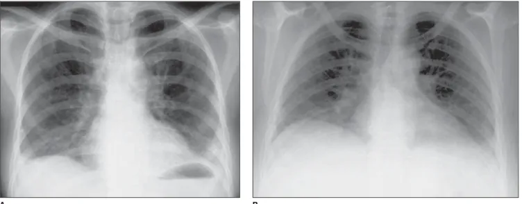
At X-ray, predominant reticular pattern on pulmonary bases was observed in 18 patients (78.2%). In some cases, more extensive lesions were observed, compromising upper portions of the lungs, besides basal segments. Among the latest ones, cases of reduction in the vertical diameters of the chest were observed as a signal of volumetric loss due to diaphragmatic retraction, a typical characteristic of pulmonary diseases coursing with fibrosis in the basal lobes (Figure 1).
patient (4.3%). The lesions observed on HRCT have predominated in the basal pulmonary segments, compromising especially the peripheral and posterior parenchymas (Figure 2). Other alterations visualized in some cases were traction bronchiectasis, bullaes and foci of pulmonary consolidation. The presence of mediastinal or hilar lymph nodes with < 10 mm in their smallest diameter was observed in five patients (21%).
DISCUSSION
Evidence of pulmonary disease has been described in chest X-rays in 20% to 65% of patients affected by systemic sclerosis(2). Our study has evidenced findings in 78.2% of cases, from subtle alterations like fine reticulations on pulmonary bases to extensive lesions with signs of pulmonary volume loss. A slightly higher frequency of radiographic findings in our study can be justified by the tertiary nature of our hospital.
The HRCT frequently demonstrates pulmonary compromising in clinically suspected patients and on normal X-ray films with subtle or doubtful findings(4). The pattern of tomographic findings has a good correlation with histopathological findings, differentiating patterns with predominance of inflammatory process (ground-glass opacities) from predominantly fibrotic lesions (reticular opacities and honeycombing), with inflammatory patterns being associated with a superior response to treatment(5). Additionally, the HRCT shows correlation with results of pulmonary function tests, evidencing restrictive pulmonary disease and diffusion disorder, mainly through reduction of pulmonary volume and diffusion capacity for carbon monoxide, respectively(6–8). There is no study correlating tomographic findings and patient’s age or disease duration time.
Our study has evidenced pulmonary lesions in the 23 patients studied, with greatest proportion of honeycombing, followed by ground-glass opacities associated with reticular opacities, and a smaller proportion of predominant reticular opacities, while the isolated predominance of ground-glass opacities was unusual. Thus, findings suggesting fibrosis were observed in the great majority of cases (honeycombing and reticular opacities corresponding to 60.8%) yet a reasonable part of them presenting patterns suggestive of inflammatory process.
Thoracic lymphadenomegaly is frequently associated with pulmonary disease in systemic sclerosis, although it shows not to be related with the pattern of HRCT findings, but rather with the extent of the pulmonary parenchyma compromising(9). Our study has demonstrated lymphadenomegaly in five (21%) of 23 cases, in four of them being associated with an extensive pulmonary compromising.
but prognosis and therapeutical response are better in patients affected by pulmonary systemic sclerosis.
Recently, interstitial pneumonias were reclassified and organized into a heterogeneous group of inflammatory diseases, with basis on their histopathological patterns and with clinical and radiological presentations different from each other(11,12). The idiopathic pulmonary fibrosis clinical presentation is pathologically described as an usual interstitial pneumonia characterized by marked interstitial fibrosis, predominating in the subpleural parenchyma of basal regions, corresponding to the honeycombing pattern seen on HRCT.
Pulmonary biopsy in cases of systemic sclerosis is not part of the clinical routine, but some studies have reported the non-specific interstitial pneumonia as a predominant histopathological pattern, finding alterations compatible with usual interstitial pneumonia, i.e., idiopathic pulmonary fibrosis in a smaller proportion of cases(13,14). As already mentioned, there is a good correlation between tomographic findings and histopathological alterations of pulmonary diseases, including in relation to the differentiation between the usual interstitial pneumonia and the non-specific interstitial pneumonia, the latest one being characterized by predominance of ground-glass and subtle reticular opacities, differently from basal and peripheral honeycombing observed in the first one(15).
Many recent studies, including this one, have demonstrated that pulmonary lesions described on HRCT of patients affected by systemic sclerosis are less extensive and with a greater proportion of findings suggestive of inflammatory process than those found in patients affected by idiopathic pulmonary fibrosis, i.e., tomographic picture of the systemic sclerosis is more similar to that of idiopathic non-specific insterstitial pneumonia(16). Therefore the HRCT importance for patients affected by systemic sclerosis should be highlighted, not only as an effective diagnosis method for early detection of alterations, but also for prognostic assessment, identifying those cases with predominance of inflammatory process which present a better therapeutical response.
REFERENCES
1. Gilliland BC. Systemic sclerosis (scleroderma). In: Braunwald E, Fauci AS, Kasper DL, Hauser SL, Longo DL, Jameson JL, editors. Harrison’s Principles of internal medicine. 15th ed. New York: McGraw-Hill, 2001.
2. Arroliga AC, Podell DN, Matthay RA. Pulmonary manifestations of scleroderma. J Thorac Imaging 1992;7:30–45.
4. Warrick JH, Bhalla M, Schabel SI, Silver RM. High resolution computed tomography in early scleroderma lung disease. J Rheumatol 1991;18:1520–1528.
5. Ooi GC, Mok MY, Tsang KWT, et al. Interstitial lung disease in systemic sclerosis: an HRCT-clinical correlative study. Acta Radiol 2003;44:258–264.
6. Remy-Jardin M, Remy J, Wallaert B, Bataille D, Hatron PY. Pulmonary involvement in progressive systemic sclerosis: sequential evaluation with CT, pulmonary function tests and bronchoalveolar lavage. Radiology 1993;188:499–506.
7. Kim EA, Johkoh T, Lee KS, et al. Interstitial pneumonia in progressive systemic sclerosis: serial high-resolution CT findings with functional correlation. J Comput Assist Tomogr 2001;25:757–763.
8. Shahin AA, Sabri YY, Mostafa HA, et al. Pulmonary function tests, high-resolution computerized tomography, alpha1-antitrypsin measurement, and early detection of pulmonary involvement in patients with systemic sclerosis. Rheumatol Int 2001;20:95–100.
9. Bhalla M, Silver RM, Shepard JA, McLoud TC. Chest CT in patients with scleroderma: prevalence of asymptomatic esophageal dilatation and mediastinal lymphadenopathy. AJR Am J Roentgenol 1993;161:269–272.
10. D’Angelo WA, Fries JF, Masi AT, Shulman LE. Pathologic observations in systemic sclerosis (scleroderma). A study of fifty-eight autopsy cases and fifty-eight matched controls. Am J Med 1969;46:428–440.
11. Müller NL, Colby TV. Idiopathic interstitial pneumonias: high resolution CT and histologic findings. RadioGraphics 1997;17:1016–1022.
12. Katzenstein AA, Myers JL. State of the art: idiopathic pulmonary fibrosis. Am J Respir Crit Care Med 1998;157:1301–1315.
13. Bouros D, Wells AU, Nicholson AG, et al. Histopathologic subsets of fibrosing alveolitis in patients with systemic sclerosis and their relationship to outcome. Am J Respir Crit Care Med 2002;165:1578–1579.
14. Kim DS, Yoo B, Lee JS, et al. The major histopathologic pattern of pulmonary fibrosis in scleroderma is nonspecific interstitial pneumonia. Sarcoidosis Vasc Diffuse Lung Dis 2002;19:121–127.
15. MacDonald SL, Rubens MB, Hansell DM, et al. Nonspecific interstitial pneumonia and usual interstitial pneumonia: comparative appearances at and diagnostic accuracy of thin-section CT. Radiology 2001;221:583–584.
COMPROMETIMENTO PULMONAR NA ESCLEROSE SISTÊMICA
Figuras
Figure 1. Examples of chest X-rays of patients with systemic sclerosis, showing pulmonary reticulation (A) that may be associated with reduction in the vertical diameter of the chest cavity (B).
Figure 2. Examples of patterns of abnormalities observed on chest high resolution CT in patients with systemic sclerosis: honeycombing (A), ground-glass opacities associated with reticular opacities (B), reticular opacities (C) and ground-glass opacities (D).
A B