Diogo Casal
, Ana Isabel Ribeiro
, Manuela Mafra
, Conceição Azeda
, Carlos Mavioso
, Maria Manuel Mendes
and Maria Manuel Mouzinho
4,5,6*Abstract
Introduction: Synovial sarcoma is a high-grade, soft-tissue sarcoma that most frequently is located in the vicinity of joints, tendons or bursae, although it can also be found in extra-articular locations. Most patients with synovial sarcoma of the hand are young and have a poor prognosis, as these tumors are locally aggressive and are associated with a relatively high metastasis rate. According to the literature, local recurrence and/or metastatic disease is found in nearly 80% of patients. Current therapy comprises surgery, systemic and limb perfusion chemotherapy, and radiotherapy. However, the 5-year survival rate is estimated to be only around 27% to 55%. Moreover, most authors agree that synovial sarcoma is one of the most commonly misdiagnosed malignancies of soft tissues because of their slow growing pattern, benign radiographic appearance, ability to change size, and the fact that they may elicit pain similar to that caused by common trauma.
Case presentation: We describe an unusual case of a large synovial sarcoma of the hand in a 63-year-old Caucasian woman followed for 12 years by a multidisciplinary team. In addition, a literature review of the most pertinent aspects of the epidemiology, diagnosis, treatment and prognosis of these patients is presented.
Conclusion: Awareness of this rare tumor by anyone dealing with hand pathology can hasten diagnosis, and this, in turn, can potentially increase survival. Therefore, a high index of suspicion for this disease should be kept in mind, particularly when evaluating young people, as they are the most commonly affected group.
Keywords: Synovial sarcoma, Hand, Surgery, Malignant tumor Introduction
Synovial sarcoma of the hand is an extremely rare entity that carries a worse prognosis than that of most soft-tissue sarcomas [1,2]. It has been estimated that, on average, even hand surgeons will encounter only one or two undiagnosed soft-tissue sarcomas of the upper ex-tremity during the entire duration of their careers [1].
In addition, the majority of authors agree that synovial sarcoma is one of the most commonly misdiagnosed malignancies of the soft tissues, owing to its slow grow-ing pattern, benign radiographic appearance, ability to change size, and their often eliciting pain similar to that caused by common trauma [3,4]. Hence, synovial sar-coma patients are often diagnosed initially as having
myositis, hematoma, synovitis, tendonitis, bursitis or other common disorders [3,4].
To make matters worse, primary synovial sarcoma size and initial status at presentation have been shown to strongly affect survival [3,5]. Therefore, a high index of suspicion for this disease should be kept in mind, par-ticularly when evaluating young people, as they are the most commonly affected group [3,4]. Herein we describe the clinical case of a large synovial sarcoma of the hand in a 63-year-old woman followed up for 12 years. More-over, a brief review of this unusual sarcoma is presented. Case presentation
A 63-year-old, right-handed Caucasian woman with an unremarkable medical history was referred to the hand clinic at our institution with a large firm mass in her left hand that had been growing steadily over the previous 3 years (Figures 1 and 2). The mass had grown to a point that it interfered with many of her daily life activities.
* Correspondence:[email protected]
4Plastic and Reconstructive Surgery Senior Consultant, São José Hospital,
Lisbon, Portugal
5Head of the Hand Surgery Clinic, São José Hospital, Lisbon, Portugal
Full list of author information is available at the end of the article
© 2012 Casal et al.; licensee BioMed Central Ltd. This is an Open Access article distributed under the terms of the Creative Commons Attribution License (http://creativecommons.org/licenses/by/2.0), which permits unrestricted use, distribution, and reproduction in any medium, provided the original work is properly cited.
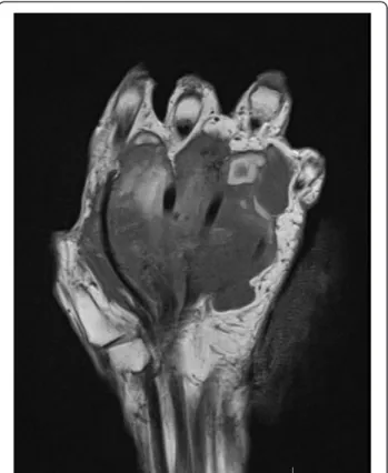
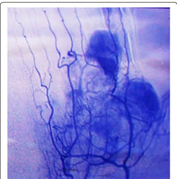
She complained of pain and occasional paresthesia in the first three rays of the hand. Ultrasound examination was inconclusive. Magnetic resonance imaging (MRI) was performed, which revealed a large mass spanning al-most the entire palmar aspect of the hand, superficially to the flexor tendons and with no evidence of bone in-volvement (Figures 3 and 4). A pre-operative angiogram showed a tumor with a rich blood supply (Figure 5). The tumor vessels were given off by the deep palmar arch, the external branch of the superficial palmar arch and the palmar digital arteries of the first, second and third fingers. With the patient under local anesthesia, an inci-sional biopsy was performed, which identified a synovial sarcoma.
Subsequently, with the patient under general anes-thesia, the mass was excised via a palmar approach, pre-serving the flexor apparatus of the fingers as well as all major vessels and nerves (Figures 6 and 7). Macro-scopically, the tumor was grayish and had a maximum
length of approximately 10cm (Figure 6). The tumor was multinodular and rubbery and seemed to be circum-scribed by a fibrous pseudocapsule. The post-operative period was uneventful. Recovery was fast, and the pa-tient had no significant functional or aesthetic impair-ment (Figure 8). She declined any physiotherapy treatments. Her only complaint was pain over the
Figure 1 Palmar aspect of the left hand showing a large nodular mass over the first four rays.
Figure 2 Dorsal aspect of the hand with the protruding mass in the first and second interdigital web spaces preventing complete finger adduction.
Figure 3 Magnetic resonance image of a coronal section of the hand showing a large mass around the palmar structures of the hand, but with no bone involvement.
Figure 4 Magnetic resonance image of a transverse section of the hand showing a large mass around the palmar structures of the hand, but with no bone involvement.
surgical scar that subsided approximately 6 months post-operatively. Pathological examination revealed the typical appearance of synovial sarcoma of the monophasic type (Figure 9). The specimen margins were found free of tumor cells. There was no evidence of systemic disease on the computed tomography (CT) scan of the thorax, abdo-men and pelvis that had been done pre-operatively. The patient underwent post-operative radiotherapy and chemo-therapy with good tolerance. Adjuvant chemochemo-therapy con-sisted of one cycle of ifosfamide and doxorubicin following the recommendations described by Kampeet al. [6]. Ifosfa-mide was given for 6 days at a dose of 14g/m2. An
equimolar dose of mesna was combined with the first dose of ifosfamide of 2g/m2. These two drugs were given as a 4-hour intravenous bolus. The subsequent ifosfamide treatment was given at the rate of 2g/m2/24 hours by con-tinuous intravenous infusion. The patient received an add-itional dose of mesna at the end of the sixth day. Doxorubicin was administered as a 48-hour continuous intravenous infusion, corresponding to a total dose of 60mg/m2. Adjuvant radiotherapy was delivered at the end of chemotherapy, including not only the tumor excision site but also a 3cm margin. A total dose of 60 Gy was ap-plied, fractioned in 2.0 Gy daily, 5 days per week, during 6 weeks.
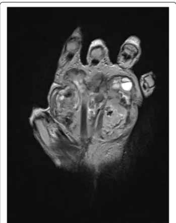
The patient was followed at the hand clinic at regular intervals for 9 years. The CT scans of the thorax, abdomen and pelvis that were done regularly during this period failed to show any evidence of systemic disease. She then decided to stop coming to the outpatient clinic, arguing that she saw no point in going as she felt perfectly well. Eleven and one-half years after surgery, the patient returned to the outpatient clinic with a recurrence of a sarcomatous mass in her left hand. MRI of the hand showed a large hypervascular mass with foci of necrosis that occupied most of the anterior compartments of the hand and encroached into the metacarpal and carpal bones (Figure 10). No evidence of metastasis was found.
A multidisciplinary team decided to propose left-hand amputation, which the patient declined. Twelve years after the initial diagnosis, she decided to stop coming to the out-patient clinic again and rebuffed any further contact. Six months later, she was found dead at her home, having had a massive hemorrhage from the recurrence in her hand. Discussion
Synovial sarcomas are malignant, high-grade, soft-tissue neoplasms that are estimated to represent between 5% and 10% of all soft-tissue sarcomas [3,7]. The estimated
Figure 5 Pre-operative angiogram showing the rich blood supply to the tumor, arising from the deep palmar arch, from the external branch of the superficial palmar arch and from the palmar digital arteries of the first, second and third fingers.
Figure 6 Intraoperative view of the excised specimen.
incidence of this tumor in the general population is 2.75 per 100,000 [3]. In fact, in adults, synovial sarcoma is the fourth most common type of sarcoma after malig-nant fibrous histiocytoma, liposarcoma and rhabdomyo-sarcoma [3]. In the United States alone, approximately 800 new cases are diagnosed each year [3,7]. In children, synovial sarcoma incidence is second only to rhabdo-myosarcoma in terms of soft-tissue sarcoma [8]. Ap-proximately one-third of synovial sarcomas occur in the first two decades of life [3,7,8]. This tumor is most prevalent in adolescents and adults between 15 and 40 years of age [3,7,8]. Concerning gender incidence, the male:female ratio is 1.2:1, with males being more fre-quently affected. Synovial sarcoma has a similar inci-dence in all ethnic groups [2,3,7,8].
This malignancy is usually located close to the large joints of the extremities, especially the lower extremities and in particular around the knee and ankle [2,3,7,8]. Other joints that are commonly affected are the shoul-der and the hip. Most often it arises in the para-articular regions, usually in close association with tendon sheaths, bursae and joint capsules [2,3,7,8]. However, it seldom involves the joints themselves [3]. In addition, contrary to what its name might suggest, synovial sarcoma also occurs in areas with no apparent relation to synovial structures, such as the heart, pericardium, pleura, lung, mediastinum, larynx, peritoneal cavity and abdominal wall [3]. In the hand, this tumor is more frequently found in the carpal region than in the fingers [2,3,7,8].
According to most authors, delay in diagnosis is very frequent [3]. In the majority of cases, the presence of a clinically detectable tumor prior to surgery is esti-mated to range from 2 to 4 years, but an insidiously growing mass or pain at the tumor site has been noted for as long as 20 years prior to initiation of proper treatment [3,4]. Recently, it has been shown that the occurrence of long-standing pain at the tumor site pre-ceding the development of a bulge is significantly more common with synovial sarcomas than with other sar-comas [9]. The imaging appearance is nonspecific, and in all cases a biopsy is necessary to confirm the diagno-sis [3,10,11].
Histologically, synovial sarcoma is typically character-ized by epithelium-like and/or spindle cell components arranged in a biphasic or monophasic pattern, although a poorly differentiated variant has also been described recently [12]. The biphasic pattern is considered the “classic” type and is generally recognizable by the coex-istence of morphologically different but genetically simi-lar epithelial cells and fibroblast-like spindle cells [3]. The monophasic type is closely related to the biphasic type and represents merely one extreme of its morpho-logical spectrum, sharing phenotypical features identical to the spindle-cell portion or the epithelium-like compo-nent, corresponding to the monophasic fibrous variant or to the monophasic epithelial variant, respectively [3]. Histologically, the poorly differentiated type is composed mostly of small, solidly packed, oval or spindle-shaped cells that seem to have an intermediate phenotype between epi-thelial and spindle cells, often with scant differentiation, simulating other neoplasms, namely, angiosarcoma or small-cell carcinoma [3].
Immunohistochemically, the majority of synovial sar-comas express cytokeratins, epithelial membrane anti-gen, calponin, B-cell lymphoma 2 (BCL-2) and CD-99. Vimentin can also be found in the spindle cells of these tumors. These markers can help differentiate synovial sarcomas from other sarcomas [12,13].
Although microscopic resemblance to the developing synovium was initially suggested, its origin from pre-formed synovial tissues remains to be proven [12,13]. Owing to the similarity between synovial sarcoma tumor cells and primitive synoviocytes, the term synovial sar-coma was coined [12,13]. However, most of these tumors occur outside the joints themselves and bear no resem-blance to synovial structures either ultrastructurally or immunohistochemically [12]. It has been proposed that synovial sarcoma arises from the pluripotential mesen-chyme of the limb bud [3].
A particular chromosomal translocation t(X;18) has been noted in over 90% of cases, both in adults and in children [12,14]. Although synovial sarcomas can be graded histologically according to mitotic index, percent-age of necrosis and tumor differentiation, almost all
tumor size (> 5cm), truncal location or proximal tumors in the limbs, male sex, bone or neurovascular invasion, incomplete excision on pathological examination, p53 overexpression, high proliferative index and, more re-cently, specific SYT-SSX fusion types [16,17]. Our pa-tient presented with only two of these risk factors, which may help explain the unusual long survival time observed. The most common site for metastasis is the
lung [4,18]. Lymph node involvement has been reported to occur in as many as 27% of patients [18].
Surgical resection is the definitive choice of treatment for primary synovial sarcomas and has been shown to both control local recurrence and prevent systemic dis-semination [2,8,12]. Unfortunately, the minimal accept-able margin has not been clearly established, and the surgeon must be aware of the possibility of microscopic infiltration of tumor cells into the pseudocapsule of the tumor [1,3,10]. Many investigators have suggested 1 cm to 2cm resection margins [1-3,10]. Because of proximity to the joints, the ablation can consist of either tenosyno-vectomy and/or post-operative radiotherapy and/or chemotherapy, or simply extremity segment amputation [3]. If ablation and tenosynovectomy are selected to re-tain maximal function, there might be a compromise of soft-tissue cover over tendons and neurovascular pedi-cles [3]. When proximity to critical anatomical struc-tures and patient desire do not allow the surgeon to obtain adequate surgical margins, isolated limb perfusion and radiotherapy must be considered, as they can poten-tially prevent amputation. In all cases, multidisciplinary discussion of adjuvant therapies that may prevent ampu-tation must occur prior to surgery [1-3,10,18]. Flaps are also an important option to bear in mind, as they can provide coverage of vital anatomical structures, as well as minimize the effects of radiation injury on these structures [19]. Discussion of the most adequate curative procedure, knowledge of the available reconstructive options, consideration of possible comorbidities and pa-tient wishes for limb preservation must all be taken into account before surgery [19].
The efficacy of adjuvant chemotherapy is still a matter of intense debate [18]. Similarly, radiotherapy is asso-ciated with a higher rate of local disease control, but not with better survival rates [3,18].
The presence of metastasis is considered the major cause of poor outcome, and several reports describing the results of current therapy showed a 5-year survival rate of around 27% to 55% [2,8,12]. Factors determining
(See figure on previous page.)
Figure 9 Histological appearance of the tumor. (A) Photomicrograph of a low-power magnification (40×) of a hematoxylin and eosin-stained section showing numerous highly packed cells forming densely cellular sheets and vague fascicles. (B) High-power magnification (400×) photomicrograph showing a hematoxylin and eosin-stained section with spindle cells with oval nuclei and scarce cytoplasm. These cells are uniform and relatively small, characterizing the monophasic“fibrous variant” of synovial sarcoma. (C) High-power magnification (400×) photomicrograph of a hematoxylin and eosin-stained section showing focal areas of a prominent hemangiopericytomatous vascular pattern, which is a frequent finding in synovial sarcoma. (D) Intermediate-power magnification (100×) photomicrograph of a hematoxylin and eosin-stained section showing focal tumor calcifications (arrows), which is also a relatively frequent finding in these tumors. (E) High-power magnification (400×) photomicrograph of an immunohistochemical section marking epithelial membrane antigen outlining the surface of the sarcomatous cells, which is typical of synovial sarcoma. (F) Intermediate-power magnification (100×) photomicrograph of an
immunohistochemical section marking cytokeratin 7 (CK7) showing CK7-positive cells either isolated or in cords. (G) High-power magnification (400×) photomicrograph of an immunohistochemical section marking CD99, which is staining the surfaces of tumor spindle cells. (H) High-power magnification (400×) photomicrograph of an immunohistochemical section marking B-cell lymphoma 2 (BCL-2), showing diffuse staining of the tumor.
Figure 10 Magnetic resonance imaging scan of the hand showing tumor recurrence with a large hypervascular mass and foci of necrosis that occupied most of the anterior
compartments of the hand and encroached into the metacarpal and carpal bones.
Awareness of this rare tumor by anyone dealing with hand pathology can hasten diagnosis, and this, in turn, can potentially increase survival [9]. Therefore, a high index of suspicion for this disease should be kept in mind, particularly when evaluating young people, as this is the most commonly affected group [3,7-9].
Consent
Written informed consent was obtained from the patient’s next-of-kin for publication of this case report and any accompanying images. A copy of the written consent is available for review by the Editor-in-Chief of this journal.
Competing interests
The authors declare that they have no competing interests. Authors’ contributions
DC played a major role in writing the manuscript and analyzed the patient data. AIR and MM played major roles in analyzing the patient data and reviewing the manuscript. CA aided in the editing of the manuscript and analyzed the patient data. CM followed up the patient, aided in the editing of the manuscript and analyzed the patient data. MM Mendes followed up the patient, edited the manuscript and analyzed the patient data. MM Mouzinho operated on the patient, participated in follow-up, played a major role in writing the manuscript and analyzed the patient data. All authors read and approved the final manuscript.
Acknowledgements
DC received a grant from The Program for Advanced Medical Education, which is sponsored by Fundação Calouste Gulbenkian, Fundação
Champalimaud, Ministério da Saúde e Fundação para a Ciência e Tecnologia, Portugal.
Author details
1Plastic and Reconstructive Surgery Resident, São José Hospital, Lisbon,
Portugal.2Pathology Resident, São José Hospital, Lisbon, Portugal.3Pathology Senior Consultant, São José Hospital, Lisbon, Portugal.4Plastic and
Reconstructive Surgery Senior Consultant, São José Hospital, Lisbon, Portugal.
5Head of the Hand Surgery Clinic, São José Hospital, Lisbon, Portugal.6Plastic
and Reconstructive Surgery Department, Hospital de São José, Rua José António Serrano, 1150-199, Lisbon, Portugal.
Received: 4 June 2012 Accepted: 5 October 2012 Published: 13 November 2012
References
1. Murray PM: Soft tissue sarcoma of the upper extremity. Hand Clin 2004, 20:325–333. vii.
2. Athanasian EA: Bone and soft tissue tumors. In Green’s Operative Surgery. Volume 2. 6th edition. Edited by Wolfe SW, Hotchkiss RN, Pederson WC, Kozin SH. Philadelphia: Churchill Livingstone; 2011:2141–2191.
3. Siegel HJ, Sessions W, Casillas MA Jr, Said-Al-Naief N, Lander PH, Lopez-Ben R: Synovial sarcoma: clinicopathologic features, treatment, and prognosis. Orthopedics 2007, 30:1020–1027.
8. Sultan I, Rodriguez-Galindo C, Saab R, Yasir S, Casanova M, Ferrari A: Comparing children and adults with synovial sarcoma in the Surveillance, Epidemiology, and End Results program, 1983 to 2005: an analysis of 1268 patients. Cancer 2009, 115:3537–3547.
9. De Silva MV, Barrett A, Reid R: Premonitory pain preceding swelling: a distinctive clinical presentation of synovial sarcoma which may prompt early detection. Sarcoma 2003, 7:131–135.
10. Wong CH, Chow L, Yen CH, Ho PC, Yip R, Hung LK: Uncommon hand tumours. Hand Surg 2001, 6:67–80.
11. Nakajima H, Matsushita K, Shimizu H, Isomi T, Nakano Y, Saito M, Aoki H: Synovial sarcoma of the hand. Skeletal Radiol 1997, 26:674–676. 12. Ferrari A, Gronchi A, Casanova M, Meazza C, Gandola L, Collini P, Lozza L,
Bertulli R, Olmi P, Casali PG: Synovial sarcoma: a retrospective analysis of 271 patients of all ages treated at a single institution. Cancer 2004, 101:627–634.
13. Miettinen M, Virtanen I: Synovial sarcoma: a misnomer. Am J Pathol 1984, 117:18–25.
14. Beck AH, West RB, van de Rijn M: Gene expression profiling for the investigation of soft tissue sarcoma pathogenesis and the identification of diagnostic, prognostic, and predictive biomarkers. Virchows Arch 2010, 456:141–151.
15. Shi W, Indelicato DJ, Morris CG, Scarborough MT, Gibbs CP, Zlotecki RA: Long-term treatment outcomes for patients with synovial sarcoma: a 40-year experience at the University of Florida. Am J Clin Oncol 2012, doi:10.1097/COC.0b013e31823fe450.
16. Wolden SL, Alektiar KM: Sarcomas across the age spectrum. Semin Radiat Oncol 2010, 20:45–51.
17. Eilber FC, Dry SM: Diagnosis and management of synovial sarcoma. J Surg Oncol 2008, 97:314–320.
18. Okcu MF, Despa S, Choroszy M, Berrak SG, Cangir A, Jaffe N, Raney RB: Synovial sarcoma in children and adolescents: thirty three years of experience with multimodal therapy. Med Pediatr Oncol 2001, 37:90–96. 19. Talbot SG, Mehrara BJ, Disa JJ, Wong AK, Pusic A, Cordeiro PG, Athanasian
EA: Soft-tissue coverage of the hand following sarcoma resection. Plast Reconstr Surg 2008, 121:534–543.
doi:10.1186/1752-1947-6-385
Cite this article as: Casal et al.: A 63-year-old woman presenting with a synovial sarcoma of the hand: a case report. Journal of Medical Case Reports 2012 6:385.
Submit your next manuscript to BioMed Central and take full advantage of:
• Convenient online submission
• Thorough peer review
• No space constraints or color figure charges
• Immediate publication on acceptance
• Inclusion in PubMed, CAS, Scopus and Google Scholar
• Research which is freely available for redistribution
Submit your manuscript at www.biomedcentral.com/submit