Early sac shrinkage predicts a low risk of late complications
after endovascular aortic aneurysm repair
F. Bastos Gonc¸alves1,3, H. Baderkhan4,5, H. J. M. Verhagen1, A. Wanhainen4, M. Bj ¨orck4, R. J. Stolker2, S. E. Hoeks2 and K. Mani4
Departments of1Vascular Surgery and2Anaesthesiology, Erasmus University Medical Centre, Rotterdam, The Netherlands,3Department of Angiology and Vascular Surgery, Hospital de Santa Marta, Centro Hospitalar de Lisboa Central, Lisbon, Portugal, and4Department of Surgical Sciences, Vascular Surgery, Uppsala University, Uppsala, and5Department of Surgery, G¨avle County Hospital, G¨avle, Sweden
Correspondence to: Dr F. Bastos Gonc¸alves, Erasmus University Medical Centre, Rotterdam, ’s-Gravendijkwal 230, 3015 CE Rotterdam, The Netherlands (e-mail: f.bastosgoncalves@erasmusmc.nl)
Background: Aneurysm shrinkage has been proposed as a marker of successful endovascular aneurysm repair (EVAR). Patients with early postoperative shrinkage may experience fewer subsequent complications, and consequently require less intensive surveillance.
Methods: Patients undergoing EVAR from 2000 to 2011 at three vascular centres (in 2 countries), who had two imaging examinations (postoperative and after 6–18 months), were included. Maximum diameter, complications and secondary interventions during follow-up were registered. Patients were categorized according to early sac dynamics. The primary endpoint was freedom from late complications. Secondary endpoints were freedom from secondary intervention, postimplant rupture and direct (type I/III) endoleaks.
Results: Some 597 EVARs (71·1 per cent of all EVARs) were included. No shrinkage was observed in 284 patients (47·6 per cent), moderate shrinkage (5–9 mm) in 142 (23·8 per cent) and major shrinkage (at least 10 mm) in 171 patients (28·6 per cent). Four years after the index imaging, the rate of freedom from complications was 84·3 (95 per cent confidence interval 78·7 to 89·8), 88·1 (80·6 to 95·5) and 94·4 (90·1 to 98·7) per cent respectively. No shrinkage was an independent risk factor for late complications compared with major shrinkage (hazard ratio (HR) 3·11; P < 0·001). Moderate compared with major shrinkage (HR 2·10; P = 0·022), early postoperative complications (HR 3·34; P < 0·001) and increasing abdominal aortic aneurysm baseline diameter (HR 1·02; P = 0·001) were also risk factors for late complications. Freedom from secondary interventions and direct endoleaks was greater for patients with major sac shrinkage. Conclusion: Early change in aneurysm sac diameter is a strong predictor of late complications after EVAR. Patients with major sac shrinkage have a very low risk of complications for up to 5 years. This parameter may be used to tailor postoperative surveillance.
Paper accepted 26 February 2014
Published online 22 April 2014 in Wiley Online Library (www.bjs.co.uk). DOI: 10.1002/bjs.9516
Introduction
Endovascular aneurysm repair (EVAR) is increasingly being used as primary mode for abdominal aortic aneurysm (AAA) repair in suitable patients owing to reduced perioperative mortality compared with open repair1. However, EVAR is associated with a significant
rate of complications over time2. Imaging surveillance is considered mandatory to identify and treat these complications before they result in life-threatening events such as postimplant rupture or graft occlusion. In
many countries the burden of post-EVAR surveillance is increasing rapidly.
Currently, there is no consensus on the frequency of post-EVAR surveillance, method of imaging or individual adaptation according to risk3 – 7. Computed tomographic angiography (CTA), which is still regarded as the standard for post-EVAR surveillance, is both costly and associated with potential risks from radiation and iodine contrast exposure, making strategies for reduction of follow-up intensity of interest8,9. Patients
intensive postoperative surveillance. Identification of individual risk factors for stent failure may allow surveillance to be tailored, focusing on patients at higher risk and reducing the surveillance costs for those at lower risk4,7,10,11.
Aneurysm sac shrinkage has been proposed as a marker of successful endovascular aneurysm exclusion11 – 13.
Con-sequently, it could be expected that patients who have significant shrinkage of the sac in the early postoper-ative phase would experience fewer complications, and consequently require less intensive imaging surveillance. The aim of this study was to evaluate the role of early AAA sac dynamics in determining long-term outcome after EVAR.
Methods
This study involved three institutions with experi-ence in EVAR, performing over 50 annual procedures each. Two institutions are university hospitals (Upp-sala University Hospital, Upp(Upp-sala, Sweden, and Erasmus University Medical Centre, Rotterdam, The Nether-lands), and the third is a county hospital (G¨avle Hos-pital, G¨avle, Sweden). The study complied with the Helsinki declaration on research ethics, and local pro-cedures for ethical clearance were followed at each participating centre.
Patients
All patients treated with EVAR from January 2000 to December 2011 at the three centres were assessed. Patients with a history of aortic reconstructive surgery or mycotic aneurysm were excluded. All three institutions used a 55-mm maximum diameter threshold for AAA intervention, and operated on smaller aneurysms only in the event of symptoms or accelerated growth. The inclusion criteria were: treatment of infrarenal aortoiliac aneurysm; and availability of two consecutive postoperative image examinations with the same technique (CTA or duplex ultrasound imaging) 6–18 months apart, with the first scan performed within the first 30 days after surgery, as recommended by the Society for Vascular Surgery (SVS) reporting standards for EVAR14. Preoperative examinations were used only when no early postoperative imaging was available and if carried out within 60 days before EVAR. The second of the two scans was considered the index examination. The measurement collected was the change in maximum aneurysm sac diameter between the first and second examinations. Patients who had two different imaging modalities during the first 18 months
after EVAR were excluded from the assessment of sac dynamics owing to variation between diameter measurements between the tests.
Postoperative image surveillance protocols
Protocols for postoperative surveillance differed between institutions, and also evolved over time. Typically, however, CTA, duplex ultrasonography or both were per-formed at regular intervals for all patients. All patients were considered eligible independently of the protocol followed, as evaluation of differences in surveillance strategy was not the aim of this study. However, preference was given to CTA-based measurements when CTA and duplex images were available, to reduce observer variability and allow
post hoc confirmation of diameters. CTA measurements
were done using outer-to-outer diameters, and duplex ultrasonography using leading-edge-to-leading-edge measurements. Each institution used the same method-ology for assessment of aneurysm diameter throughout the study.
Data management
Data from each institutional database were anonymized and entered into a study-specific database that recorded clinical and anatomical baseline characteristics, and procedural details including date, timing, intraoperative data, as well as endograft model and configuration. All image data were scrutinized by a single experienced vascular surgeon at each centre and the following endpoints were registered: diameter, and follow-up information including all registered complications and secondary interventions. All CTAs included a late arterial phase. Maximum diameter was used for assessment of sac dynamics as recommended in the SVS reporting standards for EVAR14.
Definitions
Early sac shrinkage was defined as the difference in maximum diameter between the first (within 30 days) and the second (after 6–18 months) scan. The second was considered the index examination. Intraoperative compli-cations were considered to have occurred if the device was not deployed at the intended position, if type I or III endoleak or graft obstruction was present, or if unplanned endovascular or surgical procedures were nec-essary. Clinical events (complications) were defined as any of the following occurrences after the index examination: direct (type I or III) or undetermined endoleak, endograft
occlusion, postimplantation rupture, endograft infection, migration exceeding 10 mm or device integrity failure. Undetermined endoleak was recorded if contrast was visu-alized outside the endograft and within the aneurysm sac, but the source could not be attributed to failure of a proximal or distal seal or patent aortic branch vessels. Persistent or late-onset type II endoleaks were consid-ered separately, defined as type II endoleaks being present beyond the first postoperative examination, or present-ing after a previous endoleak-free examination. Secondary interventions were those performed to resolve or pre-vent a possible complication, and included endovascular procedures (proximal cuff and stent implant, distal exten-sion implant, catheter-based thrombolysis, iliac stenting, coil or glue embolization of aortic branch vessels) as well as surgical procedures (balloon thrombectomy, femoro-femoral crossover, conversion to open repair, open or laparoscopic ligation of collaterals). Early complications were those that occurred before the second (index) exam-ination, and late complications those that occurred after this interval.
Shrinkage categories
Patients were divided into three groups, according to the observed AAA sac dynamics at 1 year. A 5-mm threshold was selected, as suggested by the SVS reporting standards for diameter changes in the aneurysm sac14. If the maximum aortic diameter increased, remained stable or decreased by less than 5 mm, patients were included in the no shrinkage group. A reduction in AAA diameter of between 5 and 9 mm was categorized as moderate shrinkage, and a reduction of 10 mm or more as major shrinkage.
Endpoints
The primary endpoint of this study was freedom from any complication. Secondary endpoints were freedom from reintervention, freedom from postimplan-tation rupture, freedom from direct or undetermined endoleaks, freedom from persistent or late-onset type II endoleaks and freedom from endograft occlusion. Only events occurring after the index imaging, which was used to categorize patients, were considered in this analysis.
Statistical analysis
Categorical variables were compared by means of χ2
linear-by-linear association tests. Continuous variables are
presented as mean(s.d.) if distributed normally and other-wise as median (range or i.q.r.), with analysis using one-way ANOVA for linearity. Estimates for the primary and sec-ondary endpoints were obtained using the Kaplan–Meier method and compared by means of the log rank test for equality. A multivariable Cox regression model was created to assess the independent influence of early sac dynamics on late complication rates; variables included were: early sac dynamics, baseline AAA diameter, rupture as surgical indication, use of aortomonoiliac endoprosthesis, occur-rence of intraoperative complications, and development of complications before the index examination. Selection bias was explored by comparing baseline characteristics, overall survival, duration of follow-up, and complication and secondary intervention rates in patients included or excluded from the present study; in this analysis the latter patients were those who survived the first 6 months, and were excluded only owing to lack of two consecutive imaging examinations. Similarly, analysis of the primary endpoint was also performed after excluding patients who had surgery for ruptured AAA. To test the validity of cat-egorization, the correlation between absolute and relative diameter changes was tested using Spearman’sρ, and com-parison between absolute and proportional shrinkage was performed for the primary endpoint. All tests were two-sided, and P < 0·050 was considered significant. Statistical analysis was done using SPSS version 20 (IBM, Armonk, New York, USA).
Results
From 2000 to 2011, 840 patients were treated with EVAR in the three participating institutions. Of these, 45 died within 6 months (27 operated on for ruptured AAA) and 198 were excluded as two equivalent consecutive scans were not available within the specified interval, leaving 597 (71·1 per cent) for assessment of early AAA sac dynamics. In 284 patients (47·6 per cent of the 597) no shrinkage was observed. Among these, growth of 5 mm or more was noted in 14 (2·3 per cent). Moderate shrinkage (5–9 mm) was registered in 142 patients (23·8 per cent) and major shrink-age (at least 10 mm) in the remaining 171 (28·6 per cent). The following endoprostheses were used in this cohort: 202 Excluder (W. L. Gore and Associates, Flagstaff, Arizona, USA), 189 Endurant (Medtronic CardioVas-cular, Santa Rosa, California, USA), 160 Zenith (Cook, Bloomington, Indiana, USA), 25 Talent (Medtronic CardioVascular) and 21 others. The median interval between the first and second (index) examination was 360 (i.q.r. 264–397) days. Baseline characteristics are described in Table 1.
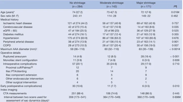
Table 1 Baseline characteristics of the three subgroups, based on early sac dynamics
No shrinkage Moderate shrinkage Major shrinkage
(n= 284) (n= 142) (n= 171) P¶
Age (years)* 74·2(7·2) 73·4(7·3) 72·1(7·6) 0·018#
Sex ratio (M : F) 243 : 41 114 : 28 149 : 22 0·462
Medical history
Ischaemic heart disease 121 of 274 (44·2) 56 of 137 (40·9) 69 of 162 (42·6) 0·757 Cerebrovascular disease 42 of 273 (15·4) 15 of 137 (10·9) 14 of 163 (8·6) 0·035 eGFR < 60‡ 47 of 184 (25·5) 20 of 86 (23) 36 of 129 (27·9) 0·525 Diabetes mellitus 44 of 274 (16·1) 17 of 137 (12·4) 21 of 163 (12·9) 0·355 Hypertension 175 of 274 (63·9) 86 of 136 (63·2) 107 of 163 (65·6) 0·676 Peripheral arterial disease 38 of 274 (13·9) 21 of 136 (15·4) 23 of 163 (14·1) 0·901 COPD 28 of 273 (10·3) 28 of 137 (20·4) 30 of 158 (19·0) 0·007 Maximum AAA diameter (mm)† 59 (36–110) 60 (32–110) 63 (35–139) < 0·001#
Operative details
Ruptured aneurysm 14 (4·9) 12 (8·5) 28 (16·4) < 0·001
Monoiliac stent configuration 11 (3·9) 7 (4·9) 6 (3·5) 0·939 Intraoperative complications 57 (20·1) 35 (24·6) 29 (17·0) 0·710
Proximal cuff/Palmaz 12 7 9
Iliac PTA/stenting 20 14 7
Iliac component extension 6 5 9
Other endovascular intervention 6 4 2
Other surgical intervention 4 5 1
Early postoperative complications§ 30 (10·6) 11 (7·7) 6 (3·5) 0·010 Index imaging
CTA measurements 251 (88·4) 106 (74·6) 148 (86·5) 0·445
Interval between two scans used for assessment of sac dynamics (days)†
359 (173–541) 364 (170–549) 360 (170–549) 0·698#
Values in parentheses are percentages unless indicated otherwise; *values are mean(s.d.) and †median (range). ‡Estimated glomerular filtration rate (eGFR) calculated using the Modification of Diet in Renal Disease formula: eGFR= 186 × serum creatinine–1·154× age–0·203× [1·212 if black] × [0·742 if female]. §Before index imaging. COPD, chronic obstructive pulmonary disease; AAA, abdominal aortic aneurysm; PTA, percutaneous transluminal angioplasty; CTA, computed tomographic angiography. Palmaz (Cordis, Bridgewater, New Jersey, USA). ¶χ2test, except #one-way ANOVA.
Freedom from late complications, according to aneurysm sac shrinkage
The total follow-up for the three groups was similar (median 3·1–3·2 years) (Table 2). The follow-up of interest for this study (after index image examination) was also similar (median 2·2 years). A total of 58 patients (9·7 per cent) developed complications during follow-up. These were more frequent in the no shrinkage group than in the moderate and major shrinkage groups (12·7, 9·9 and 4·7 per cent respectively; P = 0·038). Four years after the index imaging, the rate of freedom from complications was estimated at 84·3 (95 per cent confidence interval (c.i.) 78·7 to 89·8), 88·1 (80·6 to 95·5) and 94·4 (90·1 to 98·7) per cent respectively (Fig. S1, supporting information).
Multivariable testing for potential risk factors for late complications (occurring after the index examination) revealed that, compared with major shrinkage, moderate and no shrinkage increased the risk by 2·1 and 3·1 times respectively (Table 3). Other independent risk factors for late complications were increasing preoperative AAA diameter and a history of early postoperative complications
(Table 3). The use of discontinued endograft models did not have independent prognostic influence.
Complications in the major shrinkage group
Eight patients (4·7 per cent) with early shrinkage of the AAA sac of at least 10 mm had late complications. Three developed acute limb ischaemia owing to endograft limb thrombosis, at 1, 11 and 16 months after the index imaging (17, 18 and 26 months after EVAR). There were no imaging changes suggesting an increased risk of these events. Two patients had type I endoleaks: one type Ia 6 months after the index scan (1·0 year after EVAR) and one type Ib 2·7 years after the index scan (3·3 years after EVAR). The patient with a type Ia endoleak had a very short proximal seal zone (7 mm) at the 30-day CTA, despite a long proximal neck. The patient with a type Ib endoleak had dilatation of an iliac artery that was already wide (24 mm) before surgery. One patient developed a type III endoleak 12 months after the index examination (2 years after EVAR), owing to insufficient component overlap at implantation. One patient had an aneurysm
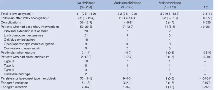
Table 2 Late outcome according to early aneurysm sac shrinkage
No shrinkage Moderate shrinkage Major shrinkage
(n= 284) (n= 142) (n= 171) P‡
Total follow-up (years)* 3·1 (0·5–11·9) 3·2 (0·5–12·2) 3·2 (0·5–12·7) 0·311§ Follow-up after index scan (years)* 2·2 (0–12·4) 2·2 (0–11·3) 2·2 (0–11·7) 0·277§
Complications 36 (12·7) 14 (9·9) 8 (4·7) 0·038
Patients who had secondary interventions 59 (20·8) 17 (12·0) 11 (6·4) < 0·001
Proximal extension cuff or stent 20 7 2
Limb component extensions 21 9 3
Coil/glue embolization 18 1 4
Open/laparoscopic collateral ligation 9 0 0
Conversion to open repair 9 2 3
Postimplantation rupture 3 (1·1) 1 (0·7) 1 (0·6) 0·819 Patients who had direct endoleak† 20 (7·0) 11 (7·7) 3 (1·8) 0·040
Type Ia 10 7 1 –
Type Ib 8 4 1 –
Type III 3 1 1 –
Undetermined type 2 1 0 –
Persistent or late-onset type II endoleak 55 (19·4) 9 (6·3) 9 (5·3) < 0·001¶
Endograft occlusion 5 (1·8) 3 (2·1) 3 (1·8) 0·979
Endograft infection 2 (0·7) 1 (0·7) 1 (0·6) 0·926
Values in parentheses are percentages unless indicated otherwise; *values are median (range). Only events after the index scan (at 6–18 months) are reported. †More than one endoleak may have occurred in the same patient. ‡Significance derived from univariable time-dependent statistical analysis (Kaplan–Meier analysis and log rank test), except §one-way ANOVA and ¶χ2test.
Table 3 Cox regression analysis of risk factors for late complications
Hazard ratio P
Moderate shrinkage (versus major shrinkage) 2·10 (1·11, 3·98) 0·022 No shrinkage (versus major shrinkage) 3·11 (1·75, 5·53) < 0·001 AAA diameter (per mm increase) 1·02 (1·01, 1·04) 0·001 Treatment of intact (versus ruptured) AAA 0·87 (0·43, 1·79) 0·712 Aortomonoiliac stent design 2·00 (0·77, 5·23) 0·156 Intraoperative complications 1·32 (0·85, 2·04) 0·219 Complications before index examination 3·34 (2·21, 5·04) < 0·001 Values in parentheses are 95 per cent confidence intervals. AAA, abdominal aortic aneurysm.
rupture 5·5 years after the index examination (6·5 years after EVAR), without a previously visible endoleak, which was treated successfully by urgent conversion to open repair. Finally, one patient had endograft infection diagnosed at the time of the index examination and died from sepsis before graft excision. This patient had low-grade fever before surgery, and was considered to have had an inflammatory aneurysm; a mycotic primary aetiology was suggested retrospectively.
Freedom from late secondary intervention, direct endoleak, persistent or late-onset type II endoleak, postimplant rupture and endograft occlusion according to early sac shrinkage
Late secondary interventions after the index imaging were needed in 87 patients overall (14·6 per cent). Patients with
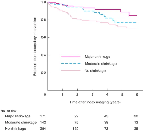
no shrinkage had significantly more secondary interven-tions than those with moderate and major shrinkage (20·8, 12·0 and 6·4 per cent respectively; P < 0·001) (Table 2). The estimated rate of freedom from secondary intervention 4 years after the index imaging examination (5 years after EVAR) was 76·6 (95 per cent c.i. 70·5 to 82·7), 81·8 (72·2 to 91·4) and 91·6 (85·9 to 97·3) per cent for no, moderate and major shrinkage groups respectively (Fig. 1).
Late direct (or undetermined) endoleaks also occurred less frequently in patients with major shrinkage, but no significant difference was observed for patients with moderate or no shrinkage. Estimates for rates of freedom of direct or undetermined endoleaks 4 years after the index imaging were 89·4 (95 per cent c.i. 84·1 to 94·6), 90·7 (83·8 to 97·6) and 97·2 (93·9 to 100) per cent for no, moderate and major shrinkage groups respectively (Fig. S2, supporting information).
Persistent or late-onset type II endoleaks were more frequent in the no shrinkage group (19·4 per cent). No difference was observed in the rate of persistent type II endoleaks for the moderate and major shrinkage groups (6·3 and 5·3 per cent respectively). Persistent or late-onset type II endoleaks were associated with sac growth in 24 patients, of whom eight also had type I endoleaks. One patient in each of the moderate and major shrinkage groups had subsequent sac growth associated with a persistent, isolated type II endoleak. No differences between groups were observed in late postimplant rupture or endograft occlusion (Table 2).
0·2 0 92 43 75 38 No. at risk Major shrinkage 171 Moderate shrinkage 142 No shrinkage 284 135 72 1 2 3
Time after index imaging (years)
Freedom from secondary intervention
4 5 20 12 38 6 0·4 0·6 0·8 1·0 Major shrinkage Moderate shrinkage No shrinkage
Fig. 1Kaplan–Meier plot for freedom from late secondary intervention, according to early sac shrinkage. P < 0·001 (log rank test)
Assessment of selection bias and sensitivity analysis
There were no differences in age and AAA diameter between patients included in, or excluded from the study (Table 4). Included patients had longer follow-up (median 3·2 versus 2·8 months after EVAR; P < 0·001), but the overall mortality rate did not differ significantly (26·1
versus 22·7 per cent; P = 0·396). However, similar numbers
of overall complications and secondary procedures were observed in both groups.
In 18 patients (3·0 per cent) a preoperative exam-ination was used to determine baseline diameter. No differences were observed in median shrinkage (5
ver-sus 4 mm; P= 0·769) or group allocations (P = 0·226)
for patients with baseline diameters measured before or after surgery.
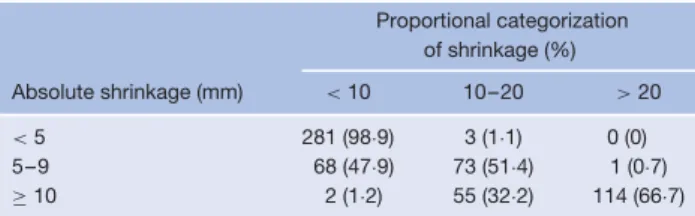
There was a strong correlation between proportional and absolute categorization of shrinkage (ρ = 0·988, P < 0·001). Proportional categorization of groups (less than 10, 10–20 and more than 20 per cent shrinkage) would have resulted in misclassification of shrinkage in 48·6 per cent of patients with moderate shrinkage defined according to absolute measurements (47·9 per cent misclassified as no shrinkage and 0·7 per cent as major shrinkage) and misclassification
Table 4 Selection bias assessment
Included Excluded‡ (n= 597) (n= 198) P§
Total follow-up after EVAR (years)*
3·2 (0·6–12·7) 2·8 (0·6–12·1) < 0·001¶ Age (years)† 73·4(7·4) 73·2(8·0) 0·658¶ Preoperative AAA diameter
(mm)†
63(14) 64(15) 0·303¶ Deaths 156 (26·1) 45 (22·7) 0·396 Total no. of secondary
complications
106 (17·7) 31 (15·7) 0·587 Total no. of secondary
interventions
109 (18·3) 32 (16·2) 0·591 Values in parentheses are percentages unless indicated otherwise; *values are median (range) and †mean(s.d.). ‡Owing to image availability. §χ2 test, except ¶one-way ANOVA.
of major shrinkage in 33·3 per cent (32·2 per cent as moderate shrinkage and 1·2 per cent as no shrinkage). Importantly, concordance in the no shrinkage group was 98·9 per cent (Table 5). Crude complication rates among patients with less than 10, 10–20 and over 20 per cent shrinkage were 12·5, 7·8 and 2·9 per cent respectively (P= 0·002). Four years after the index scan, the rate of freedom from complications was estimated at 83·5,
Table 5 Sensitivity analysis based on absolute versus proportional categorization Proportional categorization of shrinkage (%) Absolute shrinkage (mm) < 10 10–20 > 20 < 5 281 (98·9) 3 (1·1) 0 (0) 5–9 68 (47·9) 73 (51·4) 1 (0·7) ≥ 10 2 (1·2) 55 (32·2) 114 (66·7) Values in parentheses are percentages.
93·1 and 96·5 per cent respectively (Fig. S3, supporting information). In a multivariable model, less than 10 per cent shrinkage was associated with an increased risk of complications (HR 2·95, 95 per cent c.i. 1·48 to 6·16) compared with more than 20 per cent shrinkage. Had patients treated for ruptured AAA been excluded, the proportion of patients with complications in each group would not have differed (13·3 per cent for no shrinkage, 9·3 per cent for moderate shrinkage and 4·5 per cent for major shrinkage). Similarly, the Cox regression model did not change when patients with ruptured AAA were excluded from the analysis.
If the definition of shrinkage extended beyond the first year, 46 additional patients (7·7 per cent of all those eligible) would have been classified as having either moderate or major shrinkage. Intervention in one of these patients for an isolated type II endoleak, despite absence of growth, potentially interfered with the natural evolution of the aneurysm sac. One patient was identified as having a type Ib endoleak 6 months after the index procedure, with an uneventful subsequent follow-up. One patient was identified as having proximal stent migration 5 years after the index procedure, but there was no need for secondary intervention because the proximal seal was sufficient. Two patients had endograft limb occlusion during follow-up. No other complications were noted for this group of patients.
Discussion
The present study confirms that early sac shrinkage is an important prognostic factor for improved late outcome after EVAR. Patients with major sac shrinkage during the early postoperative phase have a low risk of subsequent complications for up to 5 years. Conversely, patients in whom early shrinkage does not occur are at higher risk of complications and more often require secondary interven-tions. These results may have important implications for individualization of postoperative surveillance.
The importance of sac shrinkage has been investigated previously, and contraction of the aneurysm sac has been suggested as a marker for success after EVAR. In a
recent publication involving 1450 procedures, Cieri and colleagues15reported that persistent shrinkage of the AAA sac (over 5 mm) was associated with rates of freedom from AAA-related death at 3 and 10 years of 100 and 99·7 per cent respectively. Houbballah and co-workers11
reported no postimplant ruptures or conversions, and very low rates of type I leak (2·2 per cent) and secondary intervention (3·3 per cent) at a mean follow-up of 4·2 years, for patients with significant sac contraction. Both authors concluded that significant contraction of the aneurysm sac is a robust predictor of success, which is in line with the present findings. However, these studies did not specifically investigate aneurysm sac shrinkage in the early postoperative phase, which limits the potential to apply their findings to individualized surveillance algorithms defined at an early stage.
Lee et al.12 showed that a volume reduction of greater
than 10 per cent 6 months after EVAR was a strong predictor of clinical success. The present study confirms this in a larger, contemporary series, adding that different degrees of shrinkage have different prognostic impact. Although measuring volume improved the accuracy of sac dynamics16, the added value is still undetermined and it is impractical in a clinical setting, compared with diameter measurement.
The observed differences in type and number of postoperative complications according to sac shrinkage could potentially be related to the preoperative aneurysm anatomy and the sealing length achieved at time of stent implantation. The importance of adequate seal length as verified on early postoperative imaging was confirmed previously as a strong prognostic factor for late EVAR outcome10. In the present study, early postoperative
complications were more common in the no shrinkage group. Lack of early complications may be interpreted as a surrogate for adequate implantation, which is in line with previous research. For the present study, however, this hypothesis could not be tested, as detailed baseline anatomical variables and postoperative seal length were not available for all patients.
Using sac shrinkage as a marker for success may not be applicable to limb occlusion complications. Late limb occlusion occurred uniformly in approximately 2 per cent of patients of all groups (Table 2), suggesting that shrinkage did not affect the risk of this complication in the long term. Most early limb occlusions are detectable on imaging and the result of a technical flaw, whereas late occlusions occur mostly without prior image findings17.
The present results have implications for post-EVAR surveillance. After 1 year, patients may be stratified on
the basis of sac shrinkage, and postoperative surveillance may be tailored to the expected risk of complications. Here, only 4·7 per cent of the patients with at least 10 mm sac shrinkage at 1 year had late complications, of which only three of eight were potentially preventable or predictable with surveillance imaging (2 type I and 1 type III endoleaks). Early postoperative characteristics could have predicted an increased risk of late complications in these three patients (very short seal or insufficient component overlap). Patients with major shrinkage of the aneurysm sac may benefit from adapted surveillance towards symptom-based investigations only, avoiding the need for routine investigations4,8,9.
This study has several limitations, restricting firm con-clusions. First, it is a retrospective study and may be subject to selection bias; compliance with institutional surveillance protocols is unknown. Furthermore, thresh-olds for intervention may have differed between institutions and over time. On the other hand, the results are based on a large international sample derived from prospec-tively collected data from three different hospitals, using a real-world variety of different endoprostheses. Another limitation is that the population is essentially northern European, and the results may not be generalizable to all ethnic groups. Patient categorization was based on an absolute (not proportional) reduction in diameter. As a result, patients with a smaller preoperative AAA were less likely to show sac shrinkage at 1 year, and similarly patients with ruptured AAAs were more likely to be included in the major shrinkage group (as their preoperative maximum AAA diameter was generally greater). The use of absolute reduction in sac diameter for definition of groups could potentially have resulted in misclassification of smaller or very large AAAs. The sensitivity analysis showed that classification of patients based on proportional diameter decrease would have yielded similar results. Interesting differences at baseline between groups suggest a possible prognostic influence of age, chronic obstructive pulmonary disease, maximum AAA diameter and type of presentation (intact versus ruptured AAA). Detailed anatomical base-line characteristics with known prognostic impact (such as iliac diameter or tortuosity) were not available for all patients and could not be integrated into the multi-variable model. Furthermore, as not all patients had the necessary imaging for inclusion, a chance of selec-tion bias remains. The authors assessed the risk of bias by analysing the baseline characteristics and complica-tion rates of patients excluded or included and found no differences, although the duration of follow-up differed slightly. Finally, the temporal restriction on categorization of patients may have resulted in misclassification. In the
sensitivity analysis, it was found that a further 7·7 per cent of patients could have been classified as having major sac shrinkage if the difference between scans had been extended beyond 1 year; these patients developed few complica-tions over the course of follow-up. However, the authors opted to restrict the classification interval to allow early stratification.
Acknowledgements
F.B.G. and H.B. contributed equally to this paper. This project was supported by the Swedish Research Council (grant no. K2013-64X-20406-07-3).
Disclosure: F.B.G. and S.E.H. received an unrestricted
research grant from the Lijf en Leven Fundation.
References
1 Mani K, Lees T, Beiles B, Jensen LP, Venermo M, Simo G et al. Treatment of abdominal aortic aneurysm in nine countries 2005–2009: a Vascunet report. Eur J Vasc Endovasc Surg 2011; 42: 598–607.
2 Stather PW, Sidloff D, Dattani N, Choke E, Bown MJ, Sayers RD. Systematic review and meta-analysis of the early and late outcomes of open and endovascular repair of abdominal aortic aneurysm. Br J Surg 2013; 100: 863–872.
3 Patel A, Edwards R, Chandramohan S. Surveillance of patients post-endovascular abdominal aortic aneurysm repair (EVAR). A web-based survey of practice in the UK. Clin Radiol 2013; 68: 580–587.
4 Sternbergh WC III, Greenberg RK, Chuter TA, Tonnessen BH; Zenith Investigators. Redefining postoperative surveillance after endovascular aneurysm repair: recommendations based on 5-year follow-up in the US Zenith multicenter trial. J Vasc Surg 2008; 48: 278–284. 5 Moll FL, Powell JT, Fraedrich G, Verzini F, Haulon S,
Waltham M et al.; European Society for Vascular Surgery. Management of abdominal aortic aneurysms clinical practice guidelines of the European Society for Vascular Surgery. Eur J Vasc Endovasc Surg 2011; 41(Suppl 1): S1–S58. 6 Karthikesalingam A, Page AA, Pettengell C, Hinchliffe RJ,
Loftus IM, Thompson MM et al. Heterogeneity in surveillance after endovascular aneurysm repair in the UK. Eur J Vasc Endovasc Surg 2011; 42: 585–590.
7 Dias NV, Riva L, Ivancev K, Resch T, Sonesson B, Malina M. Is there a benefit of frequent CT follow-up after EVAR? Eur J Vasc Endovasc Surg 2009; 37: 425–430.
8 Kim JK, Tonnessen BH, Noll RE Jr, Money SR, Sternbergh WC III. Reimbursement of long-term postplacement costs after endovascular abdominal aortic aneurysm repair. J Vasc Surg 2008; 48: 1390–1395.
9 Kooiman J, Pasha SM, Zondag W, Sijpkens YW, van der Molen AJ, Huisman MV et al. Meta-analysis: serum
creatinine changes following contrast enhanced CT imaging. Eur J Radiol 2012; 81: 2554–2561.
10 Bastos Gonc¸alves F, van de Luijtgaarden KM, Hoeks SE, Hendriks JM, ten Raa S, Rouwet EV et al. Adequate seal and no endoleak on the first postoperative computed
tomography angiography as criteria for no additional imaging up to 5 years after endovascular aneurysm repair. J Vasc Surg 2013; 57: 1503–1511.
11 Houbballah R, Majewski M, Becquemin JP. Significant sac retraction after endovascular aneurysm repair is a robust indicator of durable treatment success. J Vasc Surg 2010; 52: 878–883.
12 Lee JT, Aziz IN, Lee JT, Haukoos JS, Donayre CE, Walot I et al. Volume regression of abdominal aortic aneurysms and its relation to successful endoluminal exclusion. J Vasc Surg 2003; 38: 1254–1263.
13 Rhee RY, Eskandari MK, Zajko AB, Makaroun MS. Long-term fate of the aneurysmal sac after endoluminal exclusion of abdominal aortic aneurysms. J Vasc Surg 2000; 32: 689–696.
14 Chaikof EL, Blankensteijn JD, Harris PL, White GH, Zarins CK, Bernhard VM et al.; Ad Hoc Committee for Standardized Reporting Practices in Vascular Surgery of the Society for Vascular Surgery/American Association for Vascular Surgery. Reporting standards for endovas-cular aortic aneurysm repair. J Vasc Surg 2002; 35: 1048–1060.
15 Cieri E, De Rango P, Isernia G, Simonte G, Verzini F, Parlani G et al. Effect of stentgraft model on aneurysm shrinkage in 1450 endovascular aortic repairs. Eur J Vasc Endovasc Surg 2013; 46: 192–200.
16 van Keulen JW, van Prehn J, Prokop M, Moll FL, van Herwaarden JA. Potential value of aneurysm sac volume measurements in addition to diameter measurements after endovascular aneurysm repair. J Endovasc Ther 2009; 16: 506–513.
17 van Zeggeren L, Bastos Gonc¸alves F, van Herwaarden JA, Zandvoort HJ, Werson DA, Vos JA et al. Incidence and treatment results of Endurant endograft occlusion. J Vasc Surg 2013; 57: 1246–1254.
Supporting information
Additional supporting information may be found in the online version of this article:
Fig. S1 Kaplan–Meier plot for freedom from late complications, according to early sac shrinkage (Word document) Fig. S2 Kaplan–Meier plot for freedom from late direct endoleaks, according to early sac shrinkage (Word
document)
Fig. S3 Kaplan–Meier plot for freedom from late complications, according to early sac shrinkage based on