Factors involved in the migration of endoprosthesis in
patients undergoing endovascular aneurysm repair
Fatores envolvidos na migração das endopróteses em pacientes submetidos ao tratamento
endovascular do aneurisma da aorta abdominal
Marcelo José de Almeida1, Winston Bonetti Yoshida2, Ludvig Hafner3, Juliana Henrique dos Santos4, Bruno Felipe Souza5,
Flávia Fagundes Bueno5, Janaína Lopes Evangelista5, Lucas José Vaz Schiavão5
Abstract
Migration of the endoprosthesis is deined as the misplacement of its initial ixation. To assess the migration, the position of the endoprosthesis regarding a certain anatomic region is veriied. Considering the aneurysm of the infrarenal abdominal aorta, the proximal area of reference is the origin of the lowest renal artery and, at the distal region, it is located next to the internal iliac arteries. Patients should be monitored for long periods so that migrations can be identiied; these migrations usually occur 2 years after the implantation. To avoid migrations, mechanical forces that enable ixation and that are determined by the characteristics of the devices and by the incorporation of the endoprosthesis should predominate over gravitational and hemodynamic forces, which tend to drag the prosthesis toward to caudal direction. Angulation, extension, and diameter of the neck, and transversal measure of the aneurysmatic sac are important morphological aspects related to migration. In relation to the technique, endoprosthesis implantation with excessive oversizing (> 30%) is not recommended because it leads to aortic neck dilatation, folds and proximal leakage that also contribute to migration. On the other hand, endoprosthesis with additional ixation devices (hooks, barbs and suprarenal ixation) seem to be less associated with migration. he process of endoprosthesis incorporation is partial and does not seem to be enough to prevent later migrations. In this sense, experimental studies with endoprosthesis of higher porosity, as well as the use of substances that allow higher ibroplasia and adherence of the prosthesis to the artery, have been conducted and are promising. Such aspects are discussed in the present review of the literature.
Keywords: Vascular prosthesis, migration, complications, aortic aneurysm.
Resumo
A migração da endoprótese é complicação do tratamento endovascular deinida como deslocamento da ancoragem inicial. Para avaliação da migração, veriica-se a posição da endoprótese em relação a determinada região anatômica. Considerando o aneurisma da aorta abdominal infrarrenal, a área proximal de referência consiste na origem da artéria renal mais baixa e, na região distal, situa-se nas artérias ilíacas internas. Os pacientes deverão ser monitorizados por longos períodos, a im de serem identiicadas migrações, visto que estas ocorrem normalmente após 2 anos de implante. Para evitar migrações, forças mecânicas que propiciam ixação, determinadas por características dos dispositivos e incorporação da endoprótese, devem predominar sobre forças gravitacionais e hemodinâmicas que tendem a arrastar a prótese no sentido caudal. Angulação, extensão e diâmetro do colo, além da medida transversa do saco aneurismático, são importantes aspectos morfológicos do aneurisma relacionados à migração. Com relação à técnica, não se recomenda implante de endopróteses com sobredimensionamento excessivo (> 30%), por provocar dilatação do colo do aneurisma, além de dobras e vazamentos proximais que também contribuem para a migração. Por outro lado, endopróteses com mecanismos adicionais de ixação (ganchos, farpas e ixação suprarrenal) parecem apresentar menos migrações. O processo de incorporação das endopróteses ocorre parcialmente e parece não ser suiciente para impedir migrações tardias. Nesse sentido, estudos experimentais com endopróteses de maior porosidade e uso de substâncias que permitam maior ibroplasia e aderência da prótese à artéria vêm sendo realizados e parecem ser promissores. Esses aspectos serão discutidos nesta revisão.
Palavras-chave: Prótese vascular, migração, complicações, aneurisma da aorta.
Financial support: Fundação para o Amparo à Pesquisa do Estado de São Paulo – FAPESP.
1 Docente, disciplina de Cirurgia Vascular, Faculdade de Medicina de Marília (FAMEMA), Marília, SP. Pós-graduando, Faculdade de Medicina de Botucatu (FMB), Universidade Estadual de São
Paulo (UNESP), Botucatu, SP, Brazil.
2 Livre-docente. Professor adjunto, disciplina de Cirurgia Vascular, Departamento de Cirurgia e Ortopedia, FMB, UNESP, Botucatu, SP, Brazil. 3 Doutor. Docente, disciplina de Cirurgia Vascular, FAMEMA, Marília, SP, Brazil.
4 Médica. Cirurgiã Vascular, Maringá, PR, Brazil. 5 Graduandos de medicina, FAMEMA, Marília, SP, Brazil.
Introduction
Conventional treatment of abdominal aortic aneurysms consists of placement of a polyester tube grat, the ends of which are sutured to the arterial wall proximally and distally to the aneurysmal dilatation, preventing blood low from straining the wall of the aneurysm. Development of this tre-atment modality was one of the inest achievements of vascu-lar surgery, as it allowed modiication of the natural history
of the disease and reduction of rupture-associated mortality.1
Endovascular repair of abdominal aortic aneurysms (EVAR)2
is based on insertion of an endoprosthetic device through the femoral or iliac artery. he device is then deployed within the lumen of the aorta so its ends are anchored to normal artery, proximally and distally to the aneurysm. Ater the sheath is released, the elasticity of the stent grat provides ra-dial strength, which keeps the device ixated to the aneurysm neck. he endoluminally deployed stent thus excludes the aneurysmal sac from circulation. Some stents have hooks or barbs to improve ixation to the arterial wall.
EVAR is associated with decreased transfusion require-ments and avoids aortic cross-clamping, decreasing cardiac overload and the untoward efects of ischemia and reper-fusion, all of which contributes to allow shorter
postope-rative recovery times.3,4. However, long-term follow-up of
patients undergoing EVAR shows a permanent risk of grat migration and structural failure, even when treatment was successful. Close surveillance is therefore required to detect any relevant changes and, sometimes, establish the need for
reintervention.5-9 On the other hand, despite the need for
continuous postoperative monitoring, EVAR carries the beneit of lower rupture-related death rates than with no
treatment.10
Since the introduction of EVAR, expectations on short-, medium- and long-term outcomes has run high. Ten years and countless studies on, the ideal endoprosthe-tic device has yet to be developed. Despite progress in grat materials, endoprosthesis migration is still a major issue, as it may lead to type I endoleak and increased pressure wi-thin the aneurysm, culminating in rupture or collapse of the device into the aneurysmal sac, both of which indicate
emergent open repair.5,11
he present article will discuss the clinical and patho-physiological factors involved in endograt migration.
Definition and criteria
Endoprosthesis migration is defined as displace-ment of the device from its original site of attachdisplace-ment.
Assessment of migration consists of determining the position of the endoprosthesis relative to a predefined anatomical landmark. In infrarenal AAAs, the proximal landmark is the origin of the lowest renal artery, and the distal landmark is located next to the internal iliac arte-ries. The landmark for suprarenal fixation is the superior mesenteric artery (SMA). CT scanning is the imaging modality of choice for assessment of adequate placement or migration. Slices no larger than 3 mm should be ob-tained from the level of the SMA down to the common femoral arteries. The use of other anatomical landmarks, such as position of the vertebral bodies as observed on CT itself or on abdominal plain films, is unreliable, as any vertebral size changes due to osteoporosis or other bone conditions may lead to erroneous assessment of
migration.12
he Society for Vascular Surgery (SVS) deines
endo-grat miendo-gration as any displacement of 10 mm or more.13
Caudal migration of the proximal stent is most common and poses the greatest risk in EVAR. he SVS deinition is problematic in that, when the neck is small, 10 mm will cor-respond nearly to its maximum length, and diagnosis may only occur ater the patient develops complications. In light of these considerations, current practice is to regard any
migration >5 mm as clinically relevant.14
Regardless of endoprosthesis type, most migrations oc-cur ater the 13th month post-implantation, peaking at 19
months.14-16 One key inding is that the risk of migration
persists indeinitely ater EVAR; complications have been reported as late as 4 years post-treatment. Follow-up studies seeking to assess grat migration must therefore extend for no less than 24 months ater the procedure.
Although follow-up and observation for grat migration are essential, the absence of migration is no guarantee of treat-ment success. In some cases, perigrat leak may occur despite adequate proximal ixation of the endoprosthesis, leading to expansion of the aneurysm. herefore, proper device positio-ning may only be interpreted as a positive treatment outcome
if there are no perigrat leaks at the neck of the aneurysm.17-19
Current recommendations provide for CT follow-up at 1 month and 6 months post-procedure and annually there-ater. If migration is present, follow-up intervals are
shorte-ned for closer surveillance.20 In some cases, arteriography
(which allows endovascular repair if necessary) is indicated.
Importance of implantation technique
assessment due to parallax error, which is common in the tortuous aortic neck. Oblique views can minimize this issue and allow more accurate deployment.
Zarins et al.21 assessed the importance of proper
placement of the AneuRx stent graft at the origin of the renal artery. Isolated review of this factor showed that graft deployment below the intended site was directly correlated with greater risk of migration. The authors established that the greater the distance from the renal artery to the proximal end of the graft, the greater the risk of migration; each millimeter increase in distance below the renal arteries increased risk of migration by 5.8%. Likewise, each millimeter increase in length of the infrarenal neck covered by the graft decreased migration risk by 2.5%. Adequate placement of the distal end of the graft near the origin of the internal iliac arteries also cor-relates with a lower incidence of migration, as it reduces the risk of distal leakage and allows better longitudinal columnar support for the endograft, which tends to
pre-vent proximal migration.14
Close attention to proper endograt placement at the proximal and distal neck of the aneurysm is therefore of the essence, as correct placement is positively correlated with lower risk of migration-related complications.
Biomechanical forces interacting with the device
Biomechanical forces produced by oversizing, the contact area between the device and the artery, and addi-tional fixation such as hooks, barbs, or bare-metal supra-renal support extensions encourage fixation of the device to the aorta, as does the inflammatory process that
oc-curs in the arterial wall.22 Conversely, gravitational pull
and hemodynamics tend to drag the device caudally. The balance of these forces determines whether migration will occur. Complex calculations performed both in
vi-tro (in the biomechanics lab setting)23 and in vivo, with
computational analysis of the CT results of patients who
underwent EVAR,24,25 have provided important
informa-tion on the dynamic interacinforma-tions between the aortic wall, the endoprosthesis, and blood flow. Several authors have adapted mathematical formulae to experimental models of abdominal aortic aneurysms before and after stent graft deployment. These studies have concluded that, even in technically successful cases, certain areas of the endoprosthesis will always be more sensitive to hemo-dynamic changes, particularly at the aortic neck attach-ment site and at the bifurcation of the iliac extensions. In these areas, the endograft wall is subjected to strain,
producing a 1- to 2-newton drag force that pulls the de-vice distally.
These findings created a need for better understan-ding the forces involved in endograft fixation. Lambert et
al.26 assessed the mechanical behavior of endoprostheses
implanted in cadaveric aortas and found that, the greater the extent of prosthesis oversizing and the contact area between the device and the artery, the greater the load required to dislodge it. In similar experiments, Malina
et al.27 found that hooks and barbs increase fixation even
further. These studies have also provided important con-tributions by identifying the load required for dislodging endografts (3 N on average) and comparing it with mean hemodynamic drag forces (2 N). Despite their validity, however, these investigations failed to take into account several factors that interact with endoprostheses in the living body, as all experimental testing was conducted in cadaveric aortas.
Proper grat ixation to the proximal neck also reduces
the risk of migration. Wolf et al.18 reported a higher
num-ber of migration and type I endoleak events when there was poor apposition of the stent grat to the aortic wall. his is explained by decreased contact area and by leakage throu-gh folds in the grat fabric, leading to reduced friction and ixation forces.
Distal ixation was investigated by Volodos et al.,28 who
carried out in vitro assessment of straight and bifurcated polytetraluoroethylene (PTFE) grats in a specially desig-ned model: a plastic cylinder mimicking an aortic aneu-rysm was connected to a pulsatile low circuit powered by a cardiac pump, which subjected the system to diferent pres-sures. he authors found only minor changes in diameter, but signiicant changes in grat length and distal kinking, culminating in displacement of the device when load ex-ceeded 208 g (approximately 2 N), thus demonstrating the importance of distal ixation. In fact, the importance of proper distal ixation is proven by the high rate distal dis-placement-related complications in patients receiving irst-generation endoprostheses, which lacked adequate stent support in the iliac regions.
In addition to aneurysm- and endoprosthesis-re-lated aspects, certain clinical changes may destabilize graft fixation forces and encourage migration. Mohan
et al.29 studied 2,862 post-EVAR patients included in the
EUROSTAR registry. Using Massey’s formula,30 the
Smoking is an important determinant of aneurysmal wall dilatation, with an added harmful effect in
weake-ned arterial walls as a potentiator of protease activity.31-33
Hypertension was significantly correlated with migra-tion, as it increases the hemodynamic forces that push the graft caudally.
In short, an understanding of relevant biomechanical forces is a key element to be considered in the choice of endoprosthesis and EVAR technique.
Migration and device type
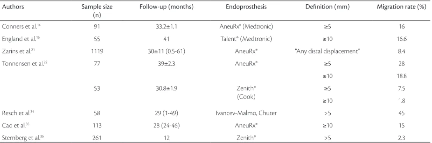
Table 1 shows diferences in complication rates de-pending on the type of endoprosthetic device. hese com-parisons must be viewed cautiously, as they represent the work of diferent teams with varying levels of experien-ce in EVAR and patients with heterogeneous aneurysm characteristics.
Diferences in migration rates may be explained by stent design – namely, by the choice of material used in
grat construction and by mode of ixation. Resch et al.,34
for instance, used Dacron-covered, Gianturco stent-based grat prototypes with proximal ixation hooks in most ca-ses, whereas other authors used third-generation commer-cial endograts, the design of which has been substantially perfected.14,16,21,22,35,36
he considerable variation in migration rates (1.8– 45%) also relects diferences in study criteria, such as length of follow-up, choice of technique and operator experience.
Tonnensem et al.22 found lower migration rates ater
use of Zenith devices. However, the authors already had ex-tensive EVAR experience when they began using this model
of endoprosthesis, which may have biased their results. At any rate, mid- and long-term assessment studies appear to support lower migration rates with use of endoprostheses
that employ auxiliary ixation systems.16 Longer follow-up
studies should deine whether this lower likelihood of mi-gration is sustained over time.
Factors involved in migration
Aneurysm neck morphology
he shorter the length of the aneu
rysm neck, the smaller the contact area between endo-prosthesis and artery, thus hampering device ixation. here is no objective deinition of the minimal area re-quired for adequate ixation, but most authors empirically
recommend a length of 15 mm.37
Aneurysm neck angle appears to inluence duration of surgery and post-EVAR complication rates. Sternberg
et al.38 measured aortic neck angulation (the angle formed
between the aortic neck and the longitudinal axis of the
aneurysm) and classiied it as severe (≥ 70º), moderate (40
to 59º) or mild (< 40º). he authors found higher rates of complications (such as type I endoleak, aneurysm expan-sion and grat migration), endovascular reintervention, and conversion to open repair in patients with severe aortic neck angulation. Furthermore, procedure duration was longer in these patients due to greater diiculty in endograt place-ment. he authors concluded that EVAR should be discou-raged in patients with aortic neck angles greater than 40º.
Albertini et al.19 assessed the risk of proximal type I
endo-leak and migration and their correlation with aortic neck size, shape, and angulation. he authors found neck angle
Authors Sample size
(n)
Follow-up (months) Endoprosthesis Definition (mm) Migration rate (%)
Conners et al.14 91 33.2±1.1 AneuRx® (Medtronic) ≥5 16
England et al.16 55 41 Talent® (Medtronic) ≥10 16.6
Zarins et al.21 1119 30±11 (0.5-61) AneuRx® “Any distal displacement” 8.4
Tonnensen et al.22 77 39±2.3 AneuRx® ≥5 28
≥10 18.8
53 30.8±1.9 Zenith® (Cook)
≥5 7.5
≥10 1.8
Resch et al.34 58 29 (1-49) Ivancev-Malmo, Chuter >5 45
Cao et al.35 113 28 (24-46) AneuRx® ≥10 15
Sternberg et al.36 261 12 Zenith® >5 2.3
to be the predominant factor associated with complications (Figure 1). Two major mechanisms explain this inding: 1) Tortuous proximal sites reduce the contact area
betwe-en the device and the arterial surface, decreasing fric-tion, which tends to anchor the device;
2) Hemodynamic studies show that the force exerted by blood low against the vessel wall at a single point is proportional to the square of the velocity of low at that point. hus, low velocity is increased in tortuous ar-teries, leading to increased drag forces. his, coupled with the fact that blood columns directly impact a lar-ger surface of the kinked endoprosthesis, would
increa-se the likelihood of distal displacement even further.38,39
Some authors have reported the use of endopros-theses in very large neck AAAs (> 28 mm diameter) as a predisposing factor for graft migration; other
stu-dies, however, have found no such correlation.36 A
gre-ater likelihood of neck dilation 10 years after repair has also been reported when proximal cuff diameter at the
time of EVAR exceeded 28 mm.40 Considering that
pos-toperative aneurysm neck expansion is a known pre-disposing factor for migration and that large neck are more likely to expand, one may infer that endovascu-lar repair of endovascu-large neck aneurysms would be inherently
complication-prone.14,41
hrombi, calciication, and other irregularities of the aortic neck wall are also associated with poor endoprosthe-sis ixation and increased likelihood of migration:
1) hrombi found at the aneurysm neck have a friable surface, which decreases the area of friction between the device and the arterial wall;
2) Irregularities and calciication of the arterial wall lead to deformities or minor kinks and folds in the prosthesis. his reduces contact area, and type I
endo-leak may also occur through these points;28
3) Calciications also harden the arterial wall, reducing complacency and decreasing device seating.
Aneurysm size and migration
Larger aneurysm size significantly correlates with parameters that predispose do endograft migration. Large aneurysms (> 55 mm) tend to have shorter, wi-der and more tortuous necks, which would thus
incre-ase the risk of migration.42,43 Ouriel et al.44 assessed 700
patients according to aneurysm size, classified as small (< 55 mm) or large (> 55 mm), and found a statistically higher rate of migration and type I endoleak in the latter group. Aneurysms with smaller diameters were deemed
more anatomically suitable for EVAR, which may explain the lower rate of complications in narrower aneurysms. In their five-year follow-up of 923 post-EVAR patients,
Zarins et al.45 grouped aneurysm diameter into three size
classes (small, < 50 mm; medium, 50 to 59 mm; large, > 60 mm) and found no significant differences in mi-gration, leakage or aneurysm expansion rates. They did, however, report significantly higher rates of conversion to open repair and rupture-related mortality in patients with larger aneurysms. Interestingly, as conversion to open repair is usually prompted by leaks, migration or aneurysm expansion, higher conversion rates may corre-late with these events.
Recent studies have shown improved outcomes and lower migration rates in patients with aneurysms smaller than 55 mm in diameter, which provide an anatomically
favorable setting for EVAR.46 However, it bears noting
that patients with small aneurysms undergoing purely clinical treatment aimed at controlling blood pressure have a low risk of rupture (0.6% per year). Indications for EVAR must therefore take life expectancy and risk of the endovascular procedure (and possible
reinterven-tion) into account.47
Oversizing and migration
Mohan et al.48 reported increased rates of type I
endo-leak when grats were oversized less than 10%, and
sugges-ted 10 to 20% oversizing as adequate. Almeida & Yoshida49
conducted to ascertain the displacement load required to dislodge the device from the aorta. In the 20% over-sizing group, displacement load was statistically higher than in the 10% oversizing group. his may be explained by the increased radial strength of the stent struts, which would penetrate the wall of the aorta and reach deeper
into the tunica media. Some authors50 have posited that
greater oversizing would produce a greater inlammatory response at the vessel wall, improving stent ixation and integration as well. In smaller vessels, such as the coronary arteries, greater oversizing would produce an even more intense inlammatory response, leading to intimal
hyper-plasia and early thrombosis.51,52 In larger arteries, however,
greater inlammation would increase prosthesis ixation, preventing grat migration. Histological examinations carried out in the Almeida & Yoshida study corroborate results reported elsewhere in the literature, showing that a ibroblastic reaction, with attending inlammation and areas of neovascularization, occurred only in the 20% oversizing group.
However, weakness of the aneurysmal aortic wall must
be taken into consideration;31-33 due to this factor, the
addi-tional strain of oversizing would produce a dilatation of the
aortic neck over time.53,54 his trend has been proven by
Sternberg et al.,36 who reported higher rates of type I
en-doleak and aneurysmal neck expansion in patients whose endograts were oversized more than 30%.
Excessive oversizing was also associated with worse aneurysmal sac outcomes, such as lower reduction rates and greater expansion, when compared with < 30% grat oversizing. Migration rates are also higher, probably due to
aneurysmal neck expansion. Schurink et al.17 carried out
experimental studies of endograt implantation in an in vitro model using cadaveric aortic segments. Ater device deployment, the authors performed vascular ultrasound, angioscopy, angiography and CT scanning. Results showed a relationship between the presence of folds in the fabric and prostate diameter; the greater the degree of oversizing, the greater the number and size of grat fabric folds asso-ciated with signiicant perigrat leaks. Although this par-ticular experimental study used water as a substitute for blood, which may have led to overestimation of leakage, the untoward efects of excessive device oversizing were clearly established.
Endograft incorporation
As mentioned above, the immediate success of EVAR is due to mechanical forces acting on the endoprosthesis and
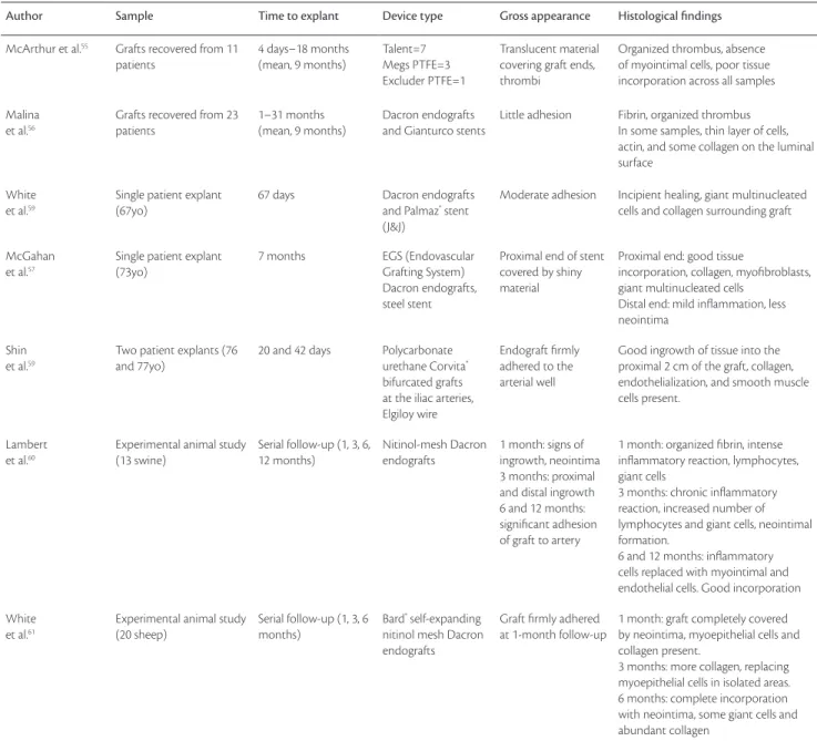
aorta. However, incorporation of the endograt fabric to the arterial wall, producing a permanent hemostatic seal, is de-sirable for medium- and long-term outcomes. he ability to obtain a healing process is absolutely critical in preventing migration and protecting against aneurysm rupture. Table 2 lists some studies of endoprosthesis incorporation.
Tissue incorporation varies depending on the material from which the device is made. Past studies have shown that tissue incorporation of PTFE devices is poor as a con-sequence of their hydrophobic surface, which would pose a limit to cell adhesion. Dacron-covered endograts have in-termediate tissue incorporation capacity, whereas polyure-thane devices induce a more intense inlammatory process and greater cell adhesion, which translate into better endo-thelialization and ixation to adjacent tissues. Limiting their use, however, is the fact that polyurethane endoprosthesis
are mechanically weak and tend to degenerate.55,56,59
Studies investigating endograts removed from pa-tients’ bodies provide conlicting accounts of the tissue
incorporation process. Some authors59,60 have reported a
good endothelialization response and good ixation into the artery, while others have reported little grat adhesion
to the vascular wall.55,56 hese dissonant indings may be
ex-plained by diferences in choice of material and by the low number of cases described in the literature. Furthermore, some patients mentioned in these reports had developed typical complications of EVAR, such as leakage or migra-tion, which interfered with histological examination of the device. Analysis of uncomplicated grats was thus limited to those removed from the small number of patients who died from non-EVAR-related causes, such as myocardial
infarction or stroke,55-59 further decreasing the number of
useful cases.
In animal experiments, the tissue incorporation pro-cess has somewhat difered from that found in human studies. In swine and sheep – the most common animal models of endoprosthesis implantation – results were far more exuberant than those found in human studies due to interspecies diferences. Furthermore, all experimental stu-dies published in the literature were performed on normal arteries with none of the typical aortic wall changes, such as calciication and thrombi. Evidence suggests that inlam-matory response and incorporation occur diferently in the
human aneurysmal aorta.60,61
In light of these results, studies assessing endograts ater their removal from human patients and those conducted in animal models must be viewed and interpreted cautiously.
substantial complication rates persist for years ater repair. hese indings suggest that the tissue incorporation process occurs only partially and is not enough to prevent late com-plications, such as endograt migration.
Future prospects
Several recent studies have been conducted with the purpose of developing endoprosthetic devices that heal
bet-ter into the arbet-terial wall.62,63
Two basic factors prevent adequate tissue incor-poration of stent grafts. The first is the use of PTFE or Dacron, which are inert materials and thus have little potential for tissue incorporation. The second is asso-ciated with a peculiar characteristic of the aneurysmal wall: depletion and decreased resistance to apoptosis of myointimal cells, which play an essential role in tissue
incorporation.62
Studies have been conducted with the aim of impro-ving the tissue incorporation process by adding coatings
Author Sample Time to explant Device type Gross appearance Histological findings
McArthur et al.55 Grafts recovered from 11
patients
4 days–18 months (mean, 9 months)
Talent=7 Megs PTFE=3 Excluder PTFE=1
Translucent material covering graft ends, thrombi
Organized thrombus, absence of myointimal cells, poor tissue incorporation across all samples Malina
et al.56
Grafts recovered from 23 patients
1–31 months (mean, 9 months)
Dacron endografts and Gianturco stents
Little adhesion Fibrin, organized thrombus In some samples, thin layer of cells, actin, and some collagen on the luminal surface
White et al.59
Single patient explant (67yo)
67 days Dacron endografts and Palmaz® stent
(J&J)
Moderate adhesion Incipient healing, giant multinucleated cells and collagen surrounding graft
McGahan et al.57
Single patient explant (73yo)
7 months EGS (Endovascular Grafting System) Dacron endografts, steel stent
Proximal end of stent covered by shiny material
Proximal end: good tissue
incorporation, collagen, myoibroblasts, giant multinucleated cells
Distal end: mild inlammation, less neointima
Shin et al.59
Two patient explants (76 and 77yo)
20 and 42 days Polycarbonate urethane Corvita®
bifurcated grafts at the iliac arteries, Elgiloy wire
Endograft irmly adhered to the arterial well
Good ingrowth of tissue into the proximal 2 cm of the graft, collagen, endothelialization, and smooth muscle cells present.
Lambert et al.60
Experimental animal study (13 swine)
Serial follow-up (1, 3, 6, 12 months)
Nitinol-mesh Dacron endografts
1 month: signs of ingrowth, neointima 3 months: proximal and distal ingrowth 6 and 12 months: signiicant adhesion of graft to artery
1 month: organized ibrin, intense inlammatory reaction, lymphocytes, giant cells
3 months: chronic inlammatory reaction, increased number of lymphocytes and giant cells, neointimal formation.
6 and 12 months: inlammatory cells replaced with myointimal and endothelial cells. Good incorporation White
et al.61
Experimental animal study (20 sheep)
Serial follow-up (1, 3, 6 months)
Bard® self-expanding
nitinol mesh Dacron endografts
Graft irmly adhered at 1-month follow-up
1 month: graft completely covered by neointima, myoepithelial cells and collagen present.
3 months: more collagen, replacing myoepithelial cells in isolated areas. 6 months: complete incorporation with neointima, some giant cells and abundant collagen
Table 2 – Tissue incorporation studies: gross and histological indings
that could potentially stimulate cell adhesion and prolife-ration, creating an environment conducive to the migra-tion of ibroblasts and pluripotent smooth muscle cells. In addition to providing a more adequate microenviron-ment, this strategy would improve collagen production by ibroblasts, decreasing rates of myointimal cell apoptosis.
Lerouge et al.62 conducted tests using nitrogen-rich
plas-ma- and chondroitin sulfate-coated stent grats and found increased adhesion of ibroblast and myointimal cell cul-tures to these surfaces, as well as decreased apoptosis, as compared to controls. he chemical properties of Nitrile
coated surfacesis believed to favor certain intracellular
signaling pathways, modulating expression of integrin receptors, which are responsible for intercellular adhe-sion. Integrins also activate the integrin-linked kinase and phosphatidylinositol pathways, which are believed to play a key role in inhibiting apoptosis. Chondroitin sulfate would decrease apoptosis by acting on a similar kinase pa-thway (speciically, the P13K papa-thway). he authors con-clude that the use of these substances may be an important option in manufacturing stent grats with added capacity to incorporate into adjacent tissues.
Device porosity appears to inluence the tissue incor-poration process. Experimental studies suggest that, in lower-porosity grats, the myointimal cells responsible for tissue ingrowth migrate from the ends of the grat and cover an intraluminal area of up to 20 mm. In microporous grats, capillaries and vascular smooth muscle cells derived from the underlying granulation tissue have been found to pe-netrate the pores present throughout the device, producing improved grat coating. Studies are currently underway to improve grat porosity and cell adhesion as a means of
in-creasing tissue incorporation.64
Van der Bas et al.63 implanted collagen- and
fibro-blast growth factor-soaked Dacron endografts in the porcine aorta and found significant improvement in tissue incorporation at 8 weeks post-implantation. The authors observed neointimal growth and, on immu-nofluorescence studies, detected an increased number smooth muscle cells consistent with myofibroblast and myointimal cell proliferation. This study proved that in vivo induction of fibroplasia is possible, despite variables such as blood pressure and the blood flow effect, which were expected to “wash away” any substances impregna-ted into the graft.
Gene therapy studies are currently investigating al-ternatives for improving the tissue incorporation process.
Eton et al.65 implanted myointimal cells transduced with
tissue plasminogen activator genes. Cells were sufused
into a dual-layer Dacron endograt and implanted into dog aortas. According to the authors, grats removed at 1, 2, 3, 4, 5 and 7 months were highly populated with gene-tically modiied smooth muscle cells, with increased t-PA antigen levels and t-PA activity, both of which were desi-red outcomes of the transduction procedure. he authors concluded that endograts can serve as an important de-livery vehicle for transduced cells. Although not its main objective, the Eton study revealed the possibility of indu-cing greater ibroplasias by employing cells transduced with genes that increase proliferation of vascular smooth muscle cells or ibroblasts, leading to improved grat in-corporation and ixation.
Almeida & Yoshida49 implanted Dacron-coated
niti-nol stent grafts in porcine thoracic aorta, applied fibrin glue to the interface between the graft and the endo-thelium, and compared the results to a control group in which no fibril glue was used. On the 14th postopera-tive day, biomechanical testing was conducted to mea-sure the displacement load required for dislodging the
device, as in the work of Malina et al.27 and Lambert et
al.26 Displacement load was significantly increased in
the fibrin glue group, and histological testing confirmed increased fibroplasia in the group. The authors conclu-ded that application of fibrin glue to the endoprosthesis/ aorta interface may become an important step in impro-ving graft adhesion and tissue incorporation to prevent migration.
In conclusion, continuous improvement of endopros-thetic devices has led to the development of improved materials, with greater wear resistance and reduced cross-sections. Current progress is moving towards development of mesh coatings that improve tissue incorporation, so as to improve long-term results and prevent endograt migra-tion, which is still a major hurdle to positive outcomes in EVAR.
References
1. Dubost C, Allary M, Oeconomos N. Resection of an aneurysm of the abdominal aorta: reestabilishment of the continuity by a pre-served human arterial graft, with result after ive months. AMA Arch Surg. 1952;64:405-8.
2. Parodi JC, Palmaz JC, Barone HD. Transfemoral intraluminal graft implantation for abdominal aortic aneurysms. Ann Vasc Surg. 1991;5:491-9
4. Matsumura JS, Brewster DC, Makaroun MS, Naftel DC. A multi-center controlled clinical trial of open versus endovascular treat-ment of abdominal aortic aneurysm. J Vasc Surg. 2003;37:262-71.
5. Rosa A, Inocentes J, da Gama AD. Rotura de aneurisma da aorta após tratamento endoluminal. A propósito de um caso clínico. Rev Port CCTV. 2001;8:30-5.
6. Riepe G, Heilberger P, Umschield T, et al. Frame dislocation of body middle rigs in endovascular stent tube grafts. Eur J Vasc Endovasc Surg. 1999;17:28-34.
7. Bohm T, Söldner J, Rott A, Kaiser WA. Perigraft leak of an aor-tic stent graft due to material fatigue. AJR Am J Roentgenol. 1999;172:1355-7.
8. Norgren L, Jernby B, Engellau L. Aotoenteric istula caused by a rup-tured stent-graft: A case report. J Endovasc Surg. 1998;5:269-72.
9. Maleux G, Rousseau H, Otal P, Colombier D, Glock Y, Jofre F. Modular component separation and reperfusion of abdominal aortic aneurysm sac after endovascular repair of the abdominal aortic aneurysm. J Vasc Surg. 1998;28:349-52.
10. Giles KA, Pomposelli F, Handar A, Wyers M, Jhaveri A, Schermerhorn ML. Decrease in total aneurysm-related deaths in the era of endo-vascular aneurysm repair. J Vasc Surg. 2009;49:543-51.
11. Tonnessen BH, Sternberg WC 3rd, Money SR. Late problems at the proximal aortic neck: migration and dilation. Semin Vasc Surg. 2004;17:288-93.
12. Greenberg RK, Turc A, Haulon S, et al. Stent graft migration: a re-appraisal of analysis methods and proposed revised deinition. J Endovasc her. 2004;11:353-63.
13. Chaikof EL, Blankensteijn JD, Harris PL, et al. Reporting standar-ds for endovascular aortic aneurysm repair. J Vasc Surg. 2002;35: 1048-60.
14. Conners MS 3rd, Sternberg WC 3rd, Carter G, Tonessen BH, Yoselevitz M, Money SR. Endograft migration one to four years after endovascular abdominal aortic aneurysm repair with the AneurRx device: a cautionary note. J Vasc Surg. 2002;36:476-84.
15. Ouriel K, Clair DG, Greenberg RK, et al. Endovascular repair of ab-dominal aortic aneurysms: device-speciic outcome. J Vasc Surg. 2003;37:991-8.
16. England A, Butterield JS, Jones N, et al. Device migration after en-dovascular abdominal aortic aneurysm repair: experience with a talent stent-graft. J Vasc Intervent Rad. 2004;15:1399-405.
17. Schurink GW, Aarts NJ, van Baalen JM, Schultze Kool LJ, van Bockel JH. Stent attachment site-related endoleakage after stent graft treatment: an in vitro study of the efects of graft size, stent type, and atherosclerotic wall changes. J Vasc Surg. 1999;30:658-67.
18. Wolf YG, Hill BB, Lee WA, Corcoran CM, Fogarty TJ, Zarins CK. Eccentric stent graft compression: An indicator of insecure proxi-mal ixation of aortic stent graft. J Vasc Surg. 2001;33:481-7.
19. Albertini JN, Kalliafas S, Travis S, et al. Anatomical risk factors for proximal perigraft endoleak and graft migration following endo-vascular repair of abdominal aortic aneurysms. Eur J Vasc Endovasc Surg. 2000;19:308-12.
20. Lifeline Registry of Endovascular Aneurysm Repair Steering Committee. Lifeline Registry: collaborative evaluation of endovas-cular aneurysm repair. J Vasc Surg. 2001;34:1139-46.
21. Zarins CK, Bloch DA, Crabtree T, Matsumoto AH, White RA, Fogarty TJ. Stent graft migration after endovascular aneurysm repair: importance of proximal ixation. J Vasc Surg. 2003;38: 1264-72.
22. Tonnessen BH, Sternberg WC 3rd, Money SR. Mid- and long-term device migration after endovascular abdominal aortic aneurysm repair: a comparison of AneuRx and Zenith endografts. J Vasc Surg. 2005;42:392-401.
23. Canic S, Ravi-Chandar K, Krajcer Z, Mirkovic D, Lapin S. Mathematical Model Analysis of Wallstent and AneurRx. Dynamic Responses of Bare-Metal Endoprosthesis compared with those of stent-graft. Tex Heart Inst J. 2005;32:502-6.
24. Li Z, Kleinstreuer C. Blood low and structure interactions in a stented abdominal aortic aneurysm model. Med Eng Phys. 2005;27:368-82.
25. Fillinger MF, Marra SP, Raghavan ML, Kennedy EF. Prediction of rupture in abdominal aortic aneurysm during observation: wall stress versus diameter. J Vasc Surg. 2003;37:724-32.
26. Lambert AW, Williams DJ, Budd JS, Horrocks M. Experimental as-sesment of proximal stent-graft (InterVascular) ixation in human cadaveric nfrarenal aortas. Eur J Vasc Endovasc Surg. 1999;17:60-5.
27. Malina M, Lindblad B, Ivancev K, Lindh M, Malina J, Brunkwall J. Endovascular AAA exclusion: will stents with hooks and barbs pre-vent stent-graft migration? J Endovasc Surg. 1998;5:310-7.
28. Volodos SM, Sayers RD, Gostelow JP, Sir Bell PR. An investigation into the cause of distal endoleaks: role of displacement force on the distal end of a stent-graft. J Endovasc her. 2005;12:115-20.
29. Mohan IV, Harris PL, van Marrewijk CJ, Laheij RJ, How TV. Factors and forces inluencing stent-graft migration after endovascular aortic aneurysm repair. J Endovasc her. 2002;9:748-55.
30. Massey B. Mechanics of luids. 7th ed. London: Stanley hornes
Publ; 2000.
31. Cohen JR, Keegan L, Sarfati I, Dana D, Ilardi C, Wise L. Neutrophil chemotaxis and neutrophil elastase in the aortic wall in patients with abdominal aortic aneurysms. J Invest Surg. 1991;4:423-30.
32. Sukhova GK, Shi GP, Simon DI, Chapman HA, Libby P. Expression of the elastolytic cathepsins S and K in human atheroma and re-gulation of their production in smooth muscle cells. J Clin Invest. 1998;102:576-83.
33. Gacko M, Chyczewski L. Activity and localization of cathepsin B, D and G in aortic aneurysm. Int Surg. 1997;82:398-402.
34. Resch T, Malina M, Lindblat B, Malina J, Brunkwall J, Ivancev K. he impact of stent design on proximal stent-graft ixation in the ab-dominal aorta: an experimental study. Eur J Vasc Endovasc Surg. 2000;20:190-5.
35. Cao P, Verzini F, Zannetti S, et al. Device migration after endo-luminal abdominal aortic aneurysm repair: analysis of 113 ca-ses with a minimum follow-up period of 2 years. J Vasc Surg. 2002;35:229-35.
36. Sternberg WC 3rd, Money SR, Greenberg RK, Chuter TA. Inluence of endograft oversizing on device migration, endoleak, aneurysm shrinkage and neck dilation: results from the Zenith Multicenter Trial. J Vasc Surg. 2004;39:20-6.
38. Sternberg WC 3rd, Carter G, York JW, Yoselevitz M, Money SR. Aortic neck angulation predicts adverse outcome with endovascular abdominal aortic aneurysm repair. J Vasc Surg. 2002;35:482-6.
39. Lawrence-Brown M, Sieunarine K, Hartley D, van Schie G, Goodman MA, Prendergast FJ. he Perth HLB bifurcated endo-luminal graft: review of the experience and intermediate results. Cardiovasc Surg. 1998;6:220-5.
40. Illig KA, Green RM, Ouriel K, Riggs P, Bartos S, DeWeese JA. Fate of the proximal aortic cuf: implications for endovascular aneurysm repair. J Vasc Surg. 1997;26:494-501.
41. Lee JT, Lee J, Aziz I, et al. Stent-graft migration following endovascu-lar repair of aneurysms with endovascu-large proximal necks: anatomical risk factors and long-term sequelae. J Endovasc her. 2002;9:652-64.
42. Greenberg R, Fairman R, Srivastava S, Criado F, Green R. Endovascular grafting in patients with short proximal necks: an analysis of short-term results. Cardiovasc Surg. 2000;8:350-4.
43. Armon MP, Yusuf SW, Whitaker SC, Gregson RH, Wenham PW, Hopkinson BR. Inluence of abdominal aortic aneurysm size on the feasibility of endovascular repair. J Endovasc Surg. 1997;4: 279-83.
44. Ouriel K, Srivastava SD, Sarac TP, et al. Disparate outcome after en-dovascular treatment of small versus large abdominal aortic aneu-rysm. J Vasc Surg. 2003;37:1206-12.
45. Zarins CK, Crabtree T, Bloch DA, Arko FR, Ouriel K, White RA. Endovascular aneurysm repair at 5 years: does aneurysm diameter predict outcome? J Vasc Surg. 2006;44:929-31.
46. Peppelenbosch N, Buth J, Harris PL, van Marrewijk C, Fransen G. Diameter of abdominal aortic aneurysm and outcome of endovas-cular aneurysm repair: does size matter? A report from EUROSTAR. J Vasc Surg. 2004;39:288-97.
47. Mortality results for randomized controlled trial of early elective surgery or ultrassonographic surveillance for small abdominal aor-tic aneurysms. he UK Small Aneurysm Trial paraor-ticipants. Lancet 1998;352:1649-55.
48. Mohan IV, Laheij JP, Harris PL. Risk factors for endoleak and the evi-dence for stent-graft oversizing in patients undergoig endovascular aneurysm repair. Eur J Vasc Endovasc Surg. 2001;21:344-9.
49. Almeida MJ, Yoshida WB. Avaliação biomecânica da ixação das endopróteses com e sem cola biológica e alterações histológicas aórticas. Estudo experimental em porcos. [dissertação]. Botucatu: Universidade Estadual Paulista; 2009.
50. Marty B. Quantiication of radial pressure caused by bare and co-vered Wallstents. In: Marty B, editor. Endovascular aneurysm re-pair: from bench to bed. Darmstadt: Steinkopt; 2005. p. 11-8.
51. Strauss Bh, Serruys PW, de Scheerder IK, et al. Relative risk analy-sis of angiographic predictors of reestenoanaly-sis within the coronary Wallstent. Circulation. 1991;84:1636-43.
52. Gravanis MB, Roubin GS. Histopathologic phenomena at the site of percutaneous transluminal coronary angioplasty: the problem of restenosis. Hum Pathol. 1989;20:477-85.
53. Sonesson B, Hansen F, Stale H, Länne T. Compliance and diameter in the human abdominal aorta – the inluence of age and sex. Eur J Vasc Surg. 1993;7:690-7.
54. Resch T, Ivancev K, Brunkwall J, Nyman U, Malina M, Lindblad B. Distal migration of stent-grafts after endovascular repair of abdominal aortic aneurysms. J Vasc Interv Radiol. 1999;10: 257-64.
55. Mc Arthur C, Teodorescu V, Eisen L, et al. Histopathologic analysis of endovascular stent grafts from patients with aortic aneurysm: does healing occur? J Vasc Surg. 2001;33:733-8.
56. Malina M, Brunkwall J, Ivancev K, Johnson J, Malina J, Lindblat B. Endovascular healing is inadequate for ixation of dacron stent-grafts in human aortoilac vessels. Eur J Vasc Endovasc Surg. 2000;19:5-11.
57. White RA, Donayre CE, de Virgilio C, Weinsten E, Tio F, Kopchok G. Deployment technique and histopathological evaluation of an endoluminal vascular prosthesis used to repair an iliac artery aneu-rysm. J Endovasc Surg. 1996;3:262-9.
58. McGahan TJ, Berry GA, McGahan SL, White GH, Yu W, May J. Results of autopsy 7 months after successful endoluminal treat-ment of an infrarenal abdominal aortic aneurysm. J Endovasc Surg. 1995;2:348-55.
59. Shin CK, Rodino W, Kiwin JD, et al. Histology and electron mi-croscopy of explanted bifurcated endovascular aortic grafts: evi-dence of early incorporation and healing. J Endovasc Surg. 1999;6: 246-50.
60. Lambert AW, Budd JS, Fox AD, Potter U, Rooney N, Horrocks M. he incorporation of a stent-graft into the porcine aorta and the inlammatory response to the endoprosthesis. Cardiovasc Surg. 1999;7:710-4.
61. White JG, Mulligan NJ, Gorin DR, D’Agostino R, Yucef EK, Menzoian JO. Response of normal aorta to endovascular grafting: a serial histopathological study. Arch Surg. 1998;133: 246-9.
62. Lerouge S, Major A, Girault-Lauriault PL, et al. Nitrogen-rich coa-tings for promoting healing around stent-grafts after endovascular aneurysm repair. Biomaterials. 2007;28:1209-17.
63. van der Bas JM, Quax PH, van den Berg AC, Visser MJ, van der Linden E, van Bockel JH. Ingrowth of aorta wall into stent grafts impregnated with basic ibroblast growth factor: a porcine in vivo study of blood vessel prosthesis healing. J Vasc Surg. 2004;39: 850-8.
64. Marois Y, Pâris E, Zhang Z, Doillon CJ, King MW, Guidoin RG. Vascugraft microporous polyesterurethane arterial pros-thesis as a thoraco-abdominal bypass in dogs. Biomaterials. 1996;17:1289-300.
65. Eton D, Hong Yu, Wang Y, Raines J, Striker G, Livingstone A. Endograft technology: a delivery vehicle for intravascular gene therapy. J Vasc Surg 2004;1066-73.
Correspondence
Marcelo José de Almeida Rua 7 de setembro, 734 CEP 17502020 – Marília, SP E-mail: [email protected], [email protected]
Author contributions