N170 asymmetry as an index of inferior occipital dysfunction in patients
with symptomatic occipital lobe epilepsy
Ricardo Lopes
a,b,*, Pedro Cabral
c, Nuno Canas
c, Paula Breia
d, John P. Foreid
e, Eulália Calado
a, Rita Silva
a,
Alberto Leal
a,fa
Department of Pediatric Neurology, Hospital Center of Central Lisbon, Rua Jacinta Marto, 1169-045 Lisbon, Portugal
bFaculty of Psychology, University of Coimbra, Coimbra, Portugal c
Department Pediatric Neurology, Hospital Center of West Lisbon, Lisbon, Portugal
d
Department Neurology, Hospital Garcia de Orta, Almada, Portugal
e
Department Neurology, Portuguese Oncology Institute, Lisbon, Portugal
f
Centre for Psychological Research and Social Intervention (Cis-IUL), Lisbon, Portugal
a r t i c l e
i n f o
Article history: Accepted 27 May 2010 Available online 20 June 2010 Keywords: Epilepsy N170 EEG Childhood Occipital lobe
a b s t r a c t
Objective: Localizing epileptic foci in posterior brain epilepsy remains a difficult exercise in surgery for epilepsy evaluation. Neither clinical manifestations, neurological, EEG nor neuropsychological evalua-tions provide strong information about the area of onset, and fast spread of paroxysms often produces mixed features of occipital, temporal and parietal symptoms. We investigated the usefulness of the N170 event-related potential to map epileptic activity in these patients.
Methods: A group of seven patients with symptomatic posterior cortex epilepsy were submitted to a high-resolution EEG (78 electrodes), with recordings of interictal spikes and face-evoked N170. Genera-tors of spikes and N170 were localized by source analysis. Range of normal N170 asymmetry was deter-mined in 30 healthy volunteers.
Results: In 3 out of 7 patients the N170 inter-hemispheric asymmetry was outside control values. Those were the patients whose spike sources were nearest (within 3 cm) to the fusiform gyrus, while foci fur-ther away did not affect the N170 ratio.
Conclusions: N170 event-related potential provides useful information about focal cortical dysfunction produced by epileptic foci located in the close neighborhood of the fusiform gyrus, but are unaffected by foci further away.
Significance: The N170 evoked by faces can improve the epileptic foci localization in posterior brain epilepsy.
Ó 2010 International Federation of Clinical Neurophysiology. Published by Elsevier Ireland Ltd. All rights reserved.
1. Introduction
Epileptic activity in occipital lobe epilepsies tends to propagate to nearby parietal, temporal and also to the distant frontal lobes (Leal et al., 2007, 2008; Williamson et al., 1992). Such phenomenon is responsible for the complex ictal manifestations involving di-verse brain areas, making it difficult to obtain a clear picture of the overall dynamics and potentially leading to important errors when surgery for epilepsy is considered. With the exception of ele-mentary visual symptoms, which are relatively rare, the clinical manifestations associated with occipital lobe epilepsy are not very informative about the particular area of onset of the epilepsy within
the posterior brain (reviewed in Sveinbjornsdottir and Duncan (1993)). Complex visual auras, hallucinations, amaurosis, version of head and eyes, eye blinking, have a poor localizing value and can be obtained by electrical stimulation in both occipital, parietal and temporal lobes (reviewed inGeller et al. (2000)). Often the ten-dency for epileptic activity to involve the temporal lobes leads to impaired consciousness and to the inability to report ictal manifes-tations with localization information. Overall the symptomatology of posterior cortex epilepsy has a poor localizing value, requiring a careful integration of both clinical and neurophysiologic informa-tion to formulate good hypothesis about the localizainforma-tion of the epileptic foci.
With the exception of hemianopia, patients with posterior brain epilepsy rarely present with deficits in the neurological examina-tion and in the subjective ability to process daily visual informa-tion. In fact classical neurological syndromes of visual agnosia
1388-2457/$36.00 Ó 2010 International Federation of Clinical Neurophysiology. Published by Elsevier Ireland Ltd. All rights reserved. doi:10.1016/j.clinph.2010.05.023
* Corresponding author at: Department of Pediatric Neurology, Hospital Center of Central Lisbon, Rua Jacinta Marto, 1169-045 Lisbon, Portugal. Tel.: +351 213126873.
E-mail address:r.lopes@clix.pt(R. Lopes).
Contents lists available atScienceDirect
Clinical Neurophysiology
such as prosopoagnosia are more often associated with bilateral le-sions (Meadows, 1974) and are rarely found in connection with epilepsy.
Despite the fact that several neuropsychological studies have uncovered an asymmetry in memory for faces (Millner, 1968), these functions have been associated with the temporal lobes and in particular with hippocampus processing (Crane and Millner, 2002). The previous studies demonstrated that the capability to discriminate faces from other objects is not affected in temporal lobe lesions, supporting the thesis that such processing is done in the ventral temporal–occipital areas (Allison et al., 1994; Haxby et al., 1996; Kanwisher et al., 1997; Steeves et al., 2006; Pitcher et al., 2007). The neuropsychological tests presently available have nevertheless been unable to detect reliably abnormalities in face recognition (Duchaine and Weidenfeld, 2003; Duchaine and Nakayama, 2004), making it difficult to produce strong inferences about the localizations of occipital lesions from the obtained results.
The visual analysis of the EEG in occipital lobe epilepsies is poorly localized (Williamson et al., 1992) and the complex dynam-ics of spike activity requires the use of sophisticated methods of data analysis (Leal et al., 2007, 2008), which have not been used very often in the surgery for epilepsy setting.
In this study, we investigate the possible contribution of the N170, a negative event-related potential peaking around 170 ms, elicited by presentation of complex pictures such as faces (Bentin et al., 1996), to discriminate inferior occipital lobe epileptic foci. The amplitudes of the N170 have been found to be approximately symmetrical (Rossion et al., 2003), despite the well-known differ-ential involvement of both hemispheres in face processing (re-viewed inDamásio et al. (1990)). We hypothesize that patients with inferior occipital epileptic foci will show a smaller N170 in the affected hemisphere when compared with the spared one and that the resulting inter-hemispheric asymmetry will provide useful neurophysiologic information about the localization of epi-leptic foci involving the inferior part of the occipital lobes.
2. Methods and subjects
We selected patients with symptomatic epilepsy of the poster-ior brain, undergoing evaluation at the program for Surgery of Epi-lepsy of the Hospital Center of West Lisbon, Portugal. The neurophysiologic studies were conducted as an add-onto the con-ventional video-EEG monitoring, and were performed before that procedure. Informed consent was obtained from the patients or from their parents. Clinical and neurophysiologic data for the pa-tients is shown inTable 1. All subjects had normal or corrected to normal vision.
The neurophysiologic study consisted in a high-density EEG recording using a cap (EasyCap, Herrsching-Breitbrunn, Germany) with 78 electrodes, including all positions in the 10–10 system plus positions FT11, FT12, TP11, TP12. Particular care was taken to ensure
a symmetrical fit of the cap on the head and that the T7/8, Fp1/2and
O1/2electrodes were at the conventional 10–20 system positions.
Ring electrodes made of sintered AgCl were applied at the cap and connected to two NuAmps 40-channels EEG amplifiers (Neuro-scan, Charlotte, USA) chained in such a way as to provide a system with 78 channels. The sampling rate was 1000 Hz and the high-and low-pass filters were 0.5 high-and 70 Hz.
The N170 was obtained by presentation of neutral emotion black and white faces taken from the Psychological Image Collection at Stir-ling (PICS) faces database (University of StirStir-ling Psychology Depart-ment, Stirling, Scotland – United Kingdom) or3=
4front view grayscale
pictures of cars, using the procedure described byRossion et al. (2003)(Fig1a).Acollectionof18differentfaces(9malesand9females) and 18 different cars were presented twice in a random sequence, using the E-Prime 1.2 software package (PST, Pittsburgh, USA). Each picture was shown for 250 ms in a CRT screen, with an inter stimulus interval of 1 s. Four of the previous block sequences were collected for each subject, with an interval of 2 min. In order to maintain atten-tion, subjects were asked to keep a mental recording of the number of female faces.
Thirty healthy volunteers, aged 19–44 years, were included in the study and used as a control group. For epileptic patients an additional sleep recording with an average duration of 30 min was done after the evoked potential acquisition. Interictal spikes with good signal/noise (S/N) ratio were visually identified by an experienced clinical neurophysiologist (AL), and averaged with synchronization by the peak of the spike.
2.1. Source analysis
In order to transform the information from the 78 channel EEG recording to a ratio expressing the relative contribution of each hemisphere, we performed source analysis with the Source2 soft-ware package (Neuroscan, Charlotte, USA), using a standard realis-tic finiteelementmodel(FEM)ofthehead,(Fig.1c).Standardelectrode coordinates for the cap were downloaded from EasyCap, Inc. (http:// www.easycap.de), which were then transformed to the Pre-Auricu-lar-Nasion (PAN) coordinate system. The conductivities were 0.33 S/ m for brain and scalp, 0.0042 S/m for the skull (Gonçalves et al., 2003).Aregionalsourcedipolewasplacedatthecenterofthefusiform gyrus(FG)ofeachhemisphere(Table2)andthemodelwasfittedtothe N170scalppotential,(Fig.1c).Theamplitudeofeachregionaldipoleat thepeak of theMean Global Field Power(MGFP)(Lehmannand Skran-dies, 1984) of the N170 was used to calculate the left/right (L/R) hemi-sphere ratio, (Fig. 1d).
The brain localizations of the sources of interictal spikes and N170 were studied in patients with the sLORETA method ( Pasc-ual-Marqui, 2002; Pascual-Marqui et al., 2002), as implemented in a free downloadable package ( http://www.uzh.ch/keyinst/lore-ta.htm), and using the previously described volume conductor model. The solution space was anatomically restricted to the cor-tex and consisted of 6239 cubic voxel elements with 5 mm side. The instantaneous solution at the spike/N170 peak was used for the mappings shown inFigs. 2 and 3.
Table 1
Clinical and neurophysiological data.
Patient ID Age Sex Age at onset MRI scan lesion location Seizure type N170 Asymmetry? ER 43 F 8 Lateral right occipital lobe Complex partial seizures No
MCR 47 F 10 Mesial right occipital lobe Complex partial seizures No ML 26 F 19 Inferior left temporal lobe Complex partial seizures No CC 7 F 5 Inferior left temporal lobe Complex partial seizures No JC 20 M 11 Lateral left occipital lobe Complex partial seizures Yes MSR 5 M 1 Lateral right temporal lobe Complex partial seizures Yes GM 9 M 7 No lesion Complex partial seizures Yes 10 R. Lopes et al. / Clinical Neurophysiology 122 (2011) 9–15
Fig. 1. (a) Examples of pictures shown to obtain the N170 potential. (b) Butterfly plot of the 78 channels N170 (above), with the Mean Global Field Power (MGFP) bellow. (c) Standard realistic volume conductor model, with the electrodes (above), and with regional dipoles at FG coordinates (bellow). (d) Source activity for the two regional dipoles at N170, right dipole above, left dipole, middle. The activities at the time of MGFP peak (bellow) were taken to determine the N170 ratio. (e) Ratio between left and right hemisphere N170 for the control group (N = 30), for the presentation of cars (left) and faces (right). The results for faces show a reduced dispersion compared the one for cars.
Table 2
Source analysis and structural lesions.
Patient Lesion (L) Fusiform gyrus (FG) Spike source (SS) DSS-L DSS-FG DL-FG Ratio
x y z x y z x y z ER 28 66 19 37 51 25 20 60 45 28 73 47 0.96 MCR 17 77 5 37 51 25 20 55 5 22 35 44 0.71 ML 30 17 30 37 51 25 20 15 20 14 40 35 0.95 CC 29 30 24 37 51 25 35 5 15 27 47 22 0.80 JC 40 85 0 37 51 25 60 55 20 41 24 42 0.66 MSR 56 28 3 37 51 25 50 50 15 25 16 37 1.29 GM 38 55 22 37 51 25 40 55 22 2 6 5 0.28
3. Results
3.1. Source analysis of interictal spikes
The preliminary visual analysis of the instantaneous scalp po-tential topographic maps of the average interictal spikes for the epileptic patients revealed spatially consistent patterns from spike onset until the peak for each patient, suggesting stable spatial configurations for the underlying generators. Source analysis was performed at the peak of the spike because this is the point with the best S/N ratio.
The visual analysis of the orientation of the field of potential over the scalp revealed very distinct patterns for the different patients
(Figs. 2 and 3), providing support for the existence of sources with diverse localizations: patients GM, JC and MSR have vertical orienta-tions with phase reversal near the inferior border of the brain, sug-gesting an origin of the abnormal activity in the inferior occipital cortex; patient ML has a similar field orientation, but shifted to a more anterior localization, suggesting an origin in the inferior cortex of the temporal lobe; patients ER and CC have an horizontal and transverse field orientation with clear maximum over the ipsilateral occipital lobe, suggesting a lateral cortex involvement; patient MR presented a horizontal and anterior-posterior orientation centered over the occipital lobe, compatible with a medial origin.
The sLORETA algorithm produced scores with clear maximum in the areas previously postulated for the different patients,
Fig. 2. (a) Patient GM, with an epileptic focus in the inferior left occipital lobe (upper left), demonstrated an asymmetrical N170 potential (lower left), outside the range of the control group (right). The sLORETA maps for the average (N = 150) interictal spikes, revealed a maximum for the statistical score in the left FG. Successful surgery for epilepsy after invasive study is shown at the lower right. For all images left is on the left. (b) Patient JC had a structural lesion in the lateral occipital cortex (arrow), but the sLORETA score for the average (N = 19) interictal spikes (upper left) localized the generator in the lower left occipital lobe (upper row). The N170 L/R ratio is decreased (right) due to the reduced left side contribution (lower left). (c) Patient MSR presents a dysplastic lateral right temporal lobe (arrow), but the maximum sLORETA score of the average interictal spikes (N = 103, upper left) localized in the inferior occipital lobe. The N170 ratio was outside the control range (right).
(Figs. 2 and 3). An analysis of the spatial localization of these sources revealed that they mostly lie in the neighborhood of the structural lesions (average distance of 2.3 cm,Table 2), suggesting that sub-lobar resolution is possible with this method in the pos-terior brain.
3.2. N170 ratio in patients
A distinct N170 potential was obtained for all healthy volun-teers and patients participating in the study, both for faces and cars. The L/R hemisphere ratio of the absolute amplitude of the two regional dipoles generating the N170 potential for the two
categories of stimuli revealed a reduced dispersion for faces in the control group (Fig. 1b). This stimulus category was then chosen for comparison with the patient data.
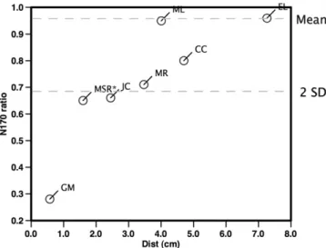
The N170 L/R ratio for the patients revealed values within the control range for four cases (ML, MR, ER and CC) and abnormal (values outside the range of the control group and more than 2 SD away from the mean) results for the other three (GM, JC and MSR) (Table 2). An analysis of the distance from the epileptic spike source to the nearest FG reveals that for the three patients with abnormal L/R ratios it is smaller than for the patients with normal ratios (Table 2). The critical distance separating the two groups of patients seems to be around 3 cm, as is suggested by the crossing
Fig. 3. Patients with symptomatic lobe epilepsies with lesions in the lower temporal lobes (a and d), middle (b) and lateral (c) occipital lobes, are shown. The sLORETA scores for the average spikes (N = 39, N = 151, N = 122 and N = 33, from (a–d) showed a maximum near the MRI lesions, but sparing the FG. The four patients demonstrated normal L/R for the N170, as demonstrated for individual ratios in comparison with the control group results (right).
of the statistical threshold of significance of the L/R N170 ratio for distances higher than this value in the plot ofFig. 4. These results suggest that only epileptic foci within the close neighborhood of the FG are able to produce an asymmetry change in the N170 po-tential for faces.
The distances from the FG to the structural lesions are also superior to 3 cm for the patients with normal L/R ratio (Table 2). This distance is nevertheless a worse predictor of N170 asymme-tries as patients JC and MSR have lesions at 4.2 and 3.7 cm away, respectively, from the FG but an abnormal L/R N170 ratio (Fig. 2b and c).
Overall these results suggest that only epileptic sources in the close proximity of the FG were able to significantly change the L/R N170 ratio, in patients with posterior cortex epilepsy. 4. Discussion
The main conclusion of this study is that an abnormal N170 ra-tio for faces is produced by epileptic sources localized near (<3 cm) the FG, but not by sources further away, and therefore can be used as a physiological marker of an epileptic source localized in the inferior occipital cortex of patients with symptomatic occipital lobe epilepsy.
Interictal epileptic activity in the occipital lobe epilepsies dem-onstrates a strong tendency to spread to nearby parietal and tem-poral lobes (Leal et al., 2007), to the contra lateral hemisphere and even to the frontal lobes (Williamson et al., 1992). This fast spread dynamics makes it difficult to localize the epileptogenic cortex not only in the raw EEG traces (Leal et al., 2008), but also using source analysis methods (Van der Meij et al., 1997). Several issues may be responsible for the poor yield of the latte techniques but the reduced spatial sampling over the posterior and inferior scalp, usu-ally used in clinical studies, seems to be a prominent one (Leal et al., 2008). Careful studies comparing the localization of sources obtained by solving the inverse problem and well-known occipital foci are lacking in the literature, but would represent a major contribution to establish the contribution of source analysis in this clinical setting.
We used a standard FEM model with standard electrode positions to solve the inverse problem, which can result in a de-creased accuracy as compared with individual realistic modeling.
Nevertheless a comparison between standard and realistic models for spike data byFuchs et al. (2002)failed to find systematic differ-ences in localization, and the mean localization differdiffer-ences were in the order of 1 cm. In the same line the effect produced by small inaccuracies in the position of the electrodes seems to have a re-duced effect on the final dipole localization error (Khosla et al., 1999; Wang and Gotman, 2001). The use of an electrode cap, ensuring a symmetrical disposition of the electrodes, strict adher-ence to the 10–20 system rules, minimizing electrode position inaccuracies, and the use of a high spatial sampling, in our view re-sulted in robust modeling of the intracranial generators. In partic-ular the calculation of the inter-hemispheric ratio of the N170 could be affected by asymmetries either in the placement of the electrodes or by large anatomical head shape differences, neither of which are present in our data. Since we used symmetrical regio-nal dipoles to express the activity of the inferior occipital cortex, the N170 ratio is not expected to be significantly affected by small inaccuracies in the postulated localization.
In our patients, we used anatomical localization of cortical le-sions as a surrogate marker for the unknown localization of intra-cranial epileptic foci. Although this procedure may not provide the ideal localization, in our view, it is reliable enough to provide sub-lobar resolution, allowing us to discriminate among the inferior, lateral and medial occipital cortex subdivisions of each hemi-sphere. The exception seems to be patients JC and MSR, which have lesions in the lateral occipital lobe, but topography of the epileptic spikes with a vertical orientation and phase reversal near the scalp projection of the lateral and inferior occipital cortex, suggesting an origin in the inferior occipital cortex. The latte localization agrees well with the localization of the source recovered by the sLORETA algorithm (Figs. 2b and c).
Overall the spatial distance between the calculated sources and the associated lesions for the five patients is on average 2.3 cm, which is very close to the resolution power of source analysis methods using realistic models for point sources (Fuchs et al., 2001). This result supports the suggestion that the methodology may provide sub-lobar resolution for the epileptic foci in occipital lobe epilepsy.
The N170 potential for faces has demonstrated slightly higher amplitude in the right hemisphere as compared with the left one, but in most studies the difference did not reach statistical signifi-cance (Bentin et al., 1996). In line with these results in our study the control group showed a L/R ratio slightly bellow 1, but with no significant asymmetry (Fig 1e).
None of our patients complained of difficulties in subjective visual recognition of faces, even those with an abnormal N170 ra-tio, a fact that most likely relates to the independent capability of each hemisphere to perform this function (Levy et al., 1972).
Three patients presented an age in the pediatric range, making the comparison with the control group of adults less reliable than for all the other patients. The two patients with asymmetrical N170 (MSR and GM) both presented very small distances from the spike source to FG (1.6 and 0.6 cm), while the one with normal ratio (CC) showed a larger distance (4.7 cm). Previous studies in the normal developmental aspects of the N170 throughout childhood, demonstrating preserved symmetry (Taylor et al., 1999), suggest that an abnormal left hemisphere function it is the most likely explanation for the inter-hemisphere N170 difference.
Rosburg et al. (2010)studied the effect on the vertex positive potential (VPP) of face inversion in a heterogeneous sample of epileptic patients undergoing surgery for epilepsy. Lateralization effects were not searched for, and no correlation of the findings with the particular type of epilepsy was performed. Because the VPP merges contributions from both hemispheres, no indepen-dent evaluation of those effects in the published material is possible.
Fig. 4. Representation of the relationship between the N170 L/R ratio (y-axis) and the corresponding interictal spike source (sLORETA) distance in cm to FG (x-axis). Patients with source distances to the FG less than 3 cm were more than 2 SD from the mean L/R ratio of the control group, while this did not happen for sources farther away from the FG. For patient MSR the N170 ratio was flipped about the mean, in order to improve visualization.
Despite the small patient sample, the overall results of our study suggest that the N170 for faces can be a useful marker of inferior occipital lobe dysfunction, providing additional informa-tion on the localizainforma-tion of the epileptic focus when the L/R inter-hemisphere ratio is significantly altered. The association of this neurophysiologic parameter with the conventional neuropsycho-logical tests for face processing could provide more robust inferences on occipital lobe dysfunction in patients with focal epi-lepsy of the posterior brain.
Acknowledgements
The authors are grateful to Heloisa Silva and Daniela Dias for their technical support.
Ricardo Lopes has been supported by the Grant SFRH/BD/ 65617/2009 from the Portuguese Foundation for Science and Tech-nology (FCT).
References
Allison T, Ginter H, McCarthy G, Nobre A, Puce A, Luby M, et al. Face recognition in human extrastriate cortex. J Neurophysiol 1994;71:821–5.
Bentin S, Allison T, Puce A, Perez E, McCarthy G. Electrophysiological studies of face perception in humans. J Cogn Neurosci 1996;8:551–65.
Crane J, Millner B. Do I know you ? Face perception and memory in patients with selective amygdalo-hippocampectomy. Neuropsychologia 2002;40:530–8. Damásio AR, Tranel D, Damásio H. Face agnosia and the neural substrates of
memory. Annu Rev Neurosci 1990;13:89–109.
Duchaine B, Weidenfeld A. An evaluation of two commonly used tests of unfamiliar face. Neuropsychologia 2003;41:713–20.
Duchaine B, Nakayama K. Developmental prosopoagnosia and the Benton Facial Recognition Test. Neurology 2004;62:1219–20.
Fuchs M, Wagner M, Kastner J. Boundary element method volume conductor models for EEG source reconstruction. Clin Neurophysiol 2001;112:1400–7. Fuchs M, Kastner J, Wagner M, Hawes S, Ebersole J. A standardized boundary
element method volume conductor model. Clin Neurophysiol 2002;113: 702–12.
Geller E, Luders H, Cheek J, Comair Y. Electrical stimulation of the visual cortex. In: Luders HO, Noachtar S, editors. Epileptic seizures – pathophysiology and clinical semiology. Churchill Livingstone; 2000. p. 219–27.
Gonçalves SI, De Munck JC, Verbunt JP, Bijna F, Heethaar RM, Lopes da Silva F. In-vivo measurements of the brain and skull resistivities using an EIT-based method and realistic models for the head. IEEE Trans Biomed Eng 2003;50:754–67.
Haxby J, Ungerleider L, Horwitz B, Maisog J, Rapoport S. Face encoding and recognition in the human brain. Proc Natl Acad Sci USA 1996;93:922–7.
Kanwisher N, McDermott J, Chun M. The fusiform face area: A module in human extrastriate cortex specialized for face perception. J Neurosci 1997;17(11):4302–11.
Khosla D, Don M, Kwong B. Spatial mislocalization of EEG electrodes – effects on accuracy of dipole estimation. Clin Neurophysiol 1999;110:261–71. Leal A, Nunes S, Dias A, Vieira J, Moreira A, Calado E. Analysis of the generators of
epileptic activity in early-onset childhood benign occipital lobe epilepsy. Clin Neurophysiol 2007;118(6):1341–7.
Leal A, Ferreira J, Dias A, Calado E. Origin of frontal lobe spikes in the early onset benign occipital lobe epilepsy (Panayiotopoulos syndrome). Clin Neurophysiol 2008;119(9):1985–91.
Lehmann D, Skrandies W. Spatial analysis of evoked potentials in man–a review. Prog Neurobiol 1984;23:227–50.
Levy J, Trevarthen C, Sperry RW. Perception of bilateral chimeric figures following hemispheric deconnexion. Brain 1972;95:61–78.
Meadows J. The anatomical basis of prosopagnosia. J Neurol Neurosurg Psychiatry 1974;37:489–501.
Millner B. Visual recognition and recall after right temporal-lobe excision in man. Neuropsychologia 1968;6:191–209.
Pascual-Marqui RD. Standardized low-resolution brain electromagnetic tomography (sLORETA): technical details. Methods Find Exp Clin Pharmacol 2002;24D:5–12.
Pascual-Marqui RD, Esslen M, Kochi K, Lehmann D. Functional imaging with low-resolution brain electromagnetic tomography (LORETA): a review. Methods Find Exp Clin Pharmacol 2002;24(Suppl. C):91–5.
Pitcher D, Walsh V, Yovel G, Duchaine B. TMS evidence for the involvement of the right occipital face area in the early face processing. Curr Biol 2007;17:1568–73. Rosburg T, Ludowig E, Dumelmann M, Alba-Ferrara L, Urbach H, Elger C. The effect of face inversion on intracranial and scalp recordings of event-related potentials. Psychophysiology 2010;47:147–57.
Rossion B, Joyce C, Cottrell G, Tarr M. Early lateralization and orientation tuning for face, word and object processing in the visual cortex. Neuroimage 2003;20:1609–24.
Steeves J, Culham J, Duchaine B, Pratesi C, Valyear K, Schindler I, Humphrey G, Milner A, Goodale M. The fusiform face area is not sufficient for face recognition: evidence from a patient with dense prosopoagnosia and no occipital face area. Neuropsychologia 2006;44:594–609.
Sveinbjornsdottir S, Duncan JS. Parietal and occipital lobe epilepsy: a review. Epilepsia 1993;34:493–521.
Taylor M, McCarthy G, Saliba E, Degiovanni E. ERP evidence of development changes in processing of faces. Clin Neurophysiol 1999;110:910–5.
Wang Y, Gotman J. The influence of electrode location errors on EEG dipole source localization with a realistic head model. Clin Neurophysiol 2001;112: 1777–80.
Williamson P, Thadani V, Darcey T, Spencer D, Spencer S, Mattson R. Occipital lobe epilepsy: clinical characteristics, seizure spread patterns and results of surgery. Ann Neurol 1992;31(1):3–13.
Van der Meij W, Dussen D, Huffelen A, Wieneke G, Nieuwenhuizen O. Dipole source analysis may differentiate benign focal epilepsy of childhood with occipital paroxysms from symptomatic occipital lobe epilepsy. Brain Topogr 1997;10(2):115–20.