Universidade de Lisboa
Faculdade de Ciências
Departamento de Biologia Vegetal
Unraveling the antifungal activity of a newly discovered
oligomer produced by an in vitro proteolytic pathway
Mestrado em Microbiologia Aplicada
Filipe Andrade Godinho Garcia Rollo
Dissertação orientada por:
Doutora Sara Alexandra Valadas Monteiro e Doutora Ana Maria Reis
Unraveling the antifungal activity of a newly discovered
oligomer produced by an in vitro proteolytic pathway
Filipe Andrade Godinho Garcia Rollo
MASTER THESIS
2015
This thesis was fully performed at the Centro de Botânica Aplicada à Agricultura from the
Instituto Superior de Agronomia (University of Lisbon) and at the facilities of Converde, SA,
under the direct supervision of Dr. Sara Alexandra Valadas Monteiro in the scope of the
Master in Applied Microbiology from the Faculty of Sciences of the University of Lisbon.
iv
Acknowledgements
First and foremost I would like to express my sincere gratitude to my supervisor at Instituto Superior de Agronomia, Dr. Sara Monteiro for the support, guidance and help. I am forever thankful for accepting me as her student, for the vote of confidence, for the constant availability and for the knowledge given.
To Professor Ana Maria Reis, my supervisor at Faculdade de Ciências from the Universidade de Lisboa, for following this work and helping me every time I needed it.
I specially want to thank Dr. Alexandra Carreira for all the availability, support, accuracy and teachings.
To Professor Ricardo Boavida Ferreira for allowing me to integrate into his working group, for the learning opportunity, support, critics, motivation and above all, for sharing his knowledge with me.
To the entire lab team, Regina Freitas, Ana Cristina Ribeiro, Ana Lima, Ricardo Chagas, and Alexandre Borges, for all the support, suggestions and help given during this last year.
To all who are part of CEV, SA, Iliana Pereira, Ana Marques, Mário Araújo, Sérgio Soares, André Barata, João Duarte and Jorge Monteiro for accepting me and for making me feel part of the team, and for all the help and support.
To the Microbiology laboratory team at Instituto Superior de Agronomia for allowing me to perform some of the work in their facilities. Mostly to Ana Carla Silva and Professor Manuel Malfeito Ferreira for all the help, support and for providing their collection of microorganisms.
On a more personal level, I must thank my patient and understanding girlfriend, Margarida, my second player, for the constant motivation, help and support. For always being there and for not letting me go down in the difficult times. For never doubting my abilities, even when I doubted. For always pushing me to the limit and ultimately for being my safe harbour when needed.
Finally, I want to thank my family and friends who helped and supported my work, outside the work environment. All the relaxation moments and motivation were essential through the past year.
v
Resumo
Os fungos patogénicos representam, à escala mundial, uma séria ameaça para a saúde humana, tendo-se vindo a registar um aumento significativo de infeções fúngicas devido a fungos patogénicos oportunistas, o que as torna num fator importante de morbidade e mortalidade.
Apesar de Candida albicans continuar a liderar as espécies de Candida causadoras de infeções da corrente sanguínea (ICS), nas últimas duas décadas tem ocorrido um desvio epidemiológico para um aumento de espécies de Candida "não-albicans". Infeções causadas por outras espécies de Candida têm vindo a aumentar, sendo responsáveis por mais de 90% das infeções fúngicas invasivas. De entre essas espécies, apenas C. glabrata pode ser verdadeiramente reconhecida como uma espécie emergente causadora de ICS, devido à sua resistência intrínseca e adquirida aos azóis e a outros agentes antifúngicos.
Nenhuma das classes de antifúngicos atualmente disponíveis possui todas as características ideais de um agente antifúngico, o que, em última análise, conduz a falhas no tratamento. Como consequência, novas formulações de antifúngicos, combinações terapêuticas e o desenvolvimento de novos compostos bioativos podem ser úteis na obtenção de melhores resultados terapêuticos.
As sementes de leguminosas são fontes bastante abundantes de proteínas, sendo o género Lupinus um dos mais ricos neste tipo de proteínas de reserva. As proteínas de reserva das sementes de leguminosas, classificadas como globulinas, compreendem duas famílias principais de proteínas: as vicilinas e as leguminas, que constituem aproximadamente 80% das proteínas totais das suas sementes, atuando não só como reservas de nutrientes durante a germinação, como também como proteínas de defesa para as plantas.
A β-conglutina é a principal proteína de reserva nas sementes das espécies de Lupinus, sendo as suas subunidades sintetizadas durante o desenvolvimento cotiledonar de L. albus, a partir de um único polipéptido precursor glicosilado. Após a germinação, as subunidades da β-conglutina sofrem uma grande alteração na sua estrutura e concentração, levando ao aparecimento de um novo conjunto de polipéptidos.
Recentemente, foi acidentalmente descoberto no nosso laboratório que a β-conglutina purificada de L. albus sofre, in vitro, um processo de proteólise. Após uma incubação de 7 dias a 25ºC, a β-conglutina sofre uma degradação controlada originando um oligómero estável e com atividade antifúngica, que denominámos PDβ.
Tendo em conta o conhecimento prévio da atividade antifúngica do PDβ, este plano de trabalho teve como objetivos principais caracterizar o oligómero e determinar a sua estabilidade e a sua atividade antifúngica, bem como tentar perceber o seu mecanismo de ação como agente antifúngico, avaliando os efeitos fisiológicos e morfológicos em C. glabrata.
A primeira fase do trabalho passou por isolar as globulinas totais dos cotilédones de L. albus seguida de purificação da fração correspondente à β-conglutina. Após purificação, a β-conglutina foi incubada a 25°C durante 7 dias, de modo a ocorrer a degradação para PDβ. Começou por se tentar perceber se o resultado desta degradação é devido a uma destruição da estrutura original da proteína dando origem a subunidades independentes. Os resultados permitiram concluir que embora a proteína sofra um processo de degradação, a sua massa molecular aumenta ligeiramente, o que
vi
permite concluir que o processo in vitro não se traduz num catabolismo com desaparecimento de sub-unidades mas sim de um arranjo estrutural diferente. O passo seguinte passou por avaliar a evolução do perfil polipeptídico e da atividade antifúngica durante o processo de degradação. Foi possível determinar que a degradação da β-conglutina começa a ocorrer a partir do terceiro dia de incubação a 25°C, sendo o pico máximo de degradação ao fim dos 7 dias. Esta degradação leva a uma acumulação progressiva de um polipéptido com 20 kDa, o que parece semelhante ao catabolismo que ocorre naturalmente nos cotilédones de L. albus. No entanto no processo in vivo a degradação origina exclusivamente um polipéptido de 20 kDa denominado Blad, que acaba por ser completamente degradado. Neste caso, o processo é interrompido sugerindo que falta uma peça chave para posterior degradação. Em relação à atividade antifúngica, o seu aparecimento é gradual: vai aumentando atingindo o pico máximo de atividade aos 7 dias de incubação.De seguida tentou caracterizar-se e identificar o mecanismo envolvido na degradação da β-conglutina em PDβ, começando por se analisar se poderia ser causada por uma reação de oxidação-redução, interferindo com o oxigénio disponível na reação. Os resultados obtidos indicam que o aumento de oxigénio disponível na reação não tem qualquer influência sobre a reação de degradação mas quando se adiciona dithiothreitol (DTT) a degradação não ocorre, ou é interrompida no momento em que este é adicionado. Uma possível explicação pode ser pelo facto de o DTT ser um eficaz desnaturante de proteínas quebrando as ligações dissulfídicas nos grupos de cisteína.
Tendo em conta estes resultados e admitindo a hipótese de haver uma protease envolvida no processo de conversão a qual poderia estar a ser inibida por um agente redutor, foram testados vários inibidores de diferentes classes de proteases, adicionando-os separadamente à β-conglutina antes do período de incubação in vitro de 7 dias. Dos inibidores de proteases testados, apenas o ácido etilenodiaminatetraacético (EDTA) teve influência na β-conglutina, impedindo a sua degradação. De acordo com este resultado é possível concluir que a degradação da β-conglutina é dependente de uma metaloprotease, inibida aquando da adição de EDTA, resultado confirmado por recurso a uma zimografia. Esta metaloprotease encontra-se sob a forma inativa, encontrando-se apenas ativa ao fim de 3 dias de incubação da β-conglutina a 25°C, altura em que se inicia o processo de degradação. Para esclarecer a origem da molécula responsável pela degradação da β-conglutina, levantou-se a hipótese de se tratar de uma contaminação externa. No entanto, esta hipótese foi abandonada uma vez que tanto a solução de β-conglutina filtrada através de filtros de fluoreto de polivinilideno (PVDF) como a β-conglutina submetida a um tratamento com antibiótico sofreram o processo normal de degradação.
Embora nesta fase do trabalho existissem resultados bastante interessantes, nenhum permitiu identificar a molécula responsável pela degradação da β-conglutina. Uma vez que os ensaios de zimografia foram os que permitiram chegar mais perto da sua identidade, foi efetuada uma nova zimografia utilizando as vicilinas totais como substrato, visto ser o substrato natural da metaloprotease em questão. As bandas com atividade proteolítica foram excisadas do gel e enviadas para sequenciação. No entanto, após receção dos resultados verificou-se que não foi possível identificar a possível metaloprotease.
vii
Em simultâneo com a caracterização bioquímica do processo de degradação, foi avaliada a atividade antimicrobiana de PDβ, o que revelou que apesar de PDβ não apresentar atividade bactericida, esta apresenta atividade fungicida em várias espécies de leveduras e de fungos filamentosos. Após o rastreio efetuado, foram avaliados os efeitos morfológicos e fisiológicos da PDβ, usando C. glabrata ISA 2163 como modelo. Começou por se comparar a actividade antifúngica de PDβ com a do itraconazole e anfotericina B, o que revelou que PDβ parece apresentar maior atividade inibitória e fungicida numa base molecular do que tanto o itraconazole como a anfotericina B.De seguida, realizaram-se curvas de crescimento de C. glabrata ISA 2163 exposta às concentrações inibitórias e letais de PDβ e de anfotericina B, de modo a estudar e comparar os seus efeitos no crescimento da levedura. As curvas obtidas permitiram concluir que a adição tanto de PDβ como de anfotericina B teve um forte efeito inibitório no crescimento de C. glabrata ISA 2163, visto que as células expostas a ambas as concentrações testadas de ambas as drogas apresentam uma diminuição na taxa de crescimento, quando comparado com a situação controlo (sem droga). Em ambos os casos, os efeitos de inibição começam a ocorrer ao fim de 4 h de incubação, verificando-se uma estabilização da DO640nm e das contagens de unidades formadoras de colónias (UFC), mais
acentuada a partir das 12 h de incubação, na fração incubada com a concentração letal de PDβ. Simultaneamente foi avaliado o efeito de PDβ na atividade metabólica, com recurso ao fluorocromo FUN-1, e na integridade da parede celular, com recurso ao calcofluor white, de C. glabrata ISA2163, recolhendo amostras ao longo da curva de crescimento. Os resultados obtidos sugerem que, mesmo ao fim de 24 h de incubação com uma concentração letal de PDβ, não ocorre perda de viabilidade celular sugerindo que as células apenas perdem a capacidade de se multiplicar, dada a estabilização do número de células. Relativamente à integridade da parede celular, existe uma diferença notória entre a fração incubada com PDβ e a fração controlo. Enquanto a integridade da parede celular da fração controlo permanece inalterada ao longo da curva, a fração exposta à PDβ apresenta falhas na marcação com calcofluor white, a partir das 8 h de incubação, tornando-se mais evidentes após as 12 h. Estes resultados sugerem perda de integridade da parede celular e podem ser a razão pela qual as células perdem a capacidade para se multiplicar.
Por último, determinou-se a localização celular da PDβ ao fim de 24 h de incubação em C. glabrata ISA 2163. Os resultados obtidos sugerem que PDβ se liga à parede celular da levedura, destabilizando-a. No entanto, não há vestígios da proteína no interior da célula.
Este trabalho permite concluir que a degradação in vitro da β-conglutina para PDβ ocorre de forma controlada, possivelmente sob a ação de uma metaloprotease. A atividade desta metaloprotease só é visível após o início da conversão da β-conglutina, aumentando de atividade à medida que o oligómero é convertido. Isto sugere que na semente seca, quando se purifica a β-conglutina, a metaloprotease se encontra inativa, sendo necessária a sua ativação para que a conversão da β-conglutina tenha início. No que respeita a atividade antimicrobiana de PDβ, é possível concluir que a mesma apresenta uma forte atividade em diferentes fungos, tanto leveduras, patogénicas e alimentares, como fungos filamentos. Relativamente ao modo de ação do oligómero, verificou-se que este tem capacidade para se ligar à parede celular da levedura em estudo, criando
viii
danos significativos e afetando a sua integridade celular. Os danos causados ao nível da parede celular parecem ser suficientes para impedir a multiplicação do microrganismo, levando à inibição do seu crescimento.ix
Abstract
Pathogenic fungi represent, worldwide, a serious threat for human’s health. Candida species are among the top ten pathogens causing bloodstream infections (BSI). C. glabrata can be described as a truly emerging pathogen that cause BSI due, in part, to its intrinsic and acquired resistance to azoles and other commonly used antifungal agents. All antifungal agents available display several disadvantages. It is therefore essential to identify new, potent and safe antifungal drugs with novel modes of action. PDβ is an in vitro breakdown oligomer resultant from the degradation of β-conglutin, with proved antifungal activity.
Based on this information and in an attempt to go deeper in knowledge, the first objective of this work was the biochemical characterization of PDβ oligomer and the understanding of the metabolic route involved in the degradation process. This process was found to occur gradually and controlled by a metalloproteinase that needs to be activated, converting an inactive protein into an oligomer with antifungal activity.
The second goal was the characterization of PDβ stability and antimicrobial activity against an array of different species. The antimicrobial activity of PDβ was shown to be quite diverse among yeasts and filamentous fungi; however no bactericidal activity was detected.
Finally, an attempt to understand PDβ mode of action by assessing its physiological and morphological effects on C. glabrata was explored. Upon exposure of C. glabrata to PDβ, the yeast is still metabolically active but loses its cell wall integrity. Furthermore, the activity of PDβ suggests binding to the cell wall, without entering into the cell. The binding of PDβ leads to damages and destabilization of the cell wall which are severe enough to prevent C. glabrata ISA 2163 multiplication.
x
Index
Acknowledgements ... iv
Resumo ... v
Abstract ... ix
List of Tables ... xii
List of Figures ... xiii
I. Introduction ... 1
II. Materials and Methods ... 8
II.1. Biological materials and growth conditions ... 8
II.1.1 Lupinus albus seeds ... 8
II.1.2. Bacteria ... 8
II.1.3. Fungi ... 8
II.1.4. Yeast ... 9
II.2. Isolation of total globulins ...10
II.3. Purification of β–conglutin ...10
II.4. Polyacrylamide gel electrophoresis in SDS-PAGE ...10
II.5. Protein Quantification ...11
II.6. Protein Staining ...11
II.7. Zymography ...11
II.8. Protein identification by mass spectra (MS) peptide mapping and sequencing analysis
...12
II.9. Assessment of the optimum conditions for the stability of PDβ ...12
II.10. Evolution of the antifungal activity of PDβ – from inactive to fully functional ...12
II.11. Degradation assays ...13
II.11.1. Temperature ...13
II.11.2. Oxidation-Reduction ...13
II.11.3. Protease inhibitor ...13
II.11.4. Microorganism ...13
II.12. Antifugal susceptibility tests ...13
II.12.1. Antifungal agents ...13
II.12.2. Yeast inhibition tests ...14
II.12.2.1. Minimum inhibitory concentrations (MICs) determination ...14
II.12.2.2. Minimum fungicidal concentrations (MFCs) determination ...14
xi
II.12.3.1. Minimum inhibitory concentrations (MICs) determination ...15
II.12.4. Fungal inhibition tests ...15
II.12.4.1. Minimum inhibitory concentrations (MICs) determination. ...15
II.13. Growth curves ...16
II.14. Morphology and viability assessments ...16
II.15. PDβ localization ...16
II.16. Staining with propidium iodide ...17
II.17. Immunofluorescency ...17
II.18. Fluorescence microscopy ...17
III. Results and discussion ... 18
III.1. Isolation and purification of β-conglutin ...18
III.2. Evolution of the antifungal activity of PDβ – from inactive to fully functional ...19
III.3. Assessment of the optimum conditions for the stability of PDβ...20
III.4. Characterization and identification of the subunit with activity ...22
III.5. Screening of the antimicrobial activity of PDβ ...30
III.6. Comparison of PDβ with two commonly used antifungal agents ...33
III.7. Growth curves ...33
III.8. Viability and cellular integrity assessments ...35
III.9. PDβ localization studies ...39
III.10. Immunofluorescence ...40
IV. General conclusion ... 42
V. References ... 45
xii
List of Tables
Table 1. Collection of bacteria used as biological material. *Strains courtesy from Instituto Superior de Agronomia. CBISA - Collection bacteria Instituto Superior de Agronomia. ... 8
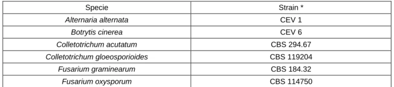
Table 2. Collection of filamental fungi used as biological material. *Strains courtesy from Converde. lda. CEV- Converde; ... 8
Table 3. Collection of yeast used as biological material. *Strains courtesy from Instituto Superior de Agronomia. ISA - Collection yeast Instituto Superior de Agronomia. ... 9
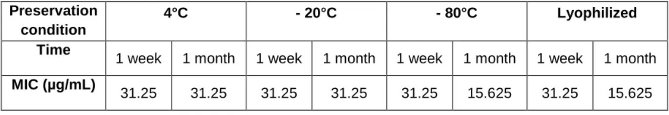
Table 4. β-conglutin MICs endpoints at different times along degradation. ... 19 Table 5. PDβ MICs endpoints in three different buffers. ... 21 Table 6. PDβ samples MICs endpoints after storage in four different preservation conditions, at different times. ... 21
Table 7. MALDI-TOF mass spectrometric sequencing analysis. ... 30
Table 8. PDβ MICs and MFCs endpoints in µg/mL in PDB medium for the collection of yeasts. (NG) – Absence of growth in PDB, (ND) - Not determined. ... 31
Table 9. PDβ MICs endpoints in µg/mL in PDB medium for the collection of fungi. ... 32 Table 10. PDβ MICs endpoints in µg/mL in Mueller-Hinton medium for the collection of bacteria. ... 32 Table 11. Itraconazole, amphotericin B and PDβ MICs and MFCs endpoints in µg/mL in PDB medium for C. glabrata ISA 2163. ... 33
xiii
List of Figures
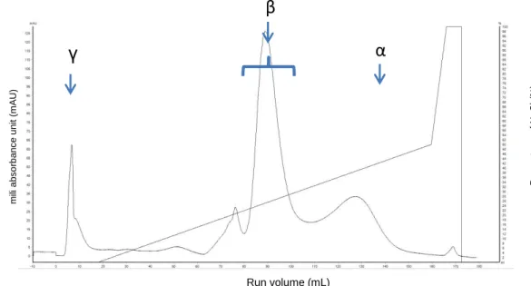
Figure 1. Chromatogram of dry seeds L. albus β-conglutin purification by anion exchange chromatography. ... 18
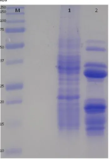
Figure 2. A - SDS-PAGE analyses. Degradation of β-conglutin day zero [1] to PDβ [2]. Precision Plus ProteinTM All Blue Standards marker (kDa) [M]. B - Size exclusion chromatogram in a Superose 12 column of β-conglutin 0 days (1) and PDβ (2). ... 18 Figure 3. SDS-PAGE analysis. Degradation of β-conglutin during 7 days. Day 0 [1]; day 1 [2]; day 2 [3]; day 3 [4];day 4[5]; day 5[6]; day6 [7] day 7 (PDβ) [8]. Precision Plus ProteinTM
All Blue Standards marker (kDa) [M]. ... 19
Figure 4. SDS-PAGE analysis. Polypeptide profile of conglutin after 1 month of degradation. β-conglutin day 0 [1]; β-β-conglutin 1 month [2] . Precision Plus ProteinTM
All Blue Standards marker (kDa) [M]. ... 20
Figure 5. SDS-PAGE analysis. Degradation of β-conglutin at different temperatures, 40°C [6A], 60°C [6B] and 25°C [6C] in different times 0 h [1;6] 3 h [2;7] 6 h [3;8] 1 day [4;9] 5 days [5;10] and 7 days [11]. Precision Plus ProteinTM All Blue Standards marker (kDa) [M] ... 22
Figure 6. SDS-PAGE analysis. Degradation of β-conglutin during 32 h at 25°C. Standard [1]; with 100 mM DTT [2]; β-conglutin after 24 h without [3] and with more oxygen [4]; β-conglutin after 26 h without [5] and with more oxygen [6]; β-conglutin after 28 h without [7] and with more oxygen [8]; β-conglutin after 30 h without [9] and with more oxygen [10] and β-conglutin after 32 h without [11] and with more oxygen [12]. Precision Plus ProteinTM All Blue Standards marker (kDa) [M] ... 23
Figure 7. SDS-PAGE analysis. Degradation of β-conglutin for 7 days at 25°C. β-conglutin 0 days [1] PDβ [2] β-conglutin with 100 mM DTT added at day 0[3] β-conglutin with 100 mM DTT added after 30 h[4] β-conglutin with 100 mM DTT added after 40 h [5] β-conglutin after with 100 mM DTT added aftter 45 h [6]. Precision Plus ProteinTM All Blue Standards marker (kDa) [M]. ... 23
Figure 8. SDS-PAGE analysis. Degradation of conglutin with different protease inhibitors. β-conglutin 0 days [1] β-β-conglutin with different protease inhibitors, 10 μM E-64 [2], 1 mM Pepstatin [3], 5 mM EDTA [4] and 1 mM Pefabloc [5]. Precision Plus ProteinTM All Blue Standards marker (kDa) [M]. 24
Figure 9. Zymography analysis with gelatin B as substrate. β-conglutin degradation during 7 days at 25°C. β-conglutin at 0 days [1] and 7 days [2]. Precision Plus ProteinTM All Blue Standards marker (kDa) [M]. ... 25
Figure 10. Zymography analysis with gelatin B as substrate. β-conglutin degradation during 7 days at 25°C. β-conglutin at 0 days [1], 1 day [2], 2 days [3], 3 days [4], 4 days [5] 6 days [6], 7 days [7]. Precision Plus ProteinTM All Blue Standards marker (kDa) [M]. ... 26
Figure 11. SDS-PAGE analysis. β-conglutin incubated for seven days at 25°C subjected to different treatments. conglutin without treatment (PDβ) [1]; conglutin filtered by PES filters 0.45 µm [2]; β-conglutin filtered by PES filters 0.22 µm [3]; β-β-conglutin filtered by acetate celulose filters 0.45 µm [4]; β-conglutin filtered by acetate cellulose filters 0.22 µm [5]; β-conglutin filtered by PVDF filters 0.45 µm [6]; β-conglutin filtered by PVDF filters 0.22 µm [7]; β-conglutin with 100 µg/mL ampicilin [8]. ... 27 Figure 12. Spectrum of bounded molecule to PES filter. ... 28
xiv
Figure 13. Zymography analysis with total vicilins as substrate. β-conglutin degradation during 7 days (PDβ) [1]; NEB Unstained Protein Ladder [M]. ... 28 Figure 14. SDS-PAGE analysis of the bands extracted from zimography. 150 plus 100 kDa [1] and 60 kDa [2]. NEB Unstained Protein Ladder [M]. ... 29Figure 15. Effect of PDβ on the growth of C. glabrata ISA 2163 in PDB medium, pH 7.5, 34°C, without agitation. Concentration of PDβ in the culture: 0 µg/mL, 37.5 µg/mL and 150 µg/mL. ... 34 Figure 16. Effect of Amphotericin B on the growth of C. glabrata ISA 2163 in PDB medium, pH 7.5, 34°C, without agitation. Concentration of Amphotericin B in the culture: 0 µg/mL, 0.25 µg/mL and 2 µg/mL. ... 34
Figure 17. Effect of PDβ on the metabolic activity and cellular integrity of C. glabrata cultivated in PDB medium, pH 7.5, at 34°C, without agitation. Samples taken after 0 h of incubation. Concentration of PDβ in the culture medium: 1 – 0 μg/mL, 2 - 37.5 μg/mL, 3 - 150 μg/mL. Bar corresponding to 10 µm. ... 37 Figure 18. Effect of PDβ on the metabolic activity and cellular integrity of C. glabrata cultivated in PDB medium, pH 7.5, at 34°C, without agitation. Samples taken after 4 h of incubation. Concentration of PDβ in the culture medium: 1 – 0 μg/mL, 2 - 37.5 μg/mL, 3 - 150 μg/mL. Bar corresponding to 10 µm. ... 37
Figure 19. Effect of PDβ on the metabolic activity and cellular integrity of C. glabrata cultivated in PDB medium, pH 7.5, at 34°C, without agitation. Samples taken after 8 h of incubation. Concentration of PDβ in the culture medium: 1 – 0 μg/mL, 2 - 37.5 μg/mL,150 μg/mL. Bar corresponding to 10 µm. ... 38 Figure 20. Effect of PDβ on the metabolic activity and cellular integrity of C. glabrata cultivated in PDB medium, pH 7.5, at 34°C, without agitation. Samples taken after 16 h of incubation. Concentration of PDβ in the culture medium: 1 – 0 μg/mL, 2 - 37.5 μg/mL, 150 μg/mL. Bar corresponding to 10 µm. .. 38 Figure 21. Effect of PDβ on the metabolic activity and cellular integrity of C. glabrata cultivated in PDB medium, pH 7.5, at 34 °C, without agitation. Samples taken after 24 h of incubation. Concentration of PDβ in the culture medium: 1 – 0 μg/mL, 2 - 37.5 μg/mL, 150 μg/mL. Bar corresponding to 10 µm. .. 39 Figure 22. Immunofluorescence in C. glabrata ISA2163 incubated with PDβ for 24 h. PDβ functions as an antigen; first antibody anti-BLAD produced in rabbit; second antibody anti-rabbit produced in goat, conjugated with FITC. Bright field microscopy (a), DAPI filter (b) and FITC filter (c). Bar corresponding to 10 µm. The first photo corresponding to the bright field microscopy was corrupted being unable to show. ... 40
1
I. IntroductionIncrease of the incidence and severity of fungal infections
Fungi are an extremely diverse group of organisms widely distributed in essentially every ecosystem with capability to grow on almost any surface, causing disease in plants, humans and animals (Ferrreira, et al., 2012). Pathogenic fungi represent, worldwide, a serious threat for human’s health with significant increase of severe fungal infections due to opportunistic fungal pathogens (Hayes, et al., 2013) being responsible for the infection of billions of people every year. Some fungal diseases have a mortality rate similar to tuberculosis or malaria (Brown, et al., 2012), and since 1980s is being reported that fungi were becoming common pathogens in nosocomial infections (Fridkin & Jarvis, 1996). Because the vast majority of life-threatening fungal infections affects people with altered immune function, the increased incidence of invasive fungal infections can be correlated with an expansion in the number of people living with conditions or treatments that affect immune function (Roemer & Krysan, 2014), like AIDS or diabetes, and/or those hospitalized with serious underlying diseases (Sardi, et al., 2013). In addition, advances in medicine that allow more patients to undergo transplantation procedures and receive aggressive immunosuppressive therapies in general create a more susceptible patient population (Klepser, 2011). Fungal infections have thus become an important factor of morbidity and mortality and represent an increasing burden on medical systems (Del Poeta, 2010), especially in seriously ill patients (Tani, et al., 2012), giving that they are difficult to treat and control because of rising problems of antifungal drug resistance and the lack of diagnostics, novel antifungal drugs, and vaccines (Drummond, et al., 2014). Knowing the complexity of the population of patients who are at risk for infection and the diversity and increasing array of fungal pathogens, opportunistic mycoses pose considerable diagnostic and therapeutic challenges (Pfaller & Diekema, 2004).
In order to effectively eliminate these infections, early diagnosis and species identification are of paramount importance. Isolation and identification of the infecting organisms are extremely important for the proper management of infections due to the less common opportunistic fungi (Pfaller & Diekema, 2004). Diagnosis depends upon clinical suspicion and the retrieval of appropriate material for culture and histopathology. Unfortunately, the current standard diagnostic methods are far from adequate. The diagnosis of invasive candidiasis, for example, requires biopsy of the involved tissue, followed by staining, culture, and histopathology. Blood cultures remain the gold standard for the diagnosis of candidemia and should be the initial diagnostic test when candidemia is suspected. However, cultures take 1 to 3 days to grow and an additional 1 to 2 days for identification of the organism, which often leads to considerable delays in initiation of targeted treatment. The impact of such delays in the case of invasive fungal infections (IFIs) is vast, with studies showing significant daily increases in mortality and hospitalization costs for every day without appropriate antifungal agents (Arvanitis, et al., 2014).
Apart from these problems, it has also been a significant increase in the emergence of resistance by some pathogenic fungi. Some microorganisms are naturally resistant to certain types of antifungal medications when other species although may be susceptible to a particular type of
2
medication, may develop resistance over time as a result of improper antifungal use (Lortholary, et al., 2011; Shah, et al., 2012).Antifungal drug resistance can be characterized as microbiological or clinical. Microbiological resistance is defined as the non-susceptibility of a fungal pathogen to an antifungal agent as determined by in vitro susceptibility testing and compared with other isolates of the same species, and can be further categorized as intrinsic or acquired. Whilst intrinsic resistance is found naturally among certain fungal strains without prior exposure to drugs, acquired resistance develops in previously susceptible fungal strains following drug exposure and can often occur as a result of altered gene expression. Clinical resistance refers to the persistence of a fungal infection despite treatment with adequate therapy. Although microbiological resistance can contribute to the development of clinical resistance, other factors may also be involved, such as impaired immune function, underlying disease, reduced drug bioavailability and increased drug metabolism (Sanguinetti, et al., 2015). Some studies have indicated that antibacterial medications may also contribute to antifungal resistance which could occur for a variety of reasons, one of which is that antibiotics reduce bacteria in the gut and create favorable conditions for Candida growth.
Most common human fungal pathogens
Candida species are among the top ten pathogens causing bloodstream infections (BSIs), and are responsible for candidaemia, an invasive fungal infection associated with substantial morbidity, mortality and healthcare costs (Orasch, et al., 2013). Candida albicans and Candida glabrata, for example, are ubiquitous commensals of humans, and can be found especially in the oral cavity and the gastrointestinal tract of most healthy humans (Cole, et al., 1996; Fidel, et al., 1999). On the other hand, they are also considered the most important pathogenic yeasts (Brunke & Hube, 2013). Although C. albicans remains the leading Candida species that causes BSIs, a shift in the epidemiology towards greater isolation of non-albicans Candida species has been a global concern in the past two decades (Won, et al., 2015). C. albicans is implicated in approximately 50% of patients with candidiasis. However, infections caused by other species, such as Candida glabrata, Candida tropicalis, Candida krusei, Candida parapsilosis, and Candida lusitaniae, are rising (Klepser, 2011), and being already responsible for more than 90% of invasive infections (Spampinato & Leonardi, 2013). Among these common species, only C. glabrata can be said to be truly emerging as a cause of BSIs, due in part to its intrinsic and acquired resistance to azoles and other commonly used antifungal agents (Pfaller, 2004). According to the ARTEMIS Global Antifungal Surveillance Program, C. glabrata is the candida cause responsible for 44% of invasive fungal infections, being only defeated by C. albicans (63–70%) (Miceli, et al., 2011). C. glabrata has emerged as an important and potentially antifungal-resistant pathogen, especially since the introduction of fluconazole in 1990 for the treatment of candidiasis, including invasive candidiasis (IC) (Pfaller, et al., 2012). C. glabrata is innately less susceptible to fluconazole and amphotericin B than most other species of Candida and displays the capacity to rapidly develop resistance to all azoles via induction of CgCDR1 and PDH1 efflux pumps. Exposure of C. glabrata to sub-therapeutic concentrations of fluconazole may result in resistance, and
3
clinical studies have shown that the frequency of colonization and infection of patients with C. glabrata may be increased in populations subjected to fluconazole prophylaxis (Pfaller & Diekema, 2004).C. glabrata is capable of colonizing host tissues as well as abiotic surfaces, where it develops as a multilayered biofilm structure, despite not being able to form filaments. In opposition to the rest of the Candida species, C. glabrata do not produce proteases. It produces phospholipases which hydrolyze phospholipids into fatty acids, contributing to host cell membrane damage, promoting cell damage or exposition of receptors that facilitate adherence. This promotes a greater and powerful interface with the host mucosae, destroying it and endorsing an effective invasion of the tissues involved. Also C. glabrata is able to degrade hemoglobin using hemolysins in order to obtain iron from host cells for metabolic processes (Rodrigues, et al., 2014). Apart from that, C. glabrata also displays several virulence factors (adherence, biofilm formation, and secretion of hydrolytic enzymes), swelling their persistence within the host, triggering host cell damage, and, finally, resulting in clinical and microbiological failure (Rodrigues et al., 2014). As a consequence, candidemia due to C. glabrata is becoming increasingly common (Pfaller, 2012), being the systemic infections due to yeast characterized by a high mortality rate giving its reduced susceptibility to azole drugs, especially to fluconazole (Spreghini, et al., 2012). Leclerc et al., (2010), reported a case of IC due to C. glabrata in which the infecting strain acquired resistance to flucytosine, fluconazole, voriconazole, and caspofungin through successive independent events following prolonged exposure to each class of antifungal agent.
Most common used antifungal agents
Four main classes of antifungal agents are presently available: polyenes, azoles, echinocandins and nucleoside analogues (Tani, et al., 2012). Polyenes, like amphotericin B, are broad-spectrum antifungal agents (Denning & Hope, 2010). Although the development of many antifungal agents in the last years, amphotericin B is still considered standard therapy for serious fungal infections (Cornely, et al., 2012; Mesa-Arango et al., 2014), even though multiple studies have established not only the unacceptable toxicity of this compound for serious infections but also demonstrate its lack of efficacy in high-risk patients with these infections (Patterson, 2006). Polyenes bind ergosterol and disrupt the major lipidic component of the fungal cell membrane resulting in the production of aqueous pores. Consequently, the cellular permeability is altered and leads to the leakage of cytosolic components and, therefore, fungal death (Spampinato & Leonardi, 2013). Although, the structural difference between ergosterol and cholesterol, the major sterol in mammalian membranes, is sufficient to explain the greater binding affinity of amphotericin B for ergosterol over cholesterol, this selectivity is low and suggests potential toxicity for mammalian cells (Odds, et al., 2003).
The antifungal activity of azoles, like polyenes is by targeting ergosterol in the fungal cell membrane and were first approved in 1980. Their main mode of action is to disrupt the cell membrane by inhibiting the activity of the lanosterol 14- -demethylase enzyme (Odds, et al., 2003) involved in the biosynthesis of ergosterol (Spampinato & Leonardi, 2013). Its activity leads ergosterol to be
4
replaced with unusual sterols and the normal permeability and fluidity of the fungal membrane is impaired (Odds, et al., 2003).The third class of antifungal agents with FDA (Food and Drug Administration) approval was the echinocandins in 2001. These agents are lipopeptidic antifungal agents that inhibit the synthesis of fungal wall by noncompetitive blockage of the (1,3)- -Dglucan synthase. This inhibition leads to the formation of fungal cell walls with impaired structural integrity, resulting in cell vulnerability to osmotic lysis (Spampinato & Leonardi, 2013). Echinocandins are being use as the first-line therapy for invasive candidiasis because of their favorable safety profile and broad-spectrum anti-Candida activity. Nevertheless, echinocandin resistance is increasing, especially among fluconazole-resistant isolates (Alexander, et al., 2013).
Azoles, unlike the other two classes of antifungal agents, present higher solubility and lower toxicity (Chapman, et al., 2008), on the other hand, clinical use of azoles is limited because of the increase of resistant strains, particularly during long-term treatments (Bendaha, et al., 2011).
Flucytosine, a pyrimidine analogue, it is a compound that mostly has some value as adjunctive treatment with amphotericin B in clinically difficult infections (Odds, 2003). It is transported into fungal cells by cytosine permeases, and then converted to fluorouracil and phosphorylated to 5-fluorodeoxyuridine monophosphate. This fluorinated nucleotide inhibits thymidylate synthase thereby interferes with DNA synthesis (Spampinato & Leonardi, 2013), preventing cell proliferation(Odds, 2003).
The frequency of both invasive fungal infections (IFIs) and resistance to antifungal therapy continue to increase despite the introduction of new antifungal agents(Pfaller, 2012b) and the clinical outcomes for most invasive fungal infections are far from ideal (Roemer & Krysan, 2014). As a consequence, new formulations of antifungals, combination therapies and development of new bioactive compounds might be useful for a better therapeutic outcome (Spampinato & Leonardi, 2013).
Antifungal proteins
All organisms have evolved several defense systems in order to protect themselves against bacteria, fungi and viruses.
Both proteins and polypeptides with proved antifungal activity have been isolated from diverse groups of organisms, including plants, fungi, bacteria, insects, and animals (both vertebrates and invertebrates). The mechanisms of action of these proteins are as varied as their sources and include fungal cell wall polymer degradation, membrane channel and pore formation, damage to cellular ribosomes, inhibition of DNA synthesis, and inhibition of the cell cycle. There are hundreds of polypeptides described in the literature which exhibit fungicide activity, with more being discovered almost daily (Selitrennikoff, 2001).
Antifungal proteins, as their name imply, serve a protective function against fungal invasion. They are involved in both constitutive and induced resistance to fungal attack and are produced by a multitude of organisms. Antifungal proteins have been categorized according to their enzymatic
5
properties (e.g. β-glucanases, chitinases), their structure (e.g. cysteine rich) or their similarity to a known type of protein (e.g. thaumatin-like protein) (Theis & Stahl, 2004). Several proteins may be classified into more than one family and some are included in the group of pathogenesis-related (PR) proteins.The most prominent group within the antifungal proteins with a close structural relationship constitutes defensins from plants, insects and mammals. Antifungal defensins and defensin-like antifungal proteins exhibit complex biological functions in addition to antimicrobial toxicity. The multitude of biological functions undoubtedly underscores the great impact of these proteins on our life (Hegedüs & Marx, 2013).
Several applications of natural occurring antifungal proteins have been discussed during the last 2 decades. They seem an attractive alternative for chemical food additives, and may also be a new source of clinically useful therapeutics. For example, heliomycin is currently examined in preclinical tests for antifungal treatment (Zasloff, 2002).
A main hurdle that has hindered the development of antimicrobial and antifungal proteins as therapeutic agents is the fact that many naturally occurring proteins with antifungal activity in vitro (e.g. magainin) are only effective in vivo at very high doses, often close to the toxic doses of the peptide (Darveau, et al., 1991; Zasloff, 2002).
A prerequisite for any application of antifungal proteins is the lack of effects on the host cells. For that reason, and despite antifungal proteins may be powerful tools in clinical treatment of pathogens, several aspects have to be thoroughly examined for a possible application (Theis & Stahl, 2004).
Seed storage proteins
Legume seeds are an abundant source of proteins and, among them, lupin is one of the richest. Lupin is a non-starch leguminous seed with a high protein content, almost as high as that of soybean (about 35% of the dry weight), and a relatively low oil content (Duranti, et al., 2008). The main proteins of legume seeds are located in the storage vacuoles of the cotyledonary tissues and mostly, but not exclusively, belong to the family of the storage proteins which contain a vast repertoire of proteolytic enzymes involved in the degradation of protein reserves to amino acids, which serve as nitrogen and carbon skeleton source during seed germination. Some of these enzymes are already present in the mature dry seed and are thought to be involved in early cleavage events and others are synthesized at germination and cause the massive degradation of the protein reserves (Magni, et al., 2012; Müntz, et al., 2001).
Lupin seeds contain two classes of proteins, which according to Osborne’s classification correspond to the albumin and globulin fractions. Albumins comprise a number of molecules which represent the functional proteins of a seed. Globulins are the typical salt-soluble storage proteins of the seeds (Duranti, et al., 2008).
The seed storage proteins classified as 7S globulins occur in a wide range of plants and are often called vicilins because of their presence in the Viciae group of legumes (Monteiro, et al., 2010).
6
Together with legumins, they usually account for approximately 80% of the total protein in mature legume seeds. Vicilins are oligomeric proteins (150 to 170 kDa) with variable degrees of glycosylation, composed of three similar subunits of ~40 to 70 kDa, with no disulphide linkages and stabilized by non-covalent forces (Monteiro, et al., 2015). Vicilins isolated from a variety of legume seeds have been reported to bind strongly to chitin, chitosan and fully acetylated chitin. In vicilins metabolism, synthesis and degradation are temporarily separated, occurring during different developmental stages. Thus, no degradation of mature vicilins is observed at the time of synthesis and accumulation during seed maturation. Also, no synthesis of vicilins is detected when breakdown takes place at the time of germination and seedling growth. The pattern of degradation of the vicilin-type globulins during germination and seedling growth of legume species was found to be species specific and independent of the rate of proteolysis. In some cases, a transient accumulation of stable intermediates of vicilin catabolism is detected (Monteiro et al., 2010).Degradation of storage proteins is an essential stage of metabolism in growing seedlings. Storage protein degradation is a complex process involving a successive action of several proteolytic enzymes. This degradation may be carried out by proteases from both dry seeds (i.e. synthesized at maturation) and seedlings (i.e. activated or synthesized at seedling growth). Besides, the above proteases reveal their apparent specificity to the protein. Some of them attack the initial storage protein while others hydrolyze the in vivo or in vitro modified storage protein.(Dunaevsky, et al., 1993).
An understanding of proteolytic processes and the enzymes involved is paramount to a thorough knowledge of metabolism at both the cellular and tissue levels. Proteolytic enzymes are categorized into four classes based on catalytic mechanisms, serine proteases, cysteine proteases, aspartate proteases, and metalloproteases, such as matrix metalloproteinases (MMPs), based on their residue or cofactor essential in catalysis. Plant MMPs are conserved proteolytic enzymes found in a wide range of monocotyledonous and dicotyledonous plant species. MMPs are distinguished from other endopeptidases by their dependence on metal ions as cofactors, their ability to degrade extracellular matrix (ECM) and their involvement in many aspects of plant physiology including growth, development, and the response to stress such as pathogen attack (Sekton, 2010).
Magni, et al., (2012) described a metallo-endopeptidase activity from lupin seeds which by acting on twin-arginine residues, is capable of producing a limited and controlled breakdown of endogenous storage protein substrates, in both lupin 7S and 11S globulins, corresponding to the vicilin and legumin-like storage proteins, respectively. The presence of specific double arginine motifs in the seed storage proteins may represent a pre-defined cleavage site for the generation of polypeptide chains bearing a new biological activity, since by the action of lupin twin-arginine endopeptidase a stable 20 kDa polypeptide with lectin and antifungal activities is generated during seed germination (Magni, et al., 2012).
Whether these events of limited proteolysis occurring in the storage proteins are merely preparatory to a facilitated degradation at germination or they concur to liberate polypeptide fragments with specific biological activity, including defense, protective, or regulatory ones, is still to be understood.
7
From β-conglutin to PDββ-conglutin is the major seed storage protein in Lupinus species, with subunits appear to be synthesized in Lupinus albus developing cotyledons as a single glycosylated precursor polypeptide. β-conglutin is composed of 10 to 12 major types of subunits with molecular masses ranging from 15 to 72 kDa. Between days 3 and 5 following the onset of germination, β-conglutin undergoes a dramatic change in its structure and concentration, involving the appearance of a new set of polypeptides, including a higher molecular mass group, whose concentration steadily declines until complete disappearance after 11 days, and a lighter molecular mass group, whose concentration surprisingly increases from 5 to 11 days. Particularly evident is the abrupt accumulation of an abundant 20 kDa polypeptide, named Blad-oligomer, which accumulates abundantly in Lupinus albus cotyledons between days 4 and 12 after the onset of germination. This polypeptide is a stable breakdown product of β-conglutin catabolism and has been shown to display lectin activity, an observation that highlights the potential physiological roles played by this protein. In this respect, vicilins have long been considered important seed storage proteins that not only play a role as a nutrient reserve for the germinating seedling but also appear to serve a dual role as plant defense-related proteins (Monteiro, et al., 2010).
Recently, in our lab, was accidentally discovered that β-conglutin from L. albus seeds undertakes a proteolytic degradation process in an in vitro reaction. After seven days at 25°C, the β-conglutin oligomer undergoes a controlled degradation process involving the appearance of a stable breakdown oligomer, fully functional, with antifungal activity, named PDβ. After the formation of PDβ the degradation process does not proceed, may be due to possible missing elements that would lead to further degradation as the in vivo process.
Objectives
In a previous work has been observed that PDβ displays a potent antifungal activity against important human pathogens. This observation along with the total absence of anti-fungal activity of β-conglutin raised the foundations for this workplan. The major goals of this work were:
(i)The biochemical characterization of PDβ oligomer and the understanding of the metabolic route involved in the degradation process;
(ii) Characterization of PDβ stability and antifungal activity against an array of different species, both pathogenic and spoilage yeasts;
(iii) Finally, an attempt to understand PDβ mode of action by assessing their physiological and morfoplogical effects on C. glabrata was made.
8
II. Materials and MethodsII.1. Biological materials and growth conditions II.1.1 Lupinus albus seeds
Dry seeds of white lupin ( Lupinus albus L.) cv. amiga from France were utilized for the extraction of total vicilins.
II.1.2. Bacteria
All bacteria (Table 1) were grown at 34°C for 24 h in Tryptone Soy Agar (TSA) medium (3% (w/v) Tryptone Soy Broth, 1.5% (w/v) Agar). For the antifungal susceptibility tests bacteria was grown in Mueller-Hinton medium.
Table 1. Collection of bacteria used as biological material. *Strains courtesy from Instituto Superior de Agronomia. CBISA -
Collection Bacteria Instituto Superior de Agronomia.
Specie Strain *
Bacillus subtilis CBISA 4050
Listeria monocytogenes
CBISA 3001 CBISA 3998
Pseudomonas aeruginosa CBISA 4076
Staphylococcus aureus CBISA 3015
II.1.3. Fungi
All fungi (Table 2) were grown at 25°C for 1 week in Sabouraud Dextrose Agar (SDA) medium, except for Colletotrichum acutatum (CBS 294.67) and Fusarium oxysporum (CBS 114750) which were grown at 25°C for 1 week in Potato Dextrose Agar (PDA) medium (2.4% (w/v) Potato Dextrose Broth (PDB), 1.5% (w/v) Agar). For the antifugal susceptibility tests the medium used was PDB medium (2.4% (w/v) Potato Dextrose Broth), buffered at pH 7.5.
Table 2. Collection of filamental fungi used as biological material. *Strains courtesy from Converde. lda. CEV- Converde.
Specie Strain *
Alternaria alternata CEV 1
Botrytis cinerea CEV 6
Colletotrichum acutatum CBS 294.67
Colletotrichum gloeosporioides CBS 119204
Fusarium graminearum CBS 184.32
9
II.1.4. YeastAll yeasts (Table 3) were grown at 34°C for 24 h in Glucose Yeast Peptone (GYP) medium (1% (w/v) peptone, 0.5% (w/v) yeast extract, 2% (w/v) glucose, 1.5% (w/v) agar). For the antifungal susceptibility tests the medium used was PDB medium (2.4% (w/v) Potato Dextrose Broth), buffered at pH 7.5.
Table 3. Collection of yeast used as biological material. *Strains courtesy from Instituto Superior de Agronomia. ISA - Collection of Yeasts Instituto Superior de Agronomia.
Specie Strain* Specie Strain*
Candida albicans
ISA 2151 Hanseniasporaguillermondi ISA 2438 ISA 2218 Hanseniaspora uvarum ISA 2392
ISA 1862 ISA 2393
Candida apicola ISA 2424 Issatchenkia occidentalis ISA 2384
Candida cantarellii ISA 2391 Issatchenkia orientalis ISA 2382
Candida deformans ISA 2415 ISA 2383
Candida diversa ISA 2433 Issatchenkia terricola ISA 2437
Candida famata
ISA 1830 Kluyveromyces marxianus ISA 2444 ISA 1831 Lachancea thermotolerans ISA 2164
Candida frutus ISA 2390 ISA 2379
Candida dubliniensis ISA 2228 ISA 2380
Candida guilliermondii
ISA 2258 Pichia burtonii ISA 2377 ISA 2167 Pichia fermentans ISA 2435
Candida glabrata
ISA 2163 ISA 2445
ISA 2242 Pichia guilliermondii ISA 2375
ISA 2243 ISA 2376
ISA 2244 Pichia kluyveri ISA 2434 ISA 2245 Pichia manshuriea ISA 2426
ISA 2246 ISA 2427
Candida krusei
ISA 2223 Pichia sporocuriosa ISA 2374 ISA 2290 Saccharomyces cerevisiae ISA 2418
Candida neoformans ISA 2293 ISA 2425
Candida parapsilosis
ISA 2238 ISA 1000
ISA 2237 Saccheromycopsis
crataegensis ISA 2373
Candida pomicola ISA 2389 Saccharomycopsis vini ISA 2372
Candida stellimalicola
ISA 2387 Yarrowia lipolytica ISA 1718 ISA 2388 Zygoascus hellenicus ISA 2284
Candida tropicalis
ISA 2292 ISA 2367
ISA 2232 Zygoascus meyerae ISA 2365
Candida zemplinina ISA 2432 ISA 2366
Debaryomyces hansenii ISA 2416 Zygosaccharomyces bailli ISA 2283
Hanseniasporaguillermondi
ISA 2417 ISA 2419
10
II.2. Isolation of total globulinsA previously optimized methodology was used to isolate the total globulin fraction from the dry seeds of Lupinus albus (Ramos, et al., 1997; Freitas, et al., 2000). The dry seeds, after embryo removal, were milled and the resulting meal was defatted with n-hexane (34 mL/g of flour) at 4°C, for 4 h with agitation, decanted and air-dried. The albumin fraction of the proteins was extracted by stirring with 10 mM CaCl2, 10 mM MgCl2 (pH 8.0) (34 mL/g of flour) at 4°C, for 4 h with agitation. The
suspension was centrifuged at 4°C 30,000 g for 1 h. The supernatant was discarded and the total globulin fraction of the proteins was extracted by stirring the pellet with 100 mM Tris-HCl buffer, pH 7.5, containing 10% (w/v) NaCl, 10 mM (w/v) Ethylenediaminetetraacetic acid (EDTA) and 10 mM (w/v) Ethylene glycol tetraacetic acid (EGTA) (34 ml/g of flour), at 4°C for 4 h. The solution was then centrifuged at 4°C 30,000 g for 1 h. The supernatant containing the globulins was precipitated by the addition of ammonium sulphate (561 g/L) in order to induce the proteins salting-out. The precipitated globulins were centrifuged at 30,000 g at 4°C for 20 min, resuspended in 50 mM Tris-HCl, pH 7.5 (5.7 mL/g of flour) and desalted on PD-10 columns previously equilibrated with the same buffer.
II.3. Purification of β–conglutin
After obtained the total globulin fraction, the individual globulins were fractionated and purified by AKTA anion exchange chromatography on a Q-Sepharose column (GE Healthcare Life Sciences;
= 1 cm; h = 8 cm; flow rate = 1.0 mL/min) essentially as described in (Monteiro et al., 2010). The bound proteins were eluted with a gradient of NaCl (0 to 1M) and desalted on PD-10 into 50 mM Tris-HCl, pH 7.5. To obtain the PDβ, the fraction correspondent to β-conglutin, hereby named β-conglutin 0 days, was then incubated at 25°C during a period of seven days.
For size exclusion chromatography, β-conglutin 0 days and PDβ were analyzed on AKTA pure chromatography on a Superose 12 HR 10/30 column (GE Healthcare Life Sciences). The column was equilibrated with 50 mM Tris-HCl, pH 7.5, and the data were analyzed with a calibration curve utilized in our laboratory ( ). The
molecules utilized to obtain the calibration curve were ferritin (440 kDa), cytochrome C (12.4 kDa), malate dehydrogenase (73 kDa), alcohol dehydrogenase (150 kDa), aldolase (158 kDa), RUBPcase (532 kDa), trypsin (23.8 kDa) , catalase (232 kDa) and blue dextran (2000 kDa).
II.4. Polyacrylamide gel electrophoresis in SDS-PAGE
For the observation of polypeptides a polyacrylamide gel in denaturing conditions (SDS-PAGE, “sodium dodecyl sulfate polyacrylamide gel electrophoresis”) was used in a discontinuous system with a concentration and a separation gel, according to the method described by Laemmli ( 1970). The samples were precipitated with iced cooled 80% (v/v) acetone, at -20°C during 30 min and then centrifuged at 15.000 g, 10 min at 4°C. The pellet was then ressuspended in sample buffer containing 0.08 M Tris-HCl pH 6.8, 0.1% (w/v) β-2-mercaptoethanol, 2% (w/v) SDS, 15% (w/v)
11
glycerol and 0.006% (w/v) of a 1% (w/v) solution of m-cresol purple. After that, the samples were vortexed and boiled for three minutes. The electrophoresis was performed on a vertical system using mini gels, with the addition of a cathode buffer (25mM Tris-HCl pH 8.8, 192 mM glycine and 0.1% (w/v) SDS). The Precision Plus Protein™ All Blue Standards marker (kDa), a marker from BioRad, that range from 10 to 250 kDa was used as a standard. The electrophoresis ran at 30 mA constant current and variable voltage up to 200 V by a power supply EPS 500/400 (Pharmacia/LKB). The polypeptide migration was interrupted when the m-cresol purple was near the lower end of the gel.II.5. Protein Quantification
Protein content was determined according to a modification of the Lowry’s method (Bensadoun & Weinstein, 1976) using bovine serum albumin as standard. The samples absorbance was read in a spectrophotometer, at 750 nm.
II.6. Protein Staining
The gels were stained with Coomassie Brilliant Blue R250 (CBB R-250). Polypeptides were fixed in TCA 10% (w/v) for 15 min. After that the mini gels were stained for 2 to 3 h with a solution containing 0.25% (w/v) CBB R-250, 25% (v/v) 2-propanol and 10% (v/v) glacial acetic acid. The destaining solution composed of 25% (v/v) 2-propanol and 10% (v/v) glacial acetic acid was kept until the polypeptides could be visualized.
For the silver staining, the gel was incubated in a 50% (v/v) methanol, 12% (v/v) acetic acid and 0.05% (v/v) formaldehyde solution, for 20 min or overnight; after three 10 min washes in 50% (v/v) ethanol, the gel was incubated 1 min in a pre-treatment solution (0.02% (w/v) Na2S2O3.5H2O), washed
three times with Milli-Q water, and incubated in staining solution (2 g/L AgNO3 containing 0.75 mL/L
formaldehyde) for 10 min; to remove excess AgNO3, the gel was washed twice with Milli-Q water and
then the development solution (60 g/L Na2CO3, 0.5 mL/L formaldehyde, 4 mg/L Na2S2O3.5H2O) was
applied until achieving the desired color intensity; to stop the reaction, the gel was incubated with a stopping solution (50% (v/v) methanol, 12% (v/v) acetic acid), for at least 5 min.
II.7. Zymography
Zymography was performed as suggested by Lantz & Ciborowski (1994) with small modifications. The samples were precipitated with ice cooled 80% (v/v) acetone, as described previously, and treated with non-denaturing buffer (62.6 mM Tris-HCl pH 6.8, 2% (w/v) SDS, 30% (v/v) glicerol, 0.01% (w/v) bromophenol blue). A solution containing 0.5% (w/v) gelatin was added to the separation gel and the electrophoresis occurred as described in the Polyacrilamide gel electrophoresis in SDS-PAGE section.
12
Two different substrates were utilized, separately, a solution containing 0.5% (w/v) gelatin and a solution containing 0.5% (w/v) of total vicilins, was added to the separation gel and the electrophoresis occurred as described in the Polyacrilamide gel electrophoresis in SDS-PAGE section. After electrophoresis the zymogram was washed three times with 2.5% (v/v) Triton X-100 solution for 30 min with gentle stirring. This step is critical in order to remove the SDS from the gel to allow the refolding of the enzyme. After that, the zymogram was incubated in a buffer solution (50 mM Tris-HCl pH 7.4, 20% (w/v) NaCl) for 10 min at 37°C, followed by an overnight incubation, in the same buffer also at 37°C, with gentle stirring. The gel was stained in a solution of 0.5% (w/v) Comassie Brilliant Blue G250, 30% (v/v) ethanol and 10% (v/v) acetic acid, with stirring for 30 min, at room temperature. After being slightly destained, in a solution containing 30% (v/v) ethanol and 10% (v/v) acetic acid, areas of digestion should appear as clear bands against a darkly stained background, where the gelatin has been degraded by the enzyme.II.8. Protein identification by mass spectra (MS) peptide mapping and sequencing analysis
The protein samples were reduced and alkylated with iodoacetamide, i.e. carbamidomethylated, and subsequently digested with trypsin that cleaves after lysine and arginine residues. The resulting peptides were eluted onto an anchorchip target for analysis on a Bruker Autoflex Speed MALDI TOF/TOF instrument. The peptide mixture was analyzed in positive reflector mode for accurate peptide mass determination. MALDI MS/MS was performed on 15 peptides for peptide fragmentation analysis, i.e partial sequencing. The MS and MS/MS spectra were combined and used for database searching using the Mascot software. The data are searched against in-house protein databases downloaded from UniProt and NCBI containing more than 47 million known non-redundant protein sequences.
II.9. Assessment of the optimum conditions for the stability of PDβ
Once the oligomer was extracted and purified, several conditions were tested including the preservation at various temperatures (4°C, -20°C and -80°C), in two different physical status (solution versus lyophilized), in different solutions (H2O pH 7.5; 50 mM Tris-HCl, pH 7.5; and 50 mM Tris-HCl
pH 7.5 with 400 mM NaCl). The stability of the oligomer was assessed by determining the Minimum Inhibitory Concentration (MIC) in each condition throughout time, using a reference yeast specie, as described in section Yeast inhibition tests. In summary, this task determined the best set of conditions to preserve PDβ for the remaining tasks of this work.
II.10. Evolution of the antifungal activity of PDβ – from inactive to fully functional
The conversion of the inactive oligomer into a fully functional oligomer with antifungal activity was assessed by determining its MIC daily, from the day of the extraction until it reaches the highest
13
antifungal activity value. The polypeptide profile of PDβ was also evaluated daily, by SDS-PAGE, in order to identify the newly produced breakdown products responsible for the increased antifungal activity.II.11. Degradation assays
II.11.1. Temperature
β-conglutin 0 days in 50 mM Tris-HCl pH 7.5 was incubated at different temperatures, 25°C, 40°C and 60°C, during a period of seven days. The results obtained were observed by SDS-PAGE.
II.11.2. Oxidation-Reduction
After the assay with different temperatures, β -conglutin 0 days in 50 mM Tris-HCl pH 7.5 was incubated at 25°C for seven days, in two different forms. One with more oxygen by letting the tubes opened, and another with 100 mM Dithiothreitol (DTT). The results obtained were observed by SDS-PAGE.
II.11.3. Protease inhibitor
Various protease inhibitors, from different classes, were added, separately, to β-conglutin 0 days and incubated at 25°C during a period of seven days. The protease inhibitors used were 10 μM E-64 (target cysteine proteases), 1 mM Pepstatin (target aspartic proteases), 5 mM Ethylenediaminetetraacetic acid (EDTA) (target metalloproteases), and 1 mM Pefabloc (target serine protease). The results obtained were observed by SDS-PAGE.
II.11.4. Microorganism
β-conglutin 0 days was filtered in polyethersulfone (PES), polyvinylidene fluoride (PVDF), GD/X Polyethersulfone and acetate cellulose filters, with both pore sizes of 0.22 μm and 0.45 µm. After that, the filtered β-conglutin 0 days was incubated at 25°C for a period of seven days, and then evaluated by SDS-PAGE. .
II.12. Antifugal susceptibility tests
II.12.1. Antifungal agents
The antifugal agent PDβ was purified and stored lyophilized at room temperature, as described
in sections "Purification of β–conglutin", "Assessment of the optimum conditions for the stability of
14
200 μL was added to the first line of the microplate. A twofold dilution was made, twelve times, using
mili-Q sterile water, in the 96-wells microplates. The final concentration of PDβ, after the addition of
the inocula, ranged from 1000 to 480 μg/mL. BLAD, amphoterinc B and itraconazole were used as
standards. BLAD was obtained and prepared as described in (Monteiro et al., 2015) and ranged from
1000 to 480 μg/mL. Amphotericin B and itraconazole were obtained from their respective manufacturers (Discovery and Sigma respectively). Stock solutions of each antifungal agent were prepared and frozen at -20°C until used. The twofold dilution ranges were 0.03 to 16 μg/mL for amphotericin B and 0.03 to 64 μg/mL for itraconazole.
II.12.2. Yeast inhibition tests
The susceptibility tests were made according to the CLSI - Clinical and Laboratory Standards Institute (former NCCLS - National Committee for Clinical Laboratory Standards) guideline M27-A3 (Clsi, 2008a), using broth microdilution method with some modifications.
II.12.2.1. Minimum inhibitory concentrations (MICs) determination
Yeast cells were grown on GYP medium for 24 h at 34°C and the inoculum suspension was prepared by picking colonies and resuspending them in 5 mL of sterile 0.9% (w/v) saline solution (NaCl). The resulting suspension was vortexed for 15 s and the cell density was adjusted with a spectrophotometer to an OD640nm =0.05, except some strains who needed an OD higher or lower in
order to obtain the same inoculum concentration (1x10 cells/mL). The final inoculum suspension was made by a 1:50 dilution followed by a 1:20 dilution with double-strength Potato Dextrose broth medium (pH 7.5), which resulted in a final concentration of 1x10³ cells/mL. Another final inoculum concentration was tested: 1x10⁵ cells/mL, achieved by a 1:10 dilution with double-strength broth medium. The inoculum size was verified by enumeration of CFU obtained by subculturing on GYP plates.
Yeast inocula (100 μL) were added to each well of the microplate, containing 100 μL of the diluted drug solution (twofold). Final volume in each well was 200 μL.
The microplate was incubated at 35°C and examined after 72 h. The MIC endpoints were the lowest drug concentration that showed absence of growth, as recorded visually.
II.12.2.2. Minimum fungicidal concentrations (MFCs) determination
MFCs were determined according to Espinel-Ingroff, A. (1998) with small modifications. MFCs were determined with two inoculum sizes: 103 CFU/mL and 105 CFU/mL. After MIC determination, as previously described, 30 μl aliquots from each well that showed complete inhibition (no visual growth) were subcultured onto GYP plates. In both cases, the plates were incubated at 34°C for 24 h. The MFC was the lowest drug concentration that killed over 99.99% of the final inoculum.

![Figure 3. SDS-PAGE analysis. Degradation of β-conglutin during 7 days. Day 0 [1]; day 1 [2]; day 2 [3]; day 3 [4];day 4[5];](https://thumb-eu.123doks.com/thumbv2/123dok_br/15653568.1059274/32.892.98.363.806.1102/figure-sds-page-analysis-degradation-conglutin-days-day.webp)
![Figure 5. SDS-PAGE analysis. Degradation of β-conglutin at different temperatures, 40°C [6A], 60°C [6B] and 25°C [6C] in different times 0 h [1;6] 3 h [2;7] 6 h [3;8] 1 day [4;9] 5 days [5;10] and 7 days [11]](https://thumb-eu.123doks.com/thumbv2/123dok_br/15653568.1059274/35.892.115.789.337.553/figure-page-analysis-degradation-conglutin-different-temperatures-different.webp)
![Figure 6. SDS-PAGE analysis. Degradation of β-conglutin during 32 h at 25°C. Standard [1]; with 100 mM DTT [2]; β-conglutin after 24 h without [3] and with more oxygen [4]; β-conglutin after 26 h without [5] and with more oxygen [6]; β-conglutin after 28](https://thumb-eu.123doks.com/thumbv2/123dok_br/15653568.1059274/36.892.201.690.122.361/figure-analysis-degradation-conglutin-standard-conglutin-conglutin-conglutin.webp)

