UNIVERSIDADE DE LISBOA
FACULDADE DE CIÊNCIAS
DEPARTAMENTO DE BIOLOGIA VEGETAL
“GENE DELIVERY TO NEURAL STEM CELLS USING
MINICIRCLES AND PLASMIDS WITHOUT CpG MOTIFS”
Mónica Sofia Correia dos Reis
MESTRADO EM BIOLOGIA CELULAR E BIOTECNOLOGIA
II
UNIVERSIDADE DE LISBOA
FACULDADE DE CIÊNCIAS
DEPARTAMENTO DE BIOLOGIA VEGETAL
“GENE DELIVERY TO NEURAL STEM CELLS USING
MINICIRCLES AND PLASMIDS WITHOUT CpG MOTIFS”
Mónica Sofia Correia dos Reis
Dissertação orientada por:
Doutora Teresa Catarina Páscoa Madeira
(Instituto Superior Técnico, Universidade Técnica de Lisboa)
Doutora Susana Maria Serrazina
(Faculdade de Ciências, Universidade de Lisboa)
MESTRADO EM BIOLOGIA CELULAR E BIOTECNOLOGIA
2011
Table of Contents
List of abbreviations I Acknowledgements III Abstract IV Keywords IV Resumo V Palavras-Chave IX 1. Introduction 1 1.1. Stem cells 11.2. Sources of stem cells 2
1.2.1. Embryonic neurogenesis 2
1.2.2. Neural differentiation of embryonic stem cells 2
1.2.3. Adult neural stem cells 3
1.3. Characterization of NSCs 4
1.3.1. In vitro proliferation of NSCs 4
1.3.2. In vitro differentiation of NSCs 4
1.3.3. In vitro and In vivo phenotype of NSCs 5
1.4. Therapeutical applications of NSCs 6
1.5. Gene delivery to NSCs 7
1.5.1. Viral methods 7
1.5.2. Non-viral methods 7
1.6. Minicircles: The future of gene therapy? 10
2. Aim of Studies 12
3. Matherials and Methods 13
3.1. Plasmids and Minicircles 13
3.1.1. Plasmids and Minicircles encoding GFP 13
3.1.2. Contruction of CpGfree plasmids encoding GFP 13
a) CpGfree plasmid 13
b) Reconstitution of E. coli GT115 strain 13
c) Cloning of eGFP into CpGfree plasmids 13
3.1.3. Production of Minicircles 14
a) Propagation of the Parental plasmid 14
b) Transformation of the Parental plasmid into E. coli ZYC10P3S2T 14
c) Induction assays for Minicircle production 15
3.1.4. Preparation of E. coli competent cells 15
3.1.5. Transformation of plasmids into E. coli 15
IV
3.1.7. Preparation of plasmid banks 16
3.1.8. Endotoxine free purification of plasmids 16
3.2. Cell Lines 17
3.2.1. Culture and expansion of HEK293T Cells 17
3.2.2. Culture and expansion of Neural Stem Cells 17
3.3. Gene transfection by microporation 18
3.4. Analysis after transfection 19
3.4.1. Fluorescence microscopy imaging 19
3.4.2. Flow cytometry 19
3.4.3. Cell viability, recovery, yield and transfection efficiency 19
3.4.4. Cell proliferation kinetics 20
3.4.5. Real Time PCR for quantification of vector DNA copies 20
3.5. Differentiation of Neural Stem Cells 21
4. Results 22
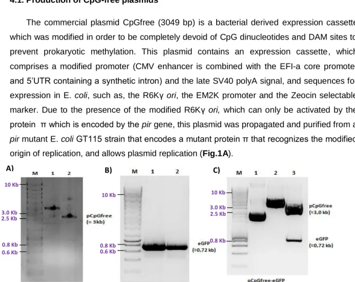
4.1. Production of CpGfree plasmids 22
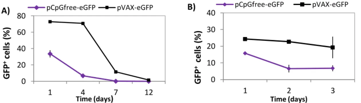
4.2. Long-Term analysis of pCpGfree-eGFP transfected cells 23
4.3. Induction assays for Minicircle production 24
4.4. Optimization of microporation conditions in NSCs 26
4.4.1. Optimization of microporation conditions using RB 26
4.4.2. Optimization of microporation conditions using HMB 28
4.4.3. Comparison of transfection efficiency using RB and HMB 29
4.5. Effect of different gene vectors on NSC transgene expression 31
4.5.1. Long-term analysis of transfected cells 31
4.5.2. Cell division kinetics analysis of transfected cells 33
4.6. Quantification of plasmids and minicircles in the nucleus 35
4.6.1. Calibration curves 35
4.6.2. DNA vector amount vs. gene expression 36
4.6.3. Dilution kinetics of DNA vector along cell division 39
4.6.4. Differentiation of NSCs 41
5. Discussion 42
6. Conclusions and Future work 48
7. References X
I
List of abbreviations
AAV Adeno-associated virus
AD Alzheimer’s disease
BDNF Brain-derived neurotrophic factor
BLBP Brain-lipid binding protein
BP Basal progenitors
CNS Central nervous system
CMV Cytomegalovirus
EGF Epidermal growth factor
eGFP Enhanced green fluorescent protein
ESC Embryonic stem cells
FGF Fibroblast growth factor
GDNF Glial cell-derived neurotrophic factor GFAP Glial fibrillary acidic protein
GFP Green fluorescent protein
GLAST Glutamate aspartate transporter
HSC Hematopoietic stem cells
HMB Homemade buffer
iPS induced pluripotent stem cell
MC Minicircles
MC-PP Minicircle Parental plasmid MSC Mesenchymal stem cells
NEP Neuroepithelial progenitors
NGF Nerve growth factor
NLS Nuclear localization sequence
NP Neural progenitors
NPC Nuclear pore complex
NSC Neural stem cells
PCR Polimerase chain reaction
PD Parkinson’s disease
PDGFα Platelet-derived growth factor
RB Resuspension Buffer
RG Radial glia
II
S/MAR Scaffold matrix attachment region
SCI Spinal cord injury
Shh Sonic hedgehog
SVZ Subventricular zone
SGZ Subgranular zone
TLR9 Toll-like receptor 9
III
Acknowledgments
“Our treasure lies in the beehive of our knowledge. We are perpetually on the way thither,
being by nature winged insects and honey gathers of the mind”
Friedrich Nietzsche
I want to express my truly gratitude to everyone who followed me during the Master Thesis year, especially to:
Professor Joaquim Cabral, for replying to my e-mail and giving me the opportunity to develop my Master Thesis at the Institute for Biotechnology and Bioengineering (IBB) at Instituto Superior Técnico (IST).
Catarina Madeira, for supervising and supporting me. For believing and trusting my work and for stimulating my scientific interest and perception. You were, without a doubt, the best supervisor I could have, and I leave the laboratory knowing that I gained not only a mentor but also a friend. Thank you for everything!
Carlos Rodrigues, for teaching me how to culture the cells and for all the useful advices. Filipa Ferreira for accompanying me.
My family, especially my parents and brother, for your support and love. It was for all of you that I wanted to pursue my goals and dreams.
IV
Abstract
Neural Stem Cells (NSC) are multipotent stem cells, capable of proliferating and differentiating in vivo and in vitro into astrocytes, oligodendrocytes and neurons. For this reason they hold a great potential for the development of gene and regenerative therapies for the treatment of neurodegenerative diseases and brain cancer. However, transfection of NSCs has proven to be difficult through conventional methods, and the disadvantages associated with the use of viral vectors make non-viral vectors more suitable for the development of gene delivery assays to NSCs. Apart from the non-viral method, one of the most important factors in gene delivery is the type of used vector. One of the factors that mostly affect the vector efficiency is the presence of CpG motifs. These motifs are responsible for the triggering of innate and acquired immune responses contributing to episomal silencing of the transgene. In this study, gene delivery to NSCs was optimized for the use of microporation technology and transfection efficiency was compared for the use of different transfection vectors with low vs. high CpG content, namely, minicircles, pCMV-GFP and pVAX-eGFP.
The optimization of microporation conditions revealed that depending on the electroporation buffer, high number of transfected cells (60 to 75%) and low cell mortality (15-10%) are obtained when using 1500V, 20 ms and 1 pulse or 1800V, 20 ms and 1 pulse as microporation conditions. When comparing the transfection efficiency using different vectors it was evident that Minicircle was the vector that allowed the obtainment of sustained and higher number of transfected cells (75%) without affecting their survival (80-90% of cell viability) and morphology. The quantification of vector copies in the nuclei revealed that the optimal dose to transfect NSCs is around 0.8 µg, and that, although a similar number of Minicircle and pCMV-GFP copies per nucleus is found, the first are the vectors that yield the highest expression levels. Long term analysis also showed Minicircles are less degraded, exhibiting higher number of copies and GFP expression than pCMV-GFP or pVAX-eGFP. Finally, microporation did not seem to affect NSCs differentiation potential.
Taken together, these results offer the first insights in the use of microporation and minicircles in non-viral transfection of NSCs, suggesting that microporation is a promising tool for NSCs transfection and that minicircles offer a new model of efficient and safe non-viral gene delivery to NSCs and have unquestionably a potential use for clinical applications and genetic engineering.
Keywords: CpG motifs; Microporatio; Minicircles; Neural stem cells; Non-viral gene delivery;
V
Resumo
Células estaminais representam um grupo específico de células indiferenciadas que apresentam a capacidade de se auto-renovar e de se diferenciar, quando estimuladas por determinadas condições, em células várias linhagens distintas. Existem dois tipos distintos de células estaminais: embrionárias e adultas. As células embrionárias apresentam a capacidade de se especificarem em vários tipos celulares no entanto, os problemas éticos muitas vezes associados com o seu isolamento e potencial cancerígeno, limitam o seu uso. Por este motivo, o interesse em células estaminais adultas, que apresentam apenas a capacidade de se especificar em tipos celulares provenientes do seu tecido de origem, tem vindo a aumentar exponencialmente.
Células estaminais neurais (Neural stem cells – NSC) representam uma população de células que pode ser isolada a partir de tecido cerebral embrionário, fetal ou adulto. No cérebro em desenvolvimento estas células existem como progenitores neuroepiteliais que se diferenciam em vários progenitores neurais que vão dar origem aos três tipos celulares característicos do sistema nervoso, astrócitos, oligodendrócitos e neurónios. No sistema nervoso adulto, estas células representam uma população de astrócitos que têm a capacidade de se diferenciar em caso de infecção ou inflamação, e habitam nichos específicos na zona subventricular do prosencéfalo e na zona subgranular do hipocampo. A sua capacidade única de se diferenciarem em células do sistema nervoso e de, após enxerto, conseguirem ultrapassar a barreira hematoencefálica, faz com que estas células apresentem um elevado potencial para o desenvolvimento de terapias regenerativas e genéticas para o tratamento de doenças neurodegenerativas e de cancro do cérebro.
Em cultura, estas células são aderentes ou formam complexos esféricos de progenitores neurais, apresentam a forma bipolar e expressam factores característicos de células neuronais, tais como nestina, Sox2 e Pax6, e astrogliais, tais como GFAP, GLAST, BLBP e RC2. A capacidade de diferenciação que estas células apresentam e o desenvolvimento de protocolos de diferenciação simples e eficazes fez com que as NSCs tenham sido alvo de diversos estudos de regeneração de tecidos e transferência de genes. Estudos recentes demonstraram que em caso de enxerto, as NSCs conseguem diferenciar-se em neurónios dopaminérgicos e substituir lesões em modelos de doença de Parkinson e conseguem reduzir o processo inflamatório em modelos de esclerose múltipla. Em termos de entrega de genes, os estudos efectuados demonstraram que as NSCs podem ser transfectadas e entregar genes terapêuticos que potenciam o melhoramento de várias doenças de foro neurológico.
VI
Um dos grandes problemas até agora tem sido o desenvolvimento de métodos de transfecção eficientes. Até muito recentemente, a transfecção de NSCs era basicamente feita através de métodos virais, no entanto os problemas patogénicos associados a estes vectores e a possibilidade de indução de mutagénese das células transfectadas fez com que a atenção dos cientistas divergisse para as metodologias não virais. Em comparação com as primeiras, estas metodologias oferecem uma série de vantagens, tais como, maior capacidade de empacotamento, menor imunogenicidade, fácil e maior segurança de manuseamento. A entrega de genes usando estes métodos pode ser feita através de tratamentos químicos, como partículas de fosfato de cálcio, biopolímeros biodegradáveis e liposomas catiónicos, e através de tratamentos físicos, tais como sonoporação, bombardeamento de partículas, magnetofecção, electroporação e microporação. Para além do método de transfecção, há que ter em atenção o tipo de vector usado. Após transfecção, os vectores têm que ultrapassar barreiras extra e intracelulares que causam silenciamento episomal do transgene. É por este motivo que têm sido desenvolvidos muitos trabalhos com o intuito de melhorar os vectores. Um dos factores que mais afecta a eficiência de um plasmídeo é o conteúdo deste em motivos CpG. Estes dinucleótidos não-metilados são característicos de genomas bacterianos e, se presentes em plasmídos, são reconhecidos pelo sistema imunitário de mamíferos, mais especificamente por células que expressam TLR9, despoletando reacções imunológicas inatas e secundárias que levam à degradação do vector e posterior silenciamento do transgene. Recentemente têm sido produzidos plasmídeos de conteúdo reduzido em motivos CpG que em relação aos pDNAs convencionais, apresentam eficiência e persistência melhoradas. Para além destes tipos de vectores, o desenvolvimento da tecnologia de minicirculos, que são pequenos vectores desprovidos de sequências bacterianas e de conteúdo CpG reduzido, permitiu um melhoramento de 10 a 10,000x das eficiências de transfecção em relação aos pDNAs convencionais. O objectivo principal deste estudo foi a optimização das condições de transfecção não viral de NSCs (CGR8-NSC) por microporação e a comparação das eficiencias de transfecção de NSCs quando transfectadas com minicirculos e pDNAs com conteúdo em motivos CpG reduzido ou mesmo inexistente.
Neste estudo, foram feitas duas optimizações das condições de microporação para o uso de dois tampões de electroporação diferentes, o RB, um tampão de formulação desconhecida, e o HMB, um tampão preparado no laboratório e que já tinha sido utilizado para transfectar células HEK293T e células estaminais mesenquimatosas. A microporação com o tampão RB foi feita transfectando pVAX-eGFP e variando a voltagem (entre 1400 e 1600V), a duração do pulso (20 e 30 ms) e o número de pulsos (1 e 2). A condição que no geral demonstrou ser mais eficaz foi 1500V, 20 ms e 1 pulso (percentagem de células
VII
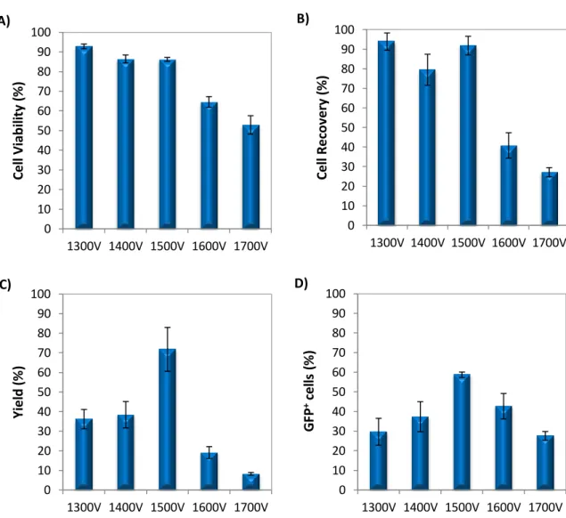
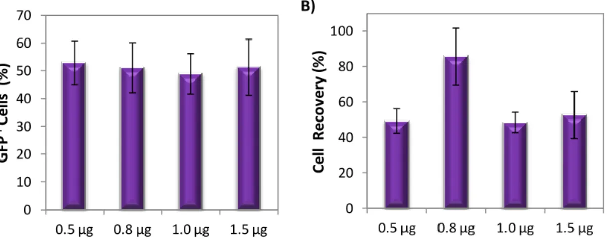
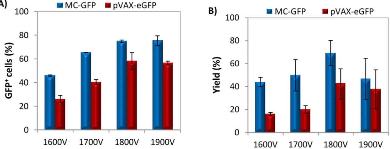
transfectadas ≈ 60%; viabilidade e recuperação celular 85-90%). A optimização com HMB foi feita microporando as NSCs com dois vectores diferentes (pVAX-eGFP e MC-GFP) e variando a voltagem entre 1600 e 1900V. Apesar de, com o aumento da voltagem se ter verificado, em ambos os casos, uma diminuição das viabilidades e recuperações celulares (mais acentuada em células transfectadas com pVAX-eGFP), no geral, a condição que obteve os melhores resultados, exibindo as percentagens de células transfectadas (50 e 75% com eGFP e PC-GFP) e rendimentos de transfecção (47 e 69% com pVAX-eGFP e MC-GFP) mais elevadas foi 1800V, 20 ms e 1 pulso. Um outro ensaio de optimização foi efectuado para determinar a quantidade ideal de DNA para transfectar NSCs. Para isto, NSCs foram ressuspendidas em RB e transfectadas com várias quantidades de pVAx-eGFP (0.5, 0.8, 1.0 e 1.5 µg) usando 1500V, 20 ms e 1 pulso como condições de microporação. Apesar de o aumento de DNA não ter afectado a percentagem de células GFP+ (≈50%) e de ter provocado uma diminuição das viabilidades e recuperações celulares, 0.8 µg foi a quantidade de pDNA que exibiu o maior rendimento de transfecção (≈40%), tendo sido por isso considerada a quantidade ideal de vector para transfectar NSCs.
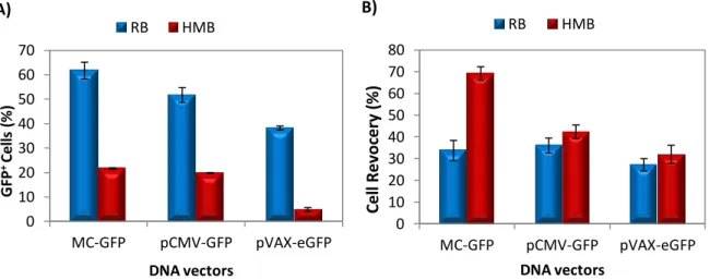
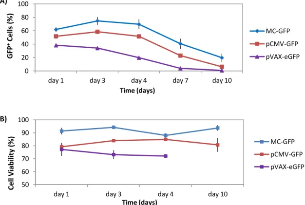
Para testar a eficiência de transfecção de minicirculos e outros pDNAs com conteúdo reduzido ou inexistente em CpGs as NSCs foram transfectadas com 2.0 x 10¹¹ moléculas de cada vector. A transfecção com plasmídeos sem motivos CpG (pCpGfree-eGFP) revelou-se muito aquém do esperado, e quando comparando com NSCs transfectadas com pVAX-eGFP, a percentagem de células GFP+ era menor e o decaimento da expressão do transgene ao longo do tempo mais rápido em células transfectadas com pCpG-eGFP, indicando que talvez, o promotor deste plasmídeo não seja tão eficiente como o do pVAX-eGFP. Comparando células transfectadas com minicirculos (MC-GFP) com outros pDNAs (pCMV-GFP – plasmídeo correspondente de MC-GFP, e pVAX-eGFP) verificou-se que usando MC-GFP é possível manter elevados níveis de células transfectadas (≈75%) sem comprometer a viabilidade celular (80-90%) e a morfologia bipolar característica das NSCs em cultura. Adicionalmente, a expressão do gene mantém-se mais alta com MC-GFP ao longo de 10 dias e não afecta a proliferação celular.
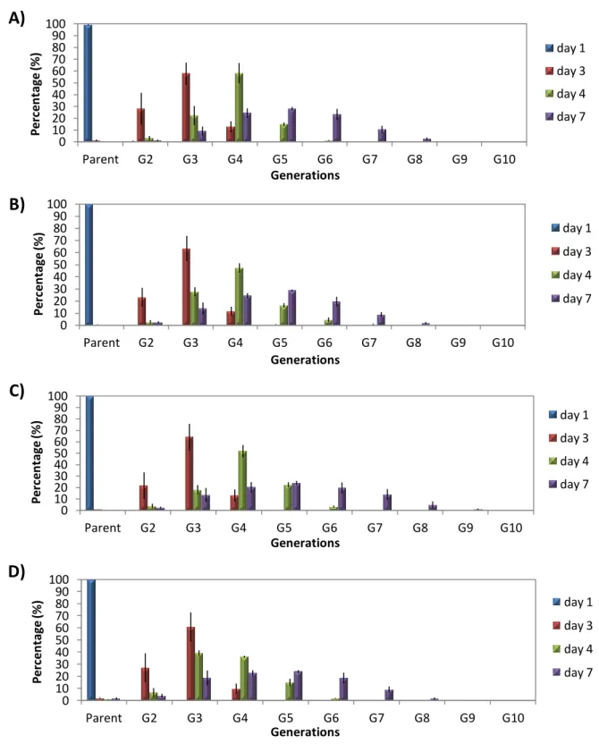
Testes adicionais foram efectuados para quantificar o número de cópias de vector no núcleo de NSCs transfectadas. Em estudos anteriores tinha-se verificado que há uma certa dose de plasmídeo a partir do qual a expressão do transgene satura. Em NSCs verificou-se que, independente do vector, a partir de 0.8 µg há uma estabilização e posterior decaimento da expressão da GFP, e, quando comparando o número de cópias de cada vector no núcleo, verificou-se que apesar da quantidade de MC-GFP e pCMV-GFP ser semelhante, o nível de expressão do primeiro era mais elevado. Adicionalmente, a análise ao longo de 10 dias da quantidade de cópias do núcleo revelou que o MC-GFP era o vector que se
VIII
encontrava em maior quantidade no núcleo e o que revelava maiores níveis de expressão do transgene, indicando que este, devido ao seu menor tamanho e conteúdo CpG mais reduzido (≈6%) é menos degradado que os outros plasmídeos. Em comparação, os resultados com pCMV-GFP e pVAX-eGFP, que apresentam conteúdo semelhante em motivos CpG (≈7%), diferem muito entre si, apresentando o pCMV-GFP eficiências e viabilidades celulares mais elevadas. Este facto é difícil de ser explicado mas há três diferenças entre estes dois plasmideos que podem justificar esses valores: i) diferente sinal de poliadenilação dos dois plasmídeos (pVAX-eGFP apresenta o sinal BGH e o pCMV-GFP apresenta o sinal SV40), que como estudos anteriores já demonstraram pode influenciar os níveis de expressão, ii) diferenças na sequência do gene reporter que pode originar uma proteína com maior ou menor intensidade de fluorescência; iii) ou ao nível de purificação de cada plasmídeo. Finalmente, nem o uso de microporação nem a transfecção com plasmídeo afectou a capacidade de diferenciação das NSCs em neurónios.
Comparando os resultados obtidos é possível concluir que o uso de microporação como método de transfecção de NSCs dá origem a elevadas eficiências de transfecção. Em comparação com os outros pDNAs, os Minicirculos foram os vectores que demonstraram as melhores eficiências de transfecção e os maiores níveis de expressão do transgene sem comprometer a morfologia e a viabilidade das células. Pode-se concluir que o tamanho destes vectores e que a diferença e 1% no conteúdo em motivos CpG é suficiente para que o silenciamento episomal do transgene seja mais fraco com MC-GFP. Como trabalho futuro, seria interessante avaliar a expressão de TLR9 em NSCs após transfecção para verificar se há realmente silenciamento devido aos motivos CpG e quantificar o RNA mensageiro para avaliar a estabilidade e frequência do transgene. Seria também muito benéfico avaliar a eficiência dos minicirculos através de transfecção por outros métodos não virais, tais como, lipofecção ou magnetofecção, e efectuar estudos in vivo para avaliar o tráfico intracelular de minicirculos, e também clonar um gene de interesse nos minicirculos para avaliar o seu potencial no desenvolvimento de futuras terapias para doenças de foro neurológico.
Até à data, ainda não tinham sido efectuados estudos de transfecção em NSCs usando microporação ou minicirculos. Este trabalho representa a primeira perspectiva para o uso destas tecnologias para a transfecção destas células e indica que os minicirculos representam um novo modelo eficiente e seguro de entrega não viral de genes para NSCs e uma alternativa muito promissora para o desenvolvimento de terapias para o tratamento de doenças neurodegenerativas ou cancro do cérebro onde uma expressão transiente de transgene pode ser necessária.
IX
Palavras-Chave: Células estaminais neurais; DNA plasmídico; Entrega de genes não-viral;
- 1 -
1. Introduction
1.1. Stem cells
A stem cell is a specific type of cell that has the potential to differentiate into one or more specialized cell type and the unique capacity of self-renewal for an expanded period of time. Unlike the majority of cells which are committed to conduct a specific function, a stem cell maintains an unspecialized state until it receives signals to differentiate into a tissue- or organ-specific cell kind [2]. Due to their special proliferative capacity and high plasticity, these cells represent an invaluable tool to basic research and an incredible resource for the repair and regeneration of diseased or damaged cells and tissue [3].
Stem cells can be found since the early stage of the embryo until the end of life and can be categorized as embryonic or adult, depending on their tissue of origin. Embryonic stem cells are derived from the inner cell mass of the blastocyst (4 to 5- day embryo) and possess the remarkable property of pluripotency, giving rise to a wide variety of cell types representative of the three primary germ layers in the embryo (mesoderm, endoderm and ectoderm) [3-4]. For this reason, they hold a great promise for tissue engineering and cell replacement therapies, however, their potential use in biomedical treatments is still controversial due to ethical concerns related to their isolation and because of their tumorigenic potential [5].
Adult stem cells are lineage-restricted undifferentiated cells, i.e. multipotent cells, with the capacity of giving rise to progeny cells of their correspondent tissue of origin [6]. These tissue-specific cells are scarce and present in a metabolically quiescent state in specialized tissues, such as the brain, bone marrow, liver, skin and gastrointestinal tract. Nowadays, on account of their high accessibility and minimally invasive harvesting procedures, there is a rising interest in these cells. The best known example of adult stem cell is the Hematopoietic stem cell (HSC) which can be isolated from the bone marrow niche and has the capacity to originate all blood cell types. Mesenchymal stem cells (MSC) and Neural stem cells (NSC) are other good examples of well studied adult multipotent cells. MSC have the potential to differentiate into different mesodermal cell lineages and are present in almost all tissues, however for therapeutic applications they are preferably isolated from the bone marrow and the umbilical cord blood [7]. NSC can be isolated from embryonic, fetal or adult brain tissue and are capable of generating neurons and glia in the developing brain. In the mammalian adult brain NSCs exist in niches that support symmetric self-renewal and fate-committed asymmetrical divisions that potentiate de replacement and recovery of lost neural cells [8].
- 2 -
1.2. Sources of Neural Stem Cells 1.2.1. Embryonic neurogenesis
The main features of Neural Stem cells (NSCs) reside in their ability to self-renewal, proliferate and generate multiple cellular lineages, such as neurons, astrocytes and oligodendrocytes both in vivo and in vitro. Their morphological and phenotypic identities vary depending on the developmental stage and region occupied by these cells during adult life. In the early embryonic stage, predetermined programs give rise to spatiotemporally different NSC populations in a process called Neurogenesis. This process begins with the induction of the neuroectoderm, which forms the neural plate that folds to originate the neural tube. In the embryonic neural tube, NSCs exist as neuroepithelial stem cells that undergo differentiation into distinct neural progenitors and posterior specification into neuronal lineages through asymmetrical cell divisions in the germinal ventricular zone (VZ). After the neurogenic phase NSCs acquire gliogenic competence and start differentiating into glia in the subventrivular zone (SVZ). This process leads to the specification of astrocytes and the disappearance of the VZ. After brain development, some parts of the brain still harbor sites where adult neurogenesis remains active with the sole purpose of maintaining homeostasis of the central nervous system (CNS) [9].
1.2.2. Neural differentiation of embryonic stem cells
The understanding of the processes involved in embryonic neurogenesis and the progress in cell culture methodologies enabled the development of protocols that induce neuralization of ESCs in vitro. The overall advantage of ESC derived NSCs is that these cells retain a developmental competence to patterning signals giving rise to regional subtypes that can be transplanted in vivo and differentiate into functional neuronal entities. Early ESC neuralization attempts reported that, in minimal inductive conditions [10], ESCs were capable of differentiating into progressive neural progenitors that expressed distinct neural markers [11], however, the resulting cultures had a heterogeneous profile characterized by the coexistence of undifferentiated ESCs and different neural progenitors. Therefore, new protocols, based on the production of embryoid bodies [12], or stromal feeder co-cultures [13] were developed. Under these conditions, the originated cell populations exhibited uniform biochemical and electrophysiological characteristics. Nowadays, with the increasing comprehension of the developmental and functional mechanisms of ESC derived neurons, more effective procedures are being established. Recently, Chambers and colleagues, based on the understanding of the involvement of the SMAD signaling pathway in neural induction, developed a new protocol in which a dual-SMAD inhibition, by the synergistic action of Noggin and SB431542, induced rapid and complete neural conversion
- 3 -
of human ESCs [14]. Apart from ESCs, induced pluripotent stem cells (iPS) represent another great source of cells for differentiation into NSCs. Many protocols for neuralization of iPS are being developed and they provide a unique opportunity for the development of autologous therapies and tissue replacement [15].
1.2.3. Adult neural stem cells
Adult Neural Stem Cells (aNSCs) are present in specific niches in the subventricular zone (SVZ) and subgranular zone (SGZ) of the adult brain where they undergo adult neurogenesis. In these areas, their regulation is managed by fate—controlling molecules and signaling pathways. These molecules are members of the fibroblast and epidermal growth factor families and permit the expansion and specialization of adult neural progenitors in vivo and in vitro. Some of the most studied mediators are: epidermal growth factor (EGF) [16] and platelet-derived growth factor (PDGF-α) [17], that induce gliogenic specification, and fibroblast growth factor (FGF) [18], Sonic hedgehog (Shh) [19] and brain-derived neurotrophic factor (BDNF) [20] that are responsible for the differentiation into neuronal cells.
In vivo, neurogenesis is regulated by the action of immune effector cells called microglia that
are abundantly present in the adult CNS. During an injury, microglia secretes neurotrophic and neurogenic factors that potentiate the recovery of the damaged regions and increase the neurogenic process [21-22].
The most active zone of adult neurogenesis is the SVZ of the forebrain. This area contains a subpopulation of astrocytes that function as aNSCs in vivo and are known as Type-B cells, which in turn, can be subdivided in Type B1 and B2 cells. Type-B1 cells are considered true aNSC and are in contact with the ventricular cavity where, in response to vascular signals, differentiate into neural progenitors (Type C cells). These progenitors undertake symmetrical divisions and give rise to neuroblasts (Type A cells) that migrate through the rostral migratory stream, aided by Type B1 and B2 cells, to the olfactory bulb where they specify as interneurons. In the SGZ in the dentate gyrus of the hippocampus, neuronal progenitors (Type-B cells) differentiate into granular neurons. However, these neuronal progenitors have a limited capacity of specification. They suffer asymmetrical divisions that instigate a regionally determined differentiation into granular neurons that act as regenerative entities in the brain [23].
- 4 -
1.3. Characterization of NSCs
1.3.1. In vitro proliferation of NSCs
NSCs can be proliferated as Neurospheres or in adherent conditions. After isolation NSCs are plated in low attachment tissue culture plastic dishes in serum-free media supplemented with the mitogens EGF and FGF [24]. Under these conditions cells that are undergoing specification die and NSCs form free-floating aggregates. The Neurosphere system is thought to provide a three dimensional environment that mimics neurogenic niches [25], however long-term cultures of neural progenitors as aggregates are difficult to maintain due to the occurrence of changes in the differentiation and self-renewal capacities, and the incidence of chromosomal instability [26-27].
In the first approaches, the adherent system involved, , expansion of NSCs on biopolymer-coated dishes in serum-free conditions [28]. These procedures permitted the exploration of NSCs multipotentiality but the cultures were short-termed and did not allow an efficient immortalization. Nowadays adherent propagation of NSCs is performed under the action of EFG and FGF-2 [29], or, as recent data show, under exposure to angiogenic factors [30]. In these conditions, cells undertake symmetrical divisions, maintain an undifferentiated state for a long period of time and retain their tripotential differentiation capacity.
1.3.2. In vitro differentiation of NSCs
Due to the potential that NSCs have to differentiate into neurons, astrocytes and oligodendrocytes, a number of protocols have been developed for controlled in vitro specification in one of the three possible fates. Differentiation within neurospheres is achieved by mitogen removal after attachment onto substrate coated coverslips [29]. Under these conditions NSCs form primary neurospheres and specify into neuronal progenitors. Upon replating the neural progenitors give rise to secondary aggregates where they differentiate into GABAergic interneurons, astrocytes and oligodendrocytes, however, with the posterior passages, the maintenance of neurogenic competence declines and the yield of gliogenesis increases thus originating an uncontrolled pattern of differentiation. In the monolayer adherent system, NSCs give rise to antigenically and electrophysiologically mature neurons, oligodencrocytes and astrocytes after prolonged expansion under optimized differentiating conditions [29, 31-32]. These in vitro differentiated cells can be efficiently transplanted into the brain and establish synaptic connection with the cells of the nervous system [33].
- 5 -
1.3.3. In vivo and In vitro phenotype of NSCs
There are currently four types of known phenotypically distinct NSCs: Neuroepithelial progenitors (NEP), Radial Glia (RG), Basal Progenitors (BPs) and Adult neural stem cells (aNSCs).
NEPs are present during the embryonic development of the CNS and express nestin, Sox1, Sox2 and Pax6, markers for immature neural cells, and some ESC markers, such as Oct4. The expression of nestin, Sox2 and Pax6 persists throughout the specification of primitive NEPs into the subsequent neural progenitors and is essential to the neurogenic process [34-36]. In vitro, these primitive NSCs lose the expression of ESC markers with the passages and become more committed to specify into neural progenitors. At the beginning of neurogenesis, NEPs give rise to RGs that serve as the main progenitors in the developing brain. RGs express astroglial markers, such as astrocyte specific glutamate aspartate transporter (GLAST), glial fibrillary acidic protein (GFAP), brain-lipid binding protein (BLBP) and RC2, and neurogenic markers like nestin, Sox2 and Pax6. Recent findings suggest that RGs can also be found in a non-ventricular niche in the adult spinal cord of mice (SCRGs) and are phenotypically similar to adult NSCs of the SVZ [37].
In the adult brain, BPs potentiate the production of neurons, however, their propagation
in vitro is difficult so little is still known about them. Adult NSCs are the main progenitors of
adult neurogenesis and combine the expression of radial glial markers, like GFAP, GLAST, BLBP and RC2; neural precursor markers like, nestin, Sox2 and Pax6; regional markers like Otx2 and Hoxb9 and others like Musashi 1 and CD133 that maintain their stemness. In vitro, aNSCs are antigenically similar to neurogenic RGs in the brain when grown in adherent conditions; however, when propagated as neurospheres, their phenotype is characterized by the presence of differentiated cells in the sphere core surrounded by RG-like neural progenitors [8, 26, 32].
- 6 -
1.4. Therapeutical applications of NSCs
In recent years, the successful production of neurons and glia from stem cells in culture fuelled efforts to develop stem-cell-based transplantation therapies for neurological disorders. It is well known that within the CNS, endogenous neural progenitors support its ‘self-repair’ during inflammatory disorders, such as spinal cord injury and stroke [38-39]. Although efficient, endogenous stem cells are not able to promote full and irreversible repair of the CNS. Adult neurogenesis is highly influenced by the surrounding microenvironment, and, upon inflammation, the supporting cells secrete molecules that attract inflammatory cells. NSCs can perceive these changes as dysfunctions and induce an uncontrolled neurogenic process [40].
Nowadays, many laboratories are diverting their attention to the development of therapies for many CNS disorders, such as, Parkinson’s disease, Huntington’s disease, multiple sclerosis, stroke, traumatic brain injury, Alzheimer’s disease, epilepsy, and others. These diseases are characterized by progressive neuronal degeneration that can ultimately be fatal. Stem-cell based approaches for these diseases involve the engraftment of in vitro propagated NSCs or differentiated neuronal cells for the restoration of a lost function. When transplanted they have low immunogenicity, are able to migrate and overcome the blood-brain barrier, and upon transfection, allow vector-directed differentiation or drug delivery [41]. Many studies have documented that neural progenitors, for instance, can be efficiently engrafted to Parkinson’s disease models, migrate to the injured areas and specify into dopamine neurons [42], can survive, migrate and differentiate under the influence of the surrounding microenvironment in stroke models [43] or can reduce brain inflammatory process in multiple sclerosis models [44].
NSCs can also be used as drug delivery vehicles. They can be transfected and produce specific neurotrophic or neuroprotective factors that promote a long-lasting amelioration and regression of the symptoms of neuronal disorders. A few authors have published results regarding, for example, the delivery of GDNF engineered NSCs to PD and amyotrophic lateral sclerosis models [45-46], the delivery of NGF or BDNF to AD models [47-48], or the delivery of VEGF to promote recovery in SCI models [49-50]. NSC based gene therapy also represents a promising tool for the treatment of brain tumors by the delivery of molecular agents that promote the conversion of prodrugs into anticancer substances, and immunostimulating cytokines [51].
- 7 -
1.5. Gene delivery to NSCs 1.5.1. Viral methods
Lately, many efforts have been made to improve the transfection efficiency of NSCs in order to provide better opportunities for future therapeutic applications. Viruses like retrovirus and lentivirus integrate into the host genome and allow stable gene transfer and long-lasting expression. In contrast, adenovirus and adeno-associated virus (AAV) are non-integrating particles that cause no side effects in humans, and allow transient expression of the transgene [52]. Although efficient, there are still some drawbacks related to their use. These vectors have limited transgene capacity, trigger cytotoxic and immunogenic reactions, cause mutagenesis due to their random integration in the host genome [52-53], and in NSCs they affect differentiation and block neurogenesis [54]. At present, a special attention is being given to AAV vectors. These vectors were initially considered nonintegrative and nonpathological however, it has been demonstrated that they integrate into the host genome and trigger malignant transformation of the cells [55-56]. Nevertheless, some of the newest methodologies for gene delivery take advantage of their demonstrated capacity and available capsid libraries to design new hybrid vectors that consist of AAV packaged plasmids [57].
1.5.2. Non-Viral methods
Regardless of having lower transfection efficiencies and low cell viabilities, there’s an increasing interest in non-viral gene delivery methods. When compared to viral methods, non-viral methods are cheaper, easier to manage, have a wider range of putative cell targets, lower immunogenicity, lesser toxicity, bigger packaging capacity and allow transient expression, a feature that might be beneficial for therapeutic strategies where a short duration of transgene expression is necessary [58]. These methods are used to transfect different molecules, such as, RNAs (e.g. siRNAs), proteins (e.g. antibodies) and DNA which is mostly delivered as plasmid vectors (pDNA) that have been cloned to contain a transgene expression cassette [59], and rely on a physical treatment or on chemical or biological particles that are used as carriers.
Some of the most used chemical DNA delivery methods are:
Calcium phosphate-mediated transfection which involves incubation of the cells with calcium phosphate coated pDNA. This method is very efficient yet restricted to some cells and sensitive to the amount of input pDNA [60].
Biodegradable cationic polymers that are used for the controlled release of DNA into the cell or tissue of interest. These polymers have a reduced toxicity and avoid polymer accumulation in the cells, however, their slow degradation rate and the inefficient
- 8 -
membrane destabilizing peptides available influence the effectiveness of these structures [61]
Cationic liposomes which are vesicular structures of positively charged lipids that once surrounding DNA form lipoplexes. These systems have a low toxicity in vitro, but can exhibit some toxicity and low transfection efficiencies in vivo due to the physiochemical properties of the cationic lipid/DNA complexes [62].
The most popular physical DNA delivery systems are:
Sonoporation that consists on the permeabilization of the cell membrane through exposure to low-field ultrasounds that induce the uptake of large molecules into the cell. This technique is used in targeted gene transfer both in vivo and in vitro, however, extended exposure to low-field ultrasounds results in increased cell mortality, thus the procedure needs to be carefully applied [62]; Gene gun which is based on the precipitation of DNA on small particles of a heavy metal, usually gold, and posterior injection, using a pistol. At present there are many listed gene gun clinical trials for Hepatitis B, malaria and influenza yet, this method is only applicable on superficial tissues (e.g. skin) due to the high level of cell damage caused by the velocity of the particles [62];
Magnetofection that involves the association of the DNA with magnetic nanoparticles which are transferred to the cells by the application of a magnetic field. Although promising, the use of magnetofection in vivo is still limited due to the inaccessibility of the targeted tissues to magnetic field and hydrodynamic forces. Ongoing improvements of this procedure involve, in some cases, the combination of the magnetic complexes with cationic lipids [62-63];
Electroporation, which is a powerful tool that relies on the application of a short electric pulse that leads to a permeable state of the cell membrane so that the molecules can be driven into the cell through elecrophoretic and electro-osmotic forces. This technique is used to transfer molecules of different sizes (e.g. genes, proteins, ionic dyes and drugs) into the cell, however, there are still some problems needing to be solved such as low viabilities and transfections caused the massive cell mortality triggered by the electric pulse [62]; To overcome some of the problems associated with electroporation, a new technology called Microporation was developed. This new layout provides high transfection efficiencies and cell viabilities and is based on the use of a pipette tip as electroporation area and a capillary electrode which allows maintenance of pH and temperature, minimizes turbulence and avoids ion metal generation [64]. Recent studies have proven that microporation is an easy and efficient method for gene delivery to a wide variety of cells, such as, Human bone marrow mesenchymal stem cells [65],
- 9 -
Human astrocytoma cells [66] and Human umbilical cord blood-derived mesenchymal stem cells (hUCB-MSCs) [67].
Even though these non-viral methods have shown to be effective and advantageous, it is important to optimize these technologies to allow better in vivo and in vitro transfection efficiency and permit the development of new therapeutic strategies. These improvements involve not only the development of new gene transfer technologies, but also the enhancement of transfection vectors that need to overcome many extra- and intracellular barriers.
- 10 -
1.6. Minicircles: The future of gene therapy?
Upon administration there are a number of extra- and intracellular systems that can impair gene delivery. Extracellularly, the vectors can be sequestrated in first pass organs (e.g. lung and liver) or affected by the extracellular matrix [68]. Intracellularly, the vectors encounter two distinct cytosolic barriers: a diffusional and a metabolic barrier. The diffusional barrier is related to the size-dependent deceleration of the vectors which might lead to the emergence of plasmid aggregates and an increase of the cytoplasm viscosity. The metabolic barrier is related to the presence of calcium-sensitive cytosolic nucleases that degrade foreign DNA [68-69]. The nuclear envelope represents the last barrier to be surpassed and to diffuse through the nuclear envelope the vectors must carry a nuclear localization sequence (NLS) to pass through the nuclear pore complex (NPC) or be transfected in dividing cells to gain access to the expression machinery after nuclear disassembly during mitosis [70].
To overcome these barriers many laboratories have been committed to the development of more effective plasmid vectors. Conventionally, pDNA is constituted by a mammalian expression cassette and a bacterial backbone consisting of an antibiotic resistance gene and an origin of replication for production in bacteria. To avoid degradation pDNA should be stable and mainly in the supercoiled form and free of contaminating factors (e.g. bacterial DNA, RNA and proteins). Many of the most recent advances in plasmid technology involve the substitution of the conventional polyadenylation (polyA) signals with more effective polyA sequences (e.g. Bovine growth hormone (BGH) polyA and SV40 polyA), that extend the half-life of supercoiled pDNA thus preventing the transformation into a circular form and exposure of secondary structures to the host nucleases [71], and the development of CpGfree plasmids. Unmethylated CpG motifs are predominantly present in bacterial DNA and therefore, are also common in bacterial derived pDNAs. These motifs are recognized by Toll-Like receptor 9 (TLR) positive cells (e.g. Dendritic cells) which activate host defense mechanisms triggering innate and acquired immune responses [72]. Transfection of CpGfree based pDNA has proven to yield high transgene expression in vivo and in vitro hence allowing sustained expression [73].
The real breakthrough in the vector upgrading was the development of the minicircle technology. Minicircle DNAs are small sized circular molecules, derived from plasmid DNA, that lack the bacterial backbone that triggers immune responses, alters gene expression and might potentiate the silencing of the encoded transgene. This technology consists on the construction of a parental plasmid that carries the expression cassette flanked by two recognition sites of a site-specific recombinase (e.g. λ derived recombinase; Cre rec.; FLP rec.) which is expressed and divides the parental plasmid into two molecules: a miniplasmid
- 11 -
that carries the bacterial backbone and a minicircle that carries the expression unit. One of the major drawbacks of minicircle technology was, until recently, the difficult and expensive purification procedure of these vectors [62]. To overcome these hardships, Chen and coworkers developed a new method that degrades the remaining miniplasmid and parental plasmids in vivo by the action of a restriction enzyme (SceI), hence allowing a more effective purification [74], and more recently, the development of a robust minicircle production system permitted the establishment of a rapid and inexpensive protocol for production of minicircles [75]. When compared to regular plasmids, minicircles display a 10- to 10,000-fold improvement in long-term transgene expression in vivo [76-77], and, further improvements in their formulation, e.g. addition of scaffold/matrix attachment region (S/MAR), enhances transgene expression and persistence [78]. Altogether, this data confirm that minicircles offer a new concept in highly efficient and safe non-viral gene transfer, and in the future, they will represent the preferred system for gene delivery for clinical and therapeutic applications.
In this work, microporation conditions were optimized for gene delivery to NSCs for the use of two electroporation buffers, RB, the buffer provided by the manufacturer, and HMB, a buffer previously prepared at the laboratory. Furthermore, transfection efficiency was tested and compared using minicircles and pDNAs of different sizes and CpG content. Stem cell potential was investigated by evaluating the survival, proliferation, and differentiation potential of transfected cells and the number of copies of vector DNA was assessed in each case, more specifically, after Minicircle, pCMV-GFP and pVAX-eGFP transfection and the optimal dose of vector DNA for gene delivery to NSCs was determined. These results strongly suggest that there is a CpG dependent episomal transgene silencing in transfected NSCs and that minicircles are the vectors that give rise to the highest levels of gene expression without influencing the cell traits of NSCs.
- 12 -
2. Aim of Studies
This study was fully accomplished at Stem Cell Bioengineering Laboratory in Institute for Biotechnology and Bioengineering at Instituto Superior Técnico.
This work focused on the following points:
Optimization of microporation conditions for gene delivery to NSCs by evaluating the effect of voltage, pulse duration, electroporation buffer and DNA amount.
Evaluation of the GFP expression levels upon transfection of CGR8-NS cells with CpGfree plasmids.
Comparison of the use of different gene vectors with different CpG content, more specifically, minicircles, pCMV-GFP and pVAx-eGFP in NSCs transfection – evaluation of cell viability, percentage of GFP expressing cells, proliferation kinetics and morphological traits.
Quantification of the number of vector copies inside each nucleus after transfection with minicircle, pCMV-GFP and pVAX-eGFP, and determination of the ideal dose of vector after which transgene expression saturates.
Evaluation of dilution kinetics of each vector – quantification of the number of vector copies inside the nucleus after transfection with minicircles, pCMV-GFP and pVAX-eGFP at specific points for 7-10 days.
Differentiation of CGR8-NSCs – effect of transfection on differentiation potential.
The results obtained by this study will certainly be useful for future gene delivery applications in Neural Stem Cells, in particular, for the development of tissue regeneration applications and gene therapies for the treatment of neurodegenerative diseases or brain cancer where a transient expression of a gene of interest might be necessary.
- 13 -
3. Materials and Methods
3.1. Plasmids and Minicircles
3.1.1. Plasmids and Minicircles encoding GFP
The transfection vectors MC07.CMV-GFP (2257 bp), that will be referred as MC-GFP, and pCMV-GFP plasmid (3487 bp), both carrying the same GFP gene under the control of the cytomegalovirus (CMV) promoter and the SV40 polyadenylation sequence, were purchased from Plasmid factory (Bielfield, Germany). MC-GFP is a bacterial backbone vector consisting only on a mammalian expression cassette and pCMV-GFP is its correspondent plasmid which contains, apart from the expression cassette, an origin of replication and a Kanamycin resistant gene for prokaryotic selection. The pVAX-eGFP plasmid (3697 bp) was obtained by the modification of the commercial plasmid pVAX1lacZ (Invitrogen, 6050 bp) by replacement of the lacZ reporter gene with the enhanced Green Fluorescent protein gene (eGFP)(Supplementary Fig.1). The details of this construction are described elsewhere [1] and this plasmid was isolated and purified from a pVAX-eGFP transformed E. coli strain stored at -80 ºC.
3.1.2. Construction of CpGfree plasmids encoding GFP a) CpGfree plasmid
The CpGfree plasmid (3049 bp, Invivogen) is a commercial vector consisting of elements devoid of CpG dinucleotides and Dam methylation sites (GATC). This plasmid contains: a R6Kγ origin of replication, a E2MK promoter, a ZeocinTM resistance gene, and in the eukaryotic expression vector the promoter combines the mouse CMV enhancer, the human elongation factor 1 alpha core promoter and 5’UTR containing a synthetic intron, the late SV40 polyadenilation signal, matrix attachment regions (MARs) and a multiple cloning site (MCS).
b) Reconstitution of E. coli GT115 strain
The reconstitution of E. coli GT115 (provided with pCpGfree) was performed by adding 1 ml of LB medium to the tube containing the strain and incubation for 5 minutes at room temperature. The cell suspension was then mixed for 2 minutes and 100 µl of this suspension was plated in LB-Agar and incubated overnight at 37ºC. The resulting colonies were used to prepare competent cells (see below section 3.1.4).
- 14 -
The eGFP gene was amplified from the pVAX-eGFP vector. Briefly, 100 µl of E. coli transformed with pVAX-eGFP plasmids was inoculated in 5 ml falcons supplemented with 50 µg/ml Kanamycin and grown overnight at 37ºC and 250 rpm. The plasmid was then purified according to the protocol described in section 3.1.6 and its integrity verified through enzymatic digestion with HindIII and Agarose gel electrophoresis analysis. The eGFP gene was amplified by Polimerase Chain Reaction (PCR), using the following primers:
NcoI-GFP-Fwd, 5’- AAACCATGGATGGTGAGCAAGGGCGA -3’; and GFP-NheI-Rev, 5’-
TTTGCTAGCTTACTTGTACAGCTCGTCCATGCC -3’ (STAB Vida). Each 50 µl of final reaction volume contained 10 ng of pVAX-eGFP, 10 µM of each primer, 25 µl of KOD Hot Start Master Mix (Novagen ®), and 20 µl of Distilled H2O. The reaction was subjected to the following conditions: 5 min at 95ºC, 35 cycles of 30 s at 95ºC, 60 s at 60ºC and 90 s at 72ºC and 5 min at 72ºC. The amplification of the eGFP fragment was assessed through Agarose gel electrophoresis analysis and its purification was made using a DNA extraction and purification from Agarose gel kit (NZYTECH). The CpGfree-eGFP plasmid was then constructed through enzymatic restriction with NcoI and NheI and subsequent insertion of the eGFP reporter gene using T4 DNA Ligase. The construction was later confirmed through double enzymatic digestion with NcoI and NheI and Agarose gel electrophoresis analysis. The production and purification of this plasmid is described in section 3.1.8.
3.1.3. Production of Minicircles
a) Propagation of the Parental plasmid
The Minicircle DNA vector technology was purchased from System Biosciences (SBI; Mountain View, California) and the propagation of the Parental plasmid (PP-DNA, ≈7.8 Kb) was performed in an E. coli XLlgold strain and in an engineered E. coli ZYCY10P3S2T strain (provided by the manufacturer) as described below (Section 3.1.5).
b) Transformation of the Parental plasmid into E. coli ZYCY10P3S2T
The Transformation of the PP-DNA into E. coli ZYCY10P3S2T was performed as described by the manufacturer. Succinctly, 2 µg of MC-DNA was added to 100 µl of cells, incubated on ice for 30 minutes and at 42ºC for 30 seconds. The cell suspension was then placed on ice for 2 minutes after which 500 µl of S.O.C. medium supplemented with Kanamycin (50 µg/ml) was added. The mixture was then incubated for 60 minutes at 37ºC and 225 rpm. Afterwards, the cells were centrifuged for 10 minutes at 4000g, resuspended in 100 µl of S.O.C medium, plated on LB-Agar with 50 µg/ml of Kanamycin and incubated overnight at 37ºC. Some colonies were then harvested and cultured overnight, at 37ºC and
- 15 -
250 rpm, in 5 ml LB falcons with antibiotics. The presence of the PP-DNA was assessed through agarose gel electrophoresis analysis after simple enzymatic digestion with HindIII.
c) Induction assays for minicircle production
On day 1 in the morning, cells from a PP-DNA transformed colony was grown in 5 ml LB supplemented with 50 µg/ml of Kanamycin and incubated at 37ºC and 250 rpm. In the evening, 100 µl of cell suspension was inoculated in 400 ml or 25 ml of TB medium supplemented with Kanamycin. On day 2 in the morning, the OD was registered and a minicircle (MC) induction mix was prepared and 4 induction assays were performed. The first 2 assays were performed as described by the manufacturer protocol and involved the combination of the MC induction mix, composed of 400 ml or 25 ml of LB supplemented with 4% 1N NaOH and 0.1% of a 20 % L-Arabinose (Sigma) solution, with the overnight culture. In the third induction assay the 25 ml of the overnight culture was split in 2 flasks and one of the cultures was combined with 25 ml of MC induction mix while the second was combined with 12,5 ml of TB medium and 25 ml of MC induction mix. The fourth assay involved the division of the 25 ml of the overnight culture in 2 flasks and the addition of 25 ml of MC induction mix to each flask: one of the cultures was mixed with MC induction mix supplemented with 1% of 20% L-ara and the other was combined with MC induction mix supplemented with 2% of 20% L-ara. All the cultures were incubated at 30ºC with shaking at 250 rpm for 7 hours or longer. The OD was registered every 2,5 hours and the presence and integrity of the MC-DNA and MC was assessed through agarose gel analysis.
3.1.4. Preparation of E. coli competent cells
A single colony of the desired E. coli strain (GT115 and ZYCY10P3S2T) was isolated and grown overnight, at 37ºC and 250 rpm, in a falcon containing 5 ml of LB.In the morning the Optical Density measured at 600 nm (OD) was measured and the appropriate volume of cell suspension was added to 20 ml of LB medium to start growth at OD = 0.1. , followed by incubation at 37ºC and 250 rpm. Simultaneously a TSS solution was prepared as follows: 10 ml of 40 g/L LB, 1 ml of DMSO, 1 ml of 1M MgCl2, 2 g PEG8000 and H2O until 20 ml; the pH was adjusted to 6.5 and the solution was sterilized by filtration . When the OD of the cell suspension reached the value of 0.9, the content of the flask was divided into falcons and centrifuged at 1000 g for 10 minutes. The pellet was resuspended in 5 ml of cold TSS (0.1 V) and incubated on ice for 10 minutes. The suspension was divided into eppendorfs (100 µL/eppendorf), and stored at -80ºC.
- 16 -
The transformation of E. coli competent cells with the desired plasmids (E. coli GT115 with pCpGfree and pCpGfree-eGFP and E. coli XLlgold with PP-DNA) was performed using a Heat Shock protocol. Briefly, 2 µg of plasmid was added to 100 µl of cells, incubated for 15 minutes on ice and at 42ºC for 1 minute. 900 µl of sterile LB medium supplemented with antibiotics (25 µg/ml of Zeocin for pCpGfree and pCpGfree-eGFP and 50 µg/ml of Kanamycin for MC-DNA) was then added to the mixture and a 40 minutes incubation, at 37ºC and 250 rpm, was performed. Afterwards, the cells were centrifuged at 4000 rpm for 10 minutes. The pellet was resuspendend in 100 µl of LB and plated overnight, at 37ºC, in LB Agar plates with respective antibiotics. Some colonies were then harvested and cultured overnight, at 37ºC and 250 rpm, in 5 ml LB falcons with antibiotics. The presence of the plasmids and their integrity was evaluated through agarose gel electrophoresis after a simple and a double enzymatic digestion of the pCpGfree and pCpGfree-eGFP with NheI, and NcoI and NheI respectively, and a simple enzymatic digestion of MC-PP with HindIII.
3.1.6. Purification of plasmids
The purification of plasmids (pCpGfree; pCpGfree-eGFP; pVAX-eGFP; PP-DNA and respective Minicircle (MC)) was performed using a miniprep kit (Promega), according to supplier’s protocol.
3.1.7. Preparation of plasmid banks
Colonies of transformed E. coli strains with the desired plasmids (E. coli GT115 transformed with pCpGfree and pCpGfree-GFP; E. coli XLlgold and ZYCY10P3S2T transformed with PP-DNA) were harvested and inoculated in falcons containing 5 ml of LB supplemented antibiotics and grown overnight, at 37ºC and 250 rpm. Afterwards, 150 µl of cell suspension was inoculated into new 5 ml LB falcons with antibiotics in order to start the new growth with a OD=0.1. When the OD reached the value of 0.8 (after 4 hours of incubation at 37ºC and 250 rpm), the suspension was divided into criovials containing 200 µL of glycerol by adding to each vial 800 µL of the cell suspension. The vials were stored at - 80ºC.
3.1.8. Endotoxine free purification of plasmids
Aliquots containing E. coli strains transformed with the plasmids pVAX-eGFP and pCpGfree-eGFP were added to 2 L flasks with 250 ml of LB medium supplemented with antibiotics and allowed to grow overnight at 37ºC at 250 rpm. Plasmid DNA was then purified using an Endotoxin-free Plasmid DNA Purification kit (Machery-Nagel) according to manufacturer protocol and plasmid concentration was determined using a NanoDrop equipment (Digital Bio) by spectrophotometric measurements at 260 nm.
- 17 -
3.2. Cell lines
3.2.1. Culture and expansion of HEK293T cells
Criovials preserved at -80ºC, containing Human Embryonic Kidney (HEK) 293T cells (ATCC-LGC Nr: CRL-11268; Middlesex, UK) were thawed by resuspending their content in 6 ml of Dulbecco’s Modified Eagle Medium (DMEM, 1g/L Glucose, Gibco) supplemented with 10% fetal bovine serum (FBS, Gibco) and 1% (v/v) penicillin/streptomycin (Gibco). The mixture was then centrifuged at 1250 rpm for 5 minutes, and the pellet was ressuspendend in 6 ml of DMEM (10% FBS, 1% PenStrep). The number of cells and their viability was determined by Trypan blue staining (Gibco). Afterwards 7.5 x 10⁵ cells were cultured in 75 cm2 T-flasks (FALCON®) (10000 cells/cm2) and grown in adherence, under static conditions, at 37ºC and 5% CO2-humidified atmosphere. Sub-confluent cells were collected and used for transfection with pCpGfree-eGFP and pVAX-eGFP. In brief, HEK293T cells were washed with 10 ml of Phosphate buffered saline (PBS) solution (Gibco) and harvested with 4 ml of Trypsin 0.25% (Gibco) after incubation for 3 minutes at 37ºC and 5% CO2-humidified atmosphere. The cells were then resuspendend in 6 ml of complete medium and centrifuged at 1250 rpm for 5 minutes and the pellet was resuspendend in culture medium. The number of cells and their viability was determined by Trypan blue staining and the cells were transfected by microporation.
3.2.2. Culture and expansion of Neural Stem Cells
CGR8-NS cells, derived from ES cell line CGR8 (Welcome Trust Centre for Stem Cell Research in Cambridge, United Kingdom) were thawed by swiftly submerging the criovials in a water bath at 37ºC and by resuspending the cells with RHB-A (Stem Cell Sciences) medium supplemented with 15 ng/ml of EGF and FGF-2 (Peprotech) and 1% of penicillin/streptomycin (100 µg/ml) (Gibco) or with DMEM/F-12 (Gibco) supplemented with 0,8 g/ml of Glucose, 1% N-2 supplement (Gibco), 20 µg/ml insulin (Sigma), 1% of penicillin/streptomycin and 15 ng/ml of EGF, FGF-2 and B27 (Invitrogen). The mixture was centrifuged at 1000 rpm for 3 minutes and the pellet was resuspendend in 5 ml of culture medium. Cell viability was then assessed through Trypan blue assay. The cells were seeded in 25 cm2 T-flasks (10000 cells/cm2) and grown under static conditions at 37ºC and 5% CO2- humidified atmosphere. When the cells reached sub-confluence they were harvested with 1 ml of Accutase (Sigma) and incubated at 37ºC and 5% CO2-humidified atmosphere for 3 minutes. Afterwards they were resuspendend in growth factor supplemented medium and centrifuged at 1000 rpm for 3 minutes. The pellet was then resuspended in culture medium and the cell number and viability was assessed by Trypan blue staining. The cells were then used directly in transfection assays or cultured in T75 flasks and grown under static conditions
- 18 -
at 37ºC and 5% CO2-humidified atmosphere. When the T75 flasks exhibited a 70-80% cell confluence, NS cells were harvested and used for transfection.
3.3. Gene transfection by microporation
Cells were resuspendend in Resuspension buffer (RB; provided by the equipment manufacturer (Digital Bio)) or in Home Made Buffer (HMB; prepared at IBB/IST whose formulation is described elsewhere [79]) and incubated with a specific amount of vector DNA (pVAX-eGFP; pCPGfree-eGFP; MC-GFP, pCMV-GFP) on ice. Microporation was accomplished using a Microporator MP100 (DigitalBio/(Neon) Invitrogen). DNA amount is referred when appropriate and used within 200 000 CGR8-NS cells/ 10 µl of RB or HMB and 150 000 HEK293T cells/10 µl of HMB. After microporation, the 10 µl of cell suspension was seeded as follows: HET293T cells were plated into a well of a 12- well culture plate with pre-warmed medium without antibiotics; CGR8-NSCs for RT-PCR assays were seeded into a 24-well culture plate with pre-warmed medium without antibiotics and CGR8-NSCs for cell proliferation kinetics analysis were added to 100 µl of pre-warmed medium without antibiotics and equally divided into 2 wells of a 24-well culture plate containing culture medium under the same conditions. Afterwards the cells were incubated at 37ºC and 5%CO2-humidified atmosphere for 24h after which the medium was replaced by complete medium. For the long term gene expression assays the cells maintained under the previously mentioned conditions for 10 days, and harvested on days 1, 3, 4, 7 and 10. On day 4 of cell proliferation kinetics assays (section 4.5.2) or on days 4 and 7 in RT-PCR assays (section 4.6.3) cells for the subsequent days were harvested and replated into two wells of a 24- or 12- well plate, respectively. The optimization assays were made with resuspension of CGR8-NS cells in RB and HMB and incubation with pVAX-eGFP and pVAX-eGFP or MC-GFP respectively. The cells were microporated using a different combination of programs and harvested and analyzed after incubation for 24h at 37ºC and 5%CO2-humidified atmosphere. In all the experiments, non-microporated (NM) cells were used as controls and each assay was repeated at least twice.
- 19 -
3.4. Analysis after transfection
3.4.1. Fluorescence microscopy imaging
Transfected and immunostained cells were visualized using a fluorescence optical microscope Leica DMI 3000B (Leica Microsystems GMbH) and digital images were obtained with a digital camera Nikon DXM 1200F. Fluorescence images were acquired with either green or red filters, depending on the purpose, and a minimum of 2 microscopic fields at 100x, 200x and 400x magnification were scored.
3.4.2. Flow cytometry
The percentage of GFP expressing cells and level of expression were measured by flow cytometry in the FACSCalibur equipment (FACSCalibur Becton Dickinson). The cells in each well were harvested by transferring the medium to FACS tubes and adding Accutase to each well. After incubation at 37ºC and 5%CO2-humidified atmosphere, the cells were resuspendend in medium supplemented with growth factors and added to the FACS tubes. A centrifugation (1000 rpm, 3 minutes) was performed and the pellet was resuspendend in 100 µl of culture medium. After counting, cells were fixed in 500 µl of 4% Paraformadehyde (PFA) and analyzed by flow cytometry. A minimum of 1000 events were gated and statistically evaluated by CellQuest software (BD Biosciences).
3.4.3. Cell viability, recovery, yield and transfection efficiency
The number of living and dead cells was assessed by Trypan blue exclusion method and cell viability, recovery and yield of transfection values were determined using the following equations:
Cell Viability (%) = x 100
Cell Recovery (%) = x 100
Yield (%) = x 100
CAt corresponds to the number of viable transfected cells, CAc is the number of viable non-transfected cells (control) and CTc is the number of total cells (control).
Total number of Viable Cells CTc %GFP+ cells x CAt CAc CAt