1. department of radiology, centro Hospital de lisboa ocidental, lisbon, portugal
2. department of radiology, National institute of geriatrics, rheumatology and rehabilitation, poland; department of diagnostic imaging, warsaw medical university, poland 3. musculoskeletal imaging unit, radiology department, Hospital da luz, grupo luz saúde, lisbon, portugal; department of radiology, Hospital Beatriz Ângelo, loures, portugal; department of radiology, madeira medical center – Hospital particular da madeira
Keywords: Spondyloarthritis; MRI; Sacroiliitis.
IntroductIon
Spondyloarthritis (spondyloarthritides, SpA) are a group of inflammatory entities which share over lapping clinical, imaging, genetic and laboratory features, that are often associated with human leukocyte antigen (HLA)-B27 positivity and seronegativity for rheuma-toid factor1,2. Based on the dominant clinical features,
they can be subdivided into two main groups: axial SpA (where sacroiliitis is the cornerstone) or peripheral SpA (where peripheral joints arthritis, dactylitis and enthe-sitis dominate)2. These groups of diseases comprise
ankylosing spondylitis, arthritis associated with in-flammatory bowel disease, reactive arthritis, psoriatic arthritis and undifferentiated SpA3. An additional group
is juvenile spondyloarthritis (JSpA).
Clinically, the diagnosis of axial SpA is often challen -ging, particularly in its earlier stages and in the youngadult population (when it typically starts), when no evi -dent sign of disease is found on physical examination nor on radiographs or when non-specific back pain is the main symptom4. The aforementioned picture may
lead to misdiagnosis and delay access to/introduction of appropriate treatment with further disease burden5.
In this setting, imaging – particularly Magnetic Reso-nance Imaging (MRI) – is fundamental for an early SpA diagnosis. MRI is capable of detecting bone marrow edema (BME) in the SIJs, a key feature that may support the diagnosis of axSpA in the appropriate clini cal context. Active sacroiliitis on MRI is one of the ele -ments of the imaging arm of “imaging arm” of the As-sessment of Spondyloarthritis International Society (ASAS) classification criteria4,6. However, BME is not
an exclusive and specific feature of SpA-related sacroilii tis and may also be seen in asymptomatic indi-viduals and in non-SpA diseases/sacroiliitis1,7.
Further-more, non-SpA related SIJs pathologies are more com-acta reumatol port. 2019;44:29-41
AbstrAct
Diagnosing early spondyloarthritis (SpA) remains a challenge in routine practice, especially in its axial form (axSpA). Magnetic resonance imaging (MRI) is capable of detecting early bone marrow edema (BME) in the sacroiliac joints (SIJs), a key criterion for the diagnosis of active sacroiliitis according to the “imaging arm” of the Assessment of Spondyloarthritis International Society (ASAS) classification. However, despite MRI ha -ving superior reliability compared to radiographs and being recognized as a crucial imaging biomarker of SpA, it has several limitations, including its limited specificity and sensitivity. There is currently a concern about a potential “overcall” of sacroiliitis on MRIs. In this setting, differential diagnoses and their imaging features come into play.
In this twopart article, we will review both the ima -ging features that suggest a “positive” MRI in SpA and the most common differential diagnoses.
In order to understand the pathophysiology of sacroiliitis and the spectrum of developing lesions, one needs to be familiar with the complex SIJs anatomy, both on radiographs and on cross-sectional imaging studies (particularly MRI). As such, in the first part of this series of articles, we provide a brief background on the anatomy and different imaging modalities used in this clinical setting and we review the imaging criteria for a “positive” MRI of the sacroiliac joints in adults (part of the imaging arm of the ASAS classification, in addition to the modified New York criteria).
Are we overcalling sacroillitis on MRI? Differential
diagnosis that every rheumatologist should know – Part I
monly found on MRI studies than SpA-related sacroili-itis, even in patients with inflammatory back pain1.
The aim of this article is to review the SIJ anatomy, imaging modality indications, features that are sug-gestive of a “positive” MRI of sacroiliitis in adults (part I) and to review the most common differential diag-noses (part II).
1. AnAtomy oF thE sIJs
To understand the imaging features of sacroiliitis, one must first understand the complex SIJs anatomy. SIJs have a central location, between the sacrum and the iliac bones, and a vertical as well as anterolateral ori-entation in the transverse plane. Obliquely orientated undulating joint facets provide stability to the SIJs, which are surrounded and additionally empowered by ligaments and muscles.
The SIJs are composed of two main anatomic com-partments (Figure 1): a C-shaped cartilaginous por-tion, which lies inferiorly/anteriorly and resembles a symphysis with hyaline cartilage firmly attached to the bone by fibrous tissue. This portion was formerly called the “synovial portion” but, in fact, only a small
part (lower third) of this cartilaginous component has a true synovial-lined joint capsule; and a ligamentous portion (syndesmosis), which lies superiorly/poste -rior ly, contains strong interosseous ligaments and has irregular borders8,9. One may grossly think of SIJs as a
block, placed anteriorly (anteroinferior part) with a tendency to fall anteriorly and inferiorly into the pelvis, held in place by tight ligaments posteriorly (postero--superior part).
2. thE rolE oF ImAGInG
The role of imaging in the setting of axial SpA has been extensively studied and, over time, different modalities have been incorporated into several SpA classification criteria, ranging from the New York criteria (NY, 1966), the Modified New York Criteria (mNY, 1984), the AMOR criteria (1990), the European Spondy-loarthropathy Study Group criteria (1991) and the most recent and popular, the Assessment of Spondy-loarthritis International Society (ASAS) criteria10.
Accor ding to the ASAS criteria (imaging arm), both ra-diographs and MRI play a critical role in the classifi-cation of SpA4,10,11. Sacroiliitis on imaging is defined as
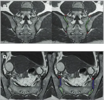
FIGurE 1. Normal SIJs anatomy on MRI. A and B) coronal-oblique T1W MR images, where A is more anterior and B is more posterior. C-E) axial oblique T1W MR images, corresponding to C) superior, D) middle and E) inferior levels of the SIJs. Red – joint capsule; green – ilio-sacral ligaments (posterior syndesmotic component of the SIJs); blue – anterior-inferior cartilaginous component of the SIJs.
A b
c
E
“definite radiographic sacroiliitis according to mNY cri-teria” and/or as active sacroiliitis on MRI (“positive” MRI)10. It important to note that the ASAS classification
criteria are not diagnostic criteria. The diagnosis of axSpA can only be made by the rheumatologist based on the combination of clinical, laboratory and imaging features. None of the above classification criteria are 100% sensitive or specific.
2.1. Radiographs
Radiographs can demonstrate structural changes, which are: erosions, subchondral sclerosis, articular space width irregularities and bone ankylosis. The 1984 mNY criteria for AS have represented the main-stay in the diagnosis of sacroiliitis for a long time12.
They comprise both three clinical criteria and a fourth, which is the radiographic criterion of unilateral sacroilii tis grade 3 or 4, or bilateral sacroiliitis grade 2 or higher4. This radiographic criterion is based on the
1966 New York 5-point-grading system of structural SIJs damage.
The radiographic evaluation of SIJs has a significant limitation resulting from the complexity of the SIJs anatomy and its double obliquity, the anterior tilt of the sacrum and the lateral tilt of the joint space - all together summated on an anteroposterior or posteroanterior im-age. In addition, significant intra- and inter-observer variability and low agreement among readers have been reported, despite a number of attempts to improve stan-dardisation13. Furthermore, a negative pelvic radiograph
cannot exclude the diagnosis of SpA due to its low sen-sitivity for early disease, mainly due to its inability to visualize BME (non-radiographic axial SpA), thus con-tributing to a delay in diagnosis14-15. Nonetheless,
ra-diographs are readily available, inexpensive, may ex-clude other pathology, and if positive, as in advanced sacroiliitis, they can be very useful. Therefore, accord-ing to EULAR (European League Against Rheumatism) and the European Society of Musculoskeletal Radiolo-gy (ESSR), radiography (anteroposterior view of the pelvis) is still considered the initial imaging modality in SpA and a useful baseline imaging technique to docu-ment progression of the structural changes3,16.
Addi-tional radiographic views (including oblique and Fer-guson views) do not improve sensitivity over the standard anteroposterior view of the pelvis3,16.
2.2. MRI
MRI allows direct visualization of the joint anatomy and abnormalities of the cartilage, capsule, synovium,
sub-chondral bone and ligaments. ASAS and ESSR both state that MRI is the most sensitive imaging modality to de-tect early sacroiliitis3,17-20and is the technique of choice
for the detection of early/active lesions in the SIJs, par-ticularly in cases of negative radiographs in a patient with suspected SpA (non-radiographic axial SpA)9,21,22. It
should be noted that some patients with negative Xrays and without active sacroiliitis on MRI can still have a di-agnosis of axSpA in the presence of the adequate clini-cal-laboratory features (e.g. a patient with inflammato-ry back pain, arthritis, uveitis, HLA-B27 positivity and elevated CRP). This possibility is also reflected in the clinical arm of the ASAS classification criteria. MRI over-comes the limitations related to superposition of struc-tures on radiographs and easily depicts periarticular BME (increasing agreement amongst rea ders) and improves follow-up by allowing monitoring of BME, assessment of response to therapy from an imaging perspective and depicting associated structural changes3,23.
MRI protocols of the SIJs are well established in the literature3,4and should be strictly followed. Even
though they are beyond the scope of this article, a brief mention should be made to the use of contrast (gadolinium). Adequate water-sensitive-sequences (fat-suppressed proton-density-weighted(W), fat-sup-pressed T2W and/or short tau inversion recovery (STIR) sequences) are used in reference centers, and, in most cases, gadolinium offers no additional benefit both in adults and in children, which is in line with the 2015 evidence-based EULAR recommendations and the 2016 update by ASAS MRI group16,24.
In children, gadolinium use has been more debated, especially given concerns about nephrotoxicity and in-tracranial gadolinium deposition25. In addition, one
needs to realize that the SIJs are usually not affected in isolation in SpA in both age groups (even more rarely in children), and the remaining locations would prob-ably require contrast (cumulative effect). A 2018 study with 99 MRI studies in patients <21 years for suspect-ed sacroiliitis showsuspect-ed abnormal enhancement in 5% of cases, but all of these had other features of active sacroili-itis that were depicted on water-sensitive sequences, and contrast did not identify additional ca ses26.
In selected cases, when joint effusion is the only finding, gadolinium may help to confirm the presence of synovitis - but interestingly, authors that state that contrast is essential to identify synovitis, do not report effusions27,28. Others report that all cases with synovial
enhancement have effusions, also depicted on water-sensitive sequences29,30. A thin rim of synovial
en-hancement may be normal in children, but this is dif-ficult to prove and more research is needed26. In adults,
synovitis alone is not sufficient to constitute a “positive MRI” for SpA-related sacroiliitis, whereas BME is well depicted on water-sensitive sequences17.
2.3. Is there any room for CT?
Computed Tomography (CT) allows for direct visual-ization of structural changes with a higher spatial res-olution than radiographs and MRI. CT has greater sen-sitivity and entails less inter-observer variability compared to radiographs, which explain why CT is useful in revealing subtle lesions and/or assessing inci-dental lesions on the radiographs23,31. In addition, CT
better depicts the osseous anatomy and is especially helpful in children and adolescents where normal os-seous SIJs structures vary considerably.
However, its inability to detect active inflammatory lesions and its higher levels of radiation exposure, par-ticularly in this young population, dissuades its rou-tine use. CT is performed mainly in equivocal cases, to either better depict small erosions and bone bridging, or to explore differential diagnosis. New emerging tech-niques such as low radiation CT (dual-energy) may have a role in the near-future, corroborated by the emerging role of structural changes, particularly, ero-sions, in the evaluation of SpA23.
3. lookInG For A “posItIvE” mrI study For spA-rElAtEd sAcroIlIItIs
According to the ASAS criteria, labelling and MRI as
suggestive of SpA in an adult is based on the evaluation of active inflammatory (“sacroiliitis”) and structural postinflammatory changes (Table I)4. A “positive” MRI
is defined by the clear presence of BME on MRI in sub-chondral bone; structural damage lesions seen on MRI may contribute to a decision by the observer that in-flammatory lesions are genuinely due to SpA but are not required to meet the definition. A limitation re-garding the definition of a positive MRI in axSpA is the age range of 18 years to 46 years of enrolled study sub-jects. Care should be taken in the extrapolation of these criteria to individuals outside this age range due to lack of data23.
3.1. Active Inflammatory Lesions on MRI
Active inflammatory findings of the SIJs are (Table I, Figure 2): BME/osteitis (primary criterion) and enthe-sitis, capsulitis and synovitis (secondary criteria)4,24.
BME, enthesitis and capsulitis are observed on water-sensitive and contrast-enhanced T1W sequences whilst synovitis may be seen on post-contrast images only; fat suppression increases their visibility. These active changes precede erosions in the initial, non-radio-graphic phase of SpA, and allow early diagnosis and treatment. Treatment with biologic agents will lead to a reduction or disappearance of BME in the majority of patients, representing a decrease in inflammatory in-filtrates, with fatty replacement and/or sclerotization remaining a sign of past inflammation.
BME (Figure 2A-B, 2F, 3B and 3F) is defined by in-creased signal intensity in the bone marrow on
water-*Primary criterion; **Secondary criteria
Abbreviations: BME: Bone marrow edema; MRI: Magnetic Resonance Imaging; SIJs: Sacroiliac Joints; STIR: Short tau inversion recovery; PDW: proton-density-weighted; FS=fat-suppressed
tAblE I. typEs oF mrI lEsIons In thE sIJs AccordInG to thE AsAs crItErIA Types of MR lesions in the SIJs
Active inflammatory lesions Chronic post-inflammatory (structural) lesions
BME/osteitis* Subchondral sclerosis
Capsulitis** Erosions
Synovitis** Backfill
Enthesitis** Fat metaplasia
Bone bridges/ankylosis
• Active inflammatory lesions are better seen on • Chronic inflammatory lesions are usually better seen fluid-sensitive (STIR, FS T2W, FS PDW) and on T1W sequences
contrast-enhanced FS T1 sequences.
• Synovitis, is the exception, which is only depicted on contrast-enhanced FS T1 sequences.
A b c d F h E G
I J
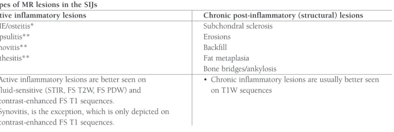
FIGurE 2. Inflammatory lesions on MRI as defined by ASAS criteria. 59-year-old male, ankylosing spondylitis: A) axial oblique fat-supressed (FS) T2W, B) coronal oblique FS T2W, C) axial oblique T1W and D) coronal oblique T1W MR images show bilateral subchondral BME, more extensive on the right SIJ (arrows in A and B). Capsulitis is also seen on the right (red asterisk in A). Note also concomitant structural changes on T1W images - subchondral sclerosis (red arrowheads in C and D) and erosions (white asterisks in D). 41-year-old male, ankylosing spondylitis: E) coronal oblique FS contrast-enhanced T1W MR image shows thickening of the right capsule with enhancement due to capsulitis (arrowhead), bilateral osteitis (arrows) as well as joint synovitis, more on the left side (asterisk). 39-year-old, female, probable psoriatic arthritis: F) axial oblique FS STIR and G) axial oblique FS T1W contrast-enhanced MR images show subtle bilateral joint effusion on the STIR image (double white arrows in F) that corresponds to joint synovitis after contrast (red arrows in G). Note also the coexisting subtle BME (asterisks in F), capsulitis (red arrowheads in G and F), enthesitis (white arrowheads in G and F) and anterior subchondral sclerosis, more on the left SIJ (white asterisks in G). 28-year-old male, Yersinia Reactive Arthritis: H and I) axial oblique FS T1W MR images before and after contrast show joint enhancement due to joint synovitis (arrows in I). Osteitis is seen bilaterally, more extensive on the sacral side (asterisks in I). J) coronal oblique T1W MR image shows erosions (white arrowheads) and fat metaplasia (asterisks). Do also note a bone bridge on the right (arrow).
sensitive and/or contrast-enhanced fat-suppressed T1 sequences in the SIJs (osteitis is the term which may be used for an equivalent-enhancing area on contrast-en-hanced T1W images (Figure 2E and 2I). According to the ASAS criteria (Table II), the presence of BME/os-teitis defines ‘active sacroiliitis’ on MRI when it is lo-cated in a typical subchondral/periarticular area and is sufficiently evident - if there is one BME lesion only, it should be present in at least two consecutive slices, if there is more than one signal abnormality on a single slice, one slice may be enough3,17. The more intense the
signal, the more likely it is to reflect active inflamma-tion32. BME is a MRI feature with moderate sensitivity
(65%) and specificity (74%) for diagnosis of SpA in
adults patients with inflammatory back pain33.
Speci-ficity increases if concomitant capsulitis, enthesitis, ero-sions or bone ankylosis are present. To avoid overcall-ing sacroiliitis, caution is advised when BME is scarce and not semilunar-shaped, and when there are no sec-ondary findings to support the diagnosis34. Also, BME
in the superior anterior part is generally related to me-chanical overload, not SpA-related inflammation (de-spite the same clinical criterion of low back pain). Fur-thermore, in the absence of additional imaging features of SpA in the SIJs (enthesitis, synovitis, capsulitis) or spine (syndesmophytes), two small lesions measuring <1cm are not sufficient to suggest axSpA, particularly when located in the proximal or distal margins of the
Abbreviations: BME: Bone marrow edema; MRI: Magnetic Resonance Imaging
tAblE II. lEArnInG poInts: “posItIvE” mrI crItErIA For sAcroIlIItIs AccordInG to thE AsAs crItErIA Learning Points - “Positive” MRI Criteria for Sacroiliitis are defined by:
• BME clearly present (in 2 consecutive slices in the same location or at least two locations in the same slice) and,
• BME in a subchondral/periarticular location.
• BME may be associated with other secondary active changes • BME may be associated with other structural changes
tAblE III. lEArnInG poInts: ActIvE InFlAmmAtory lEsIons In sAcroIlIItIs Learning Points - Active inflammatory lesions in sacroiliitis:
• Periarticular BME, even as a sole finding, is a prerequisite for a “positive” MRI for sacroiliitis in an adult.
• The sole presence of synovitis, capsulitis and/or enthesitis (secondary criteria), without concomitant BME (primary criterion) is compatible but not sufficient for the definition of active sacroiliitis on MRI in an adult.
SIJs14. The former is likely to represent overloadrela
-ted lesions, whereas the latter may represent enthe-sopathy or MRI artefacts.
Learning Point
• Periarticular BME, even as a sole fin ding, is a pre-requisite for a “positive” MRI for sacroilii tis in an adult. Other active inflammatory lesions (capsulitis, enthesi-tis and synovienthesi-tis) are suggestive of sacroillienthesi-tis, provi ded that concomitant subchondral BME is present (Table III)3,4,17. Capsulitis (Figure 2A, 2E-G and 5E) according
to 2009 ASAS definition, is defined by thickening of the SIJs capsule with signal hyperintensity on water-sensi-tive and on contrast-enhanced fat-suppressed T1 se-quences3. It involves the anterior and/or posterior
cap-sule. However, since there is no capsule or synovium in the proximal two thirds of the joint (anteriorly, the SIJs capsule gradually continues into the periosteum of the iliac and sacral bones and thus corresponds to an enthesis) periarticular inflammation in this region re presents enthesitis (Figure 2FG and 5E) which is cha -racterized by signal hyperintensity on water-sensitive and on contrast-enhanced fat-suppressed T1 images at ligaments and/or entheses (where tendons attach to bone)9. A common site to look for enthesitis is the
retroarticular space (interosseous ligaments). Enthesi-tis may present as an increased signal both within the fibrous part of the enthesis as well as BME in the area of the enthesis anchoring in the bone. Synovitis (Fi -gure 2E, 2G, 2I, 5B and 5E) is reflected by hyperin-tensity on contrast-enhanced fat-supressed T1-weight-ed images in the SIJs. Contrast is necessary for depicting synovitis, because water-sensitive sequences do not differentiate between synovitis and physiologic joint effusion, as mentioned above. Synovitis on MRI as a single feature (without BME) is a rare finding3,17
and indicates a different pathology than SpA. The name “synovitis” may be a misnomer since there is only syno
-vium in the lower part of the cartilaginous component of the SIJs.
3.2. Chronic (structural) lesions on MRI
Chronic (structural) lesions are believed to reflect pre-vious sacroiliitis. They include erosions, subchondral sclerosis, periarticular fat metaplasia, fat deposition in the intra-articular space (backfill) and ankylosis (Table I, Figure 3)4,24. These structural changes increasingly
gain importance for diagnosis and follow-up. Howev-er, they are likely to reflect a postinflammatory stage and, while having very high specificity35, they do not
suffice for the definition of a positive MRI examination for sacroiliitis.
Erosions (Figure 2D, 2J and 3C-H), probably the most important structural changes, are defined as a dis-continuity or blurring of either the cortical sacral or iliac bones, which appears with low signal intensity on both T1W and STIR sequences, together with loss of the bright signal from adjacent marrow on T1 se-quences32. Additional T2 gradient-echo or fat
sup-pressed T1W sequences (without contrast) can help to detect erosions. Erosion may appear as either single and localized, or multiple and contiguous. Due to car-tilage thickness, erosions appear initially on the iliac side (thinner cartilage), developing later on the sacral side (thicker cartilage) of the cartilaginous part of the SIJs. They may also occur in the posterior syndesmot-ic part of the joint in the course of enthesitis. Initially erosions tend to be single and, with progression, be-come confluent and cause pseudowidening of the SIJs. They may be active (filled with inflamed tissue) and consequently present with concomitant BME36.
Ero-sions may be present on MRI when radiographs are normal or inconclusive. The presence of erosions indi-cates long-lasting disease and may reflect more severe disease with greater spinal inflammation32.
Integration of structural changes may enhance diagnos tic utility23. Of all the structural features,
A b c E F G h d
sion has by far the highest positive likelihood ratio for the diagnosis of SpA – the incorporation of erosions in the MRI evaluation of the SIJs has been showed to in-crease sensitivity from 67% to 81%, without changing specificity33. As such, they man play a new or additional
role in classification systems (MORPHO proposal cri-teria)19,23,37. However, a recent update by the ASAS MRI
working group concluded that the definition of a “po -sitive” MRI should continue to primarily depend on the imaging features of “active sacroiliitis”, and not in-clude erosions as a primary feature for the time being, as there is no consensus as to how erosions should be defined on MRI or how it should be classified24.
Subchondral sclerosis (Figure 2C-D, 2G, 2J, 3A and 3C-D) is represented by areas with blurry margins which have low signal intensity on all sequences and no enhancement after gadolinium. With disease
progres-sion, they tend to become wider, as opposed to os-teoarthritis (in which they are more well-defined and narrower)22. Sclerosis attributed to SpA should extend
at least 5mm from the articular space in the iliac side and/or at least 3mm in the sacral side17.
Subchondral fatty bone marrow replacement /fat metaplasia (Figure 2J, 3F and 3I-J) represents fat con-version in inflammatory, often periarticular, bone mar-row areas. It is characterized by increased signal on T1W sequences with signal loss on fat suppressed im-ages and no enhancement17. It is the only structural
change that is visible on MRI, but not on radiographs or CT. Fat metaplasia is a non-specific finding but, like sclerosis, may indicate previous inflammation/long-standing disease and resolution of inflammation (pri-or BME) with the development of fatty metaplasia in the same area and, later, the development of bone ankylosis.
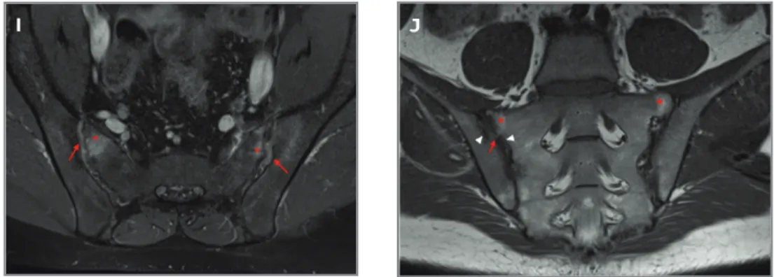
FIGurE 3. Chronic post-structural lesions on MRI as defined by ASAS criteria. 31-year-old female, ankylosing spondylitis: A) coronal oblique T1W and B) coronal oblique FS T2W MR images show subchondral sclerosis (arrows in A), with joint space narrowing, more on the left (asterisk in A). Note also still ongoing inflammation BME on left SIJ (asterisk in B). 18-year-old male, juvenile ankylosing spondylitis: C) axial CT image and D) axial oblique T1W MR image reveals bilateral erosions (arrowhead), more on the iliac side, with joint space
pseudowidening and surrounding subchondral sclerosis (asterisks). 57-year-old female, ankylosing spondylitis: E) coronal oblique T1W and F) coronal oblique STIR MR images depict multiple erosions, more marked on the right side (arrowheads in E) filled with inflamed T2-hyperintense tissue (arrowheads in F, active erosions), with surrounding fatty metaplasia and residual BME (asterisks in F). 28-year-old female, psoriatic arthritis: G) coronal oblique T1W and H) coronal oblique FS T1W MR images show erosions in the right SIJ (asterisks in G and H) and associated backfilling (arrows in
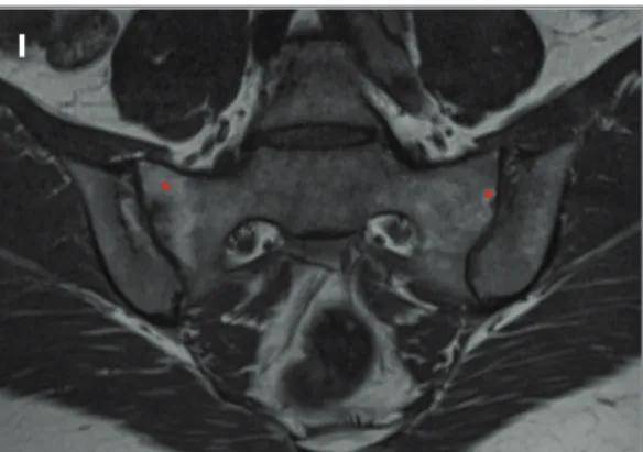
G). Erosions are nicely depicted on FS T1 MR images. Another patient, suspected for ankylosing spondylitis: I) coronal oblique T1W MR image shows periarticular fat metaplasia (asterisks). 54-year-old male, ankylosing spondylitis: J) coronal oblique T1W MR image shows bilateral joint space narrowing with bone bridging/partial ankylosis (red asterisks), surrounded by fat metaplasia (white asterisks). K) Coronal oblique T1W MR image shows almost complete bone ankylosis of the SIJs (asterisks).
I
I J
Backfill (Figure 3G) represents the presence of fat (high-signal on T1W sequences) within an erosion or erosive cavitation at the articular surface38. It may reflect
resolution of inflammation and tissue repair at sites of erosions, and is thought to be a key intermediary in the development of bone ankylosis38.
Bony ankylosis (Figure 3J-K) is characterized by confluent high T1 signal intensity across the SIJs space, with obliteration of the articular cortical margin35, and
represents the end-stage disease. It starts from single bone bridges which may progress to partial or finally a complete ankylosis of the joint35.
Learning point:
• Despite increasing importance of structural changes, particularly erosions, to date, the sole pres-ence of structural changes, without concomitant BME, is not sufficient for the definition of active sacroiliitis on MRI23-24. (Table IV).
3.3. How about MRI criteria in children?
Classification of the pediatric population remains a challenge. Unlike adults, the diagnostic value of chro -nic back pain or inflammatory back pain criterion, which in adults is a base for referring for MRI, is not so evident in children. MRI assessment of sacroiliitis in children is not well studied. Many different classifica-tion schemes have been proposed for children, but none includes imaging as a criterion4,39-41. Recent stu
-dies have postulated the usefulness of MRI in JSpA. Furthermore, adult imaging criteria (radiographic mNY and ASAS MRI criteria) have been empirically applied to the pediatric population, with children increasingly being referred for MRI. However, the adult ASAS defi-nition for sacroiliitis has a low sensitivity in children and there is still a lack for a clear definition for “posi-tive” MRI for sacroiliitis in this age group. In addition to findings seen in adults, some reported the impor-tance of the single bone marrow lesion as diagnostic for JSpA42, and others found that synovitis alone may
be specific without the need for BME in JSpA27,43. Some
also paid attention to the normal ossification process in children where BME could be seen as a normal fin -ding44- the high prevalence of ossified nuclei in the
joint space in children and adolescents could be the cause of the observed BME44. As previously mentioned,
the use of contrast for diagnosing sacroiliitis in chil-dren with JSpA is questionable and should not be ad-ministered on a regular basis26. A recent study showed
that a children-specific definition of “positive” MRI for sacroiliitis including BME visible on one slice only, syno vitis and/or capsulitis, may improve diagnostic utility and increase relevance of MRI in rheumatology guidelines in a near future45.
Learning Point:
• The adult ASAS definition for a positive MRI needs
some adjustments for children, including small BME
le-Abbreviations: BME: Bone marrow edema; MRI: Magnetic Resonance Imaging
Abbreviations: BME: Bone marrow edema; MRI: Magnetic Resonance Imaging; Juvenile SpA: JSpA
tAblE Iv. lEArnInG poInts - chronIc InFlAmmAtory lEsIons In sAcroIlIItIs: Learning Points - Active inflammatory lesions in sacroiliitis:
• Integration of the assessment of structural changes, particularly erosions, may enhance diagnostic utility of MRI; however, to date the ASAS guidelines did not add any structural change to the definition of “positive” MRI
• As such, the sole presence of erosions, sclerosis, fat metaplasia, backfill and/or ankylosis, without concomitant BME, is not sufficient for the definition of active sacroiliitis on MRI in an adult.
tAblE v. lEArnInG poInts - mrI In JuvEnIlE spA Learning Points - Active inflammatory lesions in sacroiliitis:
• The adult ASAS definition for a positive MRI needs some adjustments for children, including small BME lesions that are only visible on one slice or synovitis /capsulitis may be sufficient for a “positive” MRI in children (but more studies are needed) • The use of contrast for diagnosing sacroiliitis in children with juvenile SpA is questionable, and should not be
sions that are only visible on one slice or synovitis /cap-sulitis may be sufficient for a “positive” MRI in children
(but more studies are needed).
4. how About non-spA – rElAtEd sAcroIllItIs on mrI?
MRI features in patients with SpA may fluctuate, high-lighting the limited sensitivity of MRI for diagnostic purposes if it would be used as the only evaluation tool. Studies show that MRI is moderately sensitive (50--95%) and specific (47-90%) for the diagnosis of SpA in adults in the adequate clinical setting12,19,23,46. If BME
is used as a sole MRI criterion and the potential incre-mental contribution of structural lesions are not con-sidered, the sensitivity is lower (35-42%)47,48. This
points out the increased importance of looking for structural changes along with the clear presence of BME, and the need of clinical contextual interpretation in order to make a correct diagnosis of “sacroiliitis” in SpA. Age <45, male sex, HLAB27 positivity are among other parameters that will increase specificity of peri-articularly located BME towards SpA. If the contextu-al interpretation of MRI is not suggestive of SpA, oth-er diffoth-erential diagnosis come into play.
Recent studies estimated that 23-33% of patients re-ferred for MRI due to clinical suspicion of SpA had al-ternative non-inflammatory conditions, and that 41%-50% had normal SIJs on MRI33. Jans et al showed that
non-inflammatory disease was indeed more common than sacroiliitis on MRI of the SIJs in patients with in-flammatory type back pain33. Klang et al studied a
popu lation of patients under 40 years with low-back pain on lumbosacral CT and found some degree of SIJ changes in 14% of this population, and unequivocal sacroiliitis in 3,3%49. Slobodin et al found sacroiliitis in
3.7% of patients on abdominal CTs performed for other indications in a population aged 18-55 years--old50. As such, interpretation of MRI findings in daily
practice is critically dependent on the clinical context. Learning Point:
• Even in patients with inflammatory low-back pain, it is important to consider non-inflammatory disease because chronic or inflammatory low back pain has low specificity for axSpA.
conclusIon
Imaging modalities, particularly MRI, play a key role in
the diagnosis and follow-up of patients with axSpA. In-terpretation of MRIs of the SIJs in adult patients sus-pected to have axSpA is based on the presence of ac-tive inflammatory (BME/osteitis, capsulitis, synovitis, enthesitis) and structural postinflammatory lesions (erosions, sclerosis, fat metaplasia, backfill and anky-losis). Active lesions are particularly useful for the diagno sis and assessment of potential ongoing infla -mmation. Structural lesions gain importance for both diagno sis and follow-up. These lesions were reviewed in this article, in addition to a short description of diffe -rent imaging modalities and normal SIJs anatomy.
corrEspondEncE to
Diana Afonso
Musculoskeletal Imaging Unit, Radiology Department, Hospital da Luz, Grupo Luz Saúde
Lisbon, Portugal
E-mail: p.diana.a@gmail.com
rEFErEncEs
1. Jans L, Van Praet L, Elewaut D, et al. MRI of the SI joints com-monly shows non-inflammatory disease in patients clinically suspected of sacroiliitis. Eur J Radiol 2014; 83(1):179-184 2. Navallas M, Ares J, Beltrán B, Lisbona MP, Maymó J, Solano A.
Sacroiliitis associated with axial spondyloarthropathy: new con-cepts and latest trends. Radiographics 2013; 33(4):933–956. doi:10.1148/rg.334125025.
3. Sudoł-Szopi ska I, Jurik AG, Eshed I, et al. Recommendations of the ESSR Arthritis Subcommittee for the Use of Magnetic Res-onance Imaging in Musculoskeletal Rheumatic Diseases. Semin Musculoskelet Radiol 2015;19(4):396-411. doi: 10.1055/s-0035-1564696.
4. Sieper J, Rudwaleit M, Baraliakos X, et al. The assessment of Spondylo Artritis international Society (ASAS) handbook: a guide to assess spondyloarthritis. Ann Rheum Dis 2009; 68 (Suppl 2):ii1–44.
5. Puhakka KB, Jurik AG, Egund N, et al. Imaging of sacroiliitis in early seronegative spondylarthropathy. Assessment of abnor-malities by MR in comparison with radiography and CT. Acta Radiol 2003;44: 218–229.
6. Lambert R, Dhillon S, Jaremko JL. Advanced imaging of the axial skeleton in spondylarthropathy: techniques, interpreta-tion, and utility. Semin Musculoskelet Radiol 2012;16:389–400. 7. Marzo-Ortega H, McGonagle D, O'Connor P, et al. Baseline and 1-year magnetic resonance imaging of the sacroiliac joint and lumbar spine in very early inflammatory back pain. Relationship between symptoms, HLA-B27 and disease extent and persis-tence. Ann Rheum Dis 2009;68:1721–1727.
8. Vleeming A, Schuenke MD, Masi AT, Carreiro JE, Danneels L, Willard FH. The sacroiliac joint: an overview of its anatomy, function and potential clinical implications. J Anat 2012;221(6): 537–567.
9. Puhakka KB, Melsen F, Jurik AG, Boel LW, Vesterby A, Egund N. MR imaging of the normal sacroiliac joint with correlation to his-tology. Skeletal Radiol 2004;33(1):15–28
10. Rudwaleit M, VanderHeijde D, Landewé R, et al. The develop-ment of Assessdevelop-ment of SpondyloArthritis international Society
classification criteria for axial spondyloarthritis (part II): vali-dation and final selection. Ann Rheum Dis 2009; 68: 777–83. 11. Sudoł-Szopi ska I, Urbanik A. Diagnostic imaging of sacroiliac joint and the spine in the course of spondyloarthropathies. Pol J Radiol 2013; 78(2): 43–49.
12. Sudol-Szopinska I, Kwiatkowska B, WlodkowskaKorytkowska M, MatuszewsWlodkowskaKorytkowska G, GrochowsWlodkowskaKorytkowska E. Diagnostics of Sacroi -liitis According to ASAS Criteria: A Comparative Evaluation of Conventional Radiographs and MRI in Patients with a Clinical Suspicion of Spondyloarthropathy. Preliminary Results. Pol J Radiol 2015; 19;80:266-76. doi: 10.12659/PJR.892529. 13. Van den Berg R, Lenczner G, Feydy A, et al. Agreement between
clinical practice and trained central reading in reading of sacroil-iac joints on plain pelvic radiographs: results from the DESIR cohort. Arthritis Rheumatol 2014; 66:2403–11.
14. Schueller-Weidekamm C, Mascarenhas VV, Sudol-Szopinska I, et al. Imaging and interpretation of axial spondylarthritis: the ra-diologist's perspective—consensus of the Arthritis Subcom-mittee of the ESSR. Semin Musculoskelet Radiol 2014;18: 265–79. doi:10.1055/s-0034-1375567.
15. Heuft-Dorenbosch L, Weijers R, Landewe R, et al. Magnetic res-onance imaging changes of sacroiliac joints in patients with re-cent-onset inflammatory back pain: inter-reader reliability and prevalence of abnormalities. Arthritis Res Ther 2006; 8: R11. 16. Mandl P, Navarro-Compán V, Terslev L, et al. European League
Against Rheumatism (EULAR). EULAR recommendations for the use of imaging in the diagnosis and management of spondy-loarthritis in clinical practice. Ann Rheum Dis. 2015 Jul;74(7):1327-39. doi: 10.1136/annrheumdis-2014-206971. 17. Rudwaleit M, Jurik AG, Hermann KG, et al. Defining active sacroiliitis on magnetic resonance imaging (MRI) for classifica-tion of axial spondyloarthritis: a consensual approach by the ASAS/OMERACT MRI group. Ann Rheum Dis 2009;68: 1520–1527.
18. Lacout A, Rousselin B, Pelage J-P. CT and MRI of spine and sacroiliac involvement in spondyloartropathy. Am J Roentgenol 2008; 191: 1016–1023.
19. Weber U, Lambert RG, Ostergaard M, et al. The diagnostic uti -lity of magnetic resonance imaging in spondyloarthritis. An in-ternational multricenter evaluation of one hundred eighty se ven subjects. Arthritis Rheum 2010; 62: 3048–3058.
20. Heuft-Dorenbosch L, Landewe R, Weijers R, et al. Combining information obtained from magnetic resonance imaging and conventional radiographs to detect sacroiliitis in patients with recent onset inflammatory back pain. Ann Rheum Dis 2006; 65: 804–808.
21. Montandon C, Costa MAB, Carvalho TN, et al. Sacroiliitis: ima -ging evaluation, Radiolog Brasil 2007; vol. 40 (53-60). 22. Tuite MJ. Sacroiliac Joint Imaging. Semin Musculoskelet Radiol
2008; 12: 72–82.
23. Lukas C, Cyteval C, Dougados M, Weber U. MRI for diagnosis of axial spondyloarthritis: major advance with critical limita-tions 'Not everything that glisters is gold (standard)'. RMD Open 2018 ;Jan 12;4(1):e000586. doi:10.1136/rmdopen-2017-000586.
24. Lambert RG, Bakker PA, van der Heijde, D et al. Defining active sacroiliitis on MRI for classification of axial spondyloarthritis: update by the ASAS MRI working group. Ann Rheum Dis 2016;75(11):1958-1963. doi:10.1136/annrheumdis-2015-208642.
25. McDonald RJ, McDonald JS, Kallmes DF, et al. Intracranial
gadolinium deposition after contrast-enhanced MR imaging. Radiology 2015;275:772–782. doi: 10.1148/radiol.15150025. 26. Orr KE, Andronikou S, Bramham MJ, Holjar-Erlic I, Menegot-to F, Ramanan AV. Magnetic resonance imaging of sacroiliitis in children: frequency of findings and interobserver reliability. Pe-diatr Radiol 2018;48(11):1621-1628.
27. Lin Clara, MacKenzie John D, Courtier Jesse L, Gu Jeffrey T, Milojevic Diana. Magnetic resonance imaging findings in juve-nile spondyloarthropathy and effects of treatment observed on subsequent imaging. Pediatric Rheumatology 2014;12(1):25. doi: 10.1186/1546-0096-12-25.
28. Sheybani EF, Khanna G, White AJ, et al. Imaging of juvenile id-iopathic arthritis: a multimodality approach. Radiographics 2013;33:1253–1273. doi: 10.1148/rg.335125178.
29. Herregods N, Dehoorne J, Joos R, et al. Diagnostic value of MRI features of sacroiliitis in juvenile spondyloarthritis. Clin Radiol 2015;70:1428–1438. doi:10.1016/j.crad.2015.09.003. 30. Weiss PF, Xiao R, Biko DM, et al. Detection of inflammatory
sacroiliitis in children with magnetic resonance imaging: is gadolinium contrast enhancement necessary? Arthritis Rheuma-tol 2015;67:2250–2256. doi: 10.1002/art.39159.
31. Rueda JC, Arias-Correal S, Vasquez AY, et al. Interobserver Agree-ment in Magnetic Resonance of the Sacroiliac Joints in Patients with Spondyloarthritis. Int J Rheumatol. 2017;:3143069. 32. Maksymowych WP, Wichuk S, Dougados M, et al. MRI evidence
of structural changes in the sacroiliac joints of patients with non-radiographic axial spondyloarthritis even in the absence of MRI inflammation. Arthritis Res Ther. 2017;6;19(1):126. doi:10.1186/s13075-017-1342-9.
33. Jans L, Coeman L, Van Praet L, et al. How sensitive and specif-ic are MRI features of sacroiliitis for diagnosis of spondy-loarthritis in patients with inflammatory back pain? JBR-BTR 2014;Jul-Aug;97(4):202-205.
34. Bennett AM, McGonagle D, O’Connor P, et al. Severity of base-line magnetic resonance imaging evident sacroiliitis and HLA B27 status in early inflammatory back pain predict radio-graphically evident ankylosing spondylitis at eight years. Arthri-tis Rheum 2008;58: 3413–3418
35. Jans L, Egund N, Eshed I, Sudoł-Szopi ska I, Jurik A. Sacroili-itis in Axial SpondyloarthrSacroili-itis: Assessing Morphology and Ac-tivity. Seminars in Musculoskeletal Radiology 2018; 22(02), 180–188. doi.org/10.1055/s-0038-1639470.
36. Gong Y, Zheng N, Chen SB, et al. Ten years’ experience with needle biopsy in the early diagnosis of sacroiliitis. Arthritis Rheum 2012; 64(5):1399–1406.
37. Weber U, Maksymowych WP. Sensitivity and specificity of mag-netic resonance imaging for axial spondyloarthritis. Am J Med Sci 2011;341(04):272–277.
38. Maksymowych WP, Wichuk S, Chiowchanwisawakit P, Lambert RG, Pedersen SJ. Fat metaplasia and backfill are key intermedi-aries in the development of sacroiliac joint ankylosis in patients with ankylosing spondylitis. Arthritis Rheumatol 2014; Nov;66(11):2958-67. doi: 10.1002/art.38792.
39. Tse SM, Laxer RM. New advances in juvenile spondyloarthritis. Nat Rev Rheumatol 2012;Apr 10;8(5):269-79. doi: 10.1038/nr-rheum.2012.37.
40. Petty RE, Southwood TR, Manners P, et al. International League of Association for Rheumatology Classification of juvenile idio-pathic arthritis: second revision. Edmonton. 2001. J Rheuma-tol 2004;31:390–392.
classification criteria for juvenile spondyloarthropathies. Pedi-atr Rheumatol Online J 2014; 12 Suppl 1:45. doi:10.1186/ 1546-0096-12-S1-P45.
42. Herregods N, Dehoorne J, Jaremko J, et al. Diagnostic Value of MRI of the Sacroiliac Joints in Juvenile Spondyloarthritis. Jour-nal of the Belgian Society of Radiology 2016;100(1):95.DOI: http://doi.org/10.5334/jbr-btr.1198.
43. Hemke R, Kuijpers TW, van den Berg JM, et al. The diagnostic accuracy of unenhanced MRI in the assessment of joint abnor-malities in juvenile idiopathic arthritis. Eur Radiol 2013; 23(7):1998–2004.
44. Zejden, Anna and Anne Grethe Jurik. Anatomy of the sacro iliac joints in children and adolescents by computed tomography. Pediatric rheumatology online journal vol. 15,1 82. 25 Nov. 2017. doi:10.1186/s12969-017-0210-0.
45. Herregods N, Dehoorne J, Van den Bosch F, et al. ASAS defini-tion for sacroiliitis on MRI in SpA: applicable to children? Pe-diatr Rheumatol Online J. 2017; Apr 11;15(1):24. doi: 10.1186/s12969-017-0159-z.
46. Blum U, Buitrago-Tellez C, Mundinger A, et al. Magnetic reso-nance imaging (MRI) for detection of active sacroiliitis – a prospective study compering conventional radiography, scintig-raphy, and contrast-enhanced MRI. J Rheumatol 1996;23: 2107–2115.
47. Van den Berg R, Lenczner G, Thevenin F, et al. Classification of axial SpA based on positive imaging (radiographs and/or MRI of the sacroiliac joints) by local rheumatologists or radiologists versus central trained readers in the DESIR cohort. Ann Rheum Dis 2015;74:2016–2021.
48. Van den Berg R, de Hooge M, van Gaalen F, et al. Percentage of patients with spondyloarthritis in patients referred because of chronic back pain and performance of classification criteria: ex-perience from the Spondyloarthritis Caught Early (SPACE) co-hort. Rheumatology 2013;52:1492–1499.
49. Klang E, Lidar M, Lidar Z, Aharoni D, Eshed I. Prevalence and awareness of sacroiliac joint alterations on lumbar spine CT in low back pain patients younger than 40 years. Acta Radiol 2017;Apr;58(4):449-455. doi:10.1177/0284185116656490. 50. Slobodin G, Croitoru S, Starikov N, et al. Incidental computed tomography sacroiliitis: clinical significance and inappropri-ateness of the New York radiological grading criteria for the di-agnosis. Clin Rheumatol 2012; 31:425–428.